Correlation between Diastolic Function and Maximal Exercise
Capacity on Exercise Test
Maria Estefânia Bosco Otto, Márcio Mendes Pereira, Adenalva Lima de Souza Beck, Maurício Milani
Instituto de Cardiologia do Distrito Federal, Brasília, DF - BrazilMailing address: Maria Estefânia Otto •
AOS 02 Bloco B apto 604 - Octogonal - 70660-022 - Brasília, DF - Brazil E–mail: [email protected], [email protected] Manuscript received October 08, 2009; revised manuscript received November 16, 2009; accepted March 11, 2010.
Abstract
Background: Increased pulmonary capillary pressure (PCP) is one of the mechanisms of exercise intolerance. Assessment of the diastolic function by echocardiography (ECHO) enables estimation of PCP.
Objective: To identify variables that determine the exercise capacity in patients undergoing routine exercise test (ET), conventional ECHO, and tissue Doppler imaging (TD).
Methods: A total of 640 patients undergoing ET, ECHO, and TD were retrospectively studied. Patients with ejection fraction < 55% were excluded. Mitral annulus velocities by conventional Doppler imaging were obtained in early diastole (E) and late diastole (A), and TD of the mitral annulus measured early diastole (e’) and late diastole (a’) velocities. E/e’ > 10 was considered an estimate of increased PCP. Maximal exercise capacity was analyzed by the number of metabolic equivalents (MET). The patients were divided into two groups for analysis: MET<7 (n=48) and MET>7 (n=572). Morise score showed a population at low risk (60%) for coronary artery disease (CAD).
Results: The number of patients with E/e’ > 10 was significantly higher in the MET < 7 group in relation to the MET > 7 group (41.7% vs 9.4%, p=0.001), and so was the presence of any degree of diastolic dysfunction (76.6% vs 34.1% p=0.001). Using logistic regression analysis, age, female gender and A velocity (late diastole) were the independent variables related to a low exercise capacity (MET < 7).
Conclusion: Diastolic dysfunction as determined by ECHO, female gender, and age are associated with a lower exercise capacity in a population at low risk for CAD. (Arq Bras Cardiol 2011; 96(2): 107-113)
Keywords: Ventricular dysfunction; exercise test; exercise; diastole; echocardiography, tissue Doppler.
Introduction
Exercise capacity is influenced by innumerous factors, such as advanced age1, comorbidities (metabolic syndrome
and hypertension)2, obesity3 and previous cardiorespiratory
fitness1. Identification of other factors that modify the exercise
capacity is highly relevant, because its reversal can improve the quality of life of the patients and have an effect on survival, since exercise capacity is associated with prognosis both in healthy individuals4-6 and in patients with heart diseases7.
In an attempt to explain the cardiac mechanisms that decrease exercise capacity, ejection fraction did not show a significant correlation, perhaps for being dependent on the preload8. However, maximal exercise capacity, as well as the
symptoms triggered by exercise, are directly related to increased pulmonary capillary pressure and, therefore, to increased left ventricular filling pressures. Filling pressures, in turn, are directly related to the left ventricular diastolic function9,10.
Recent clinical studies observed a significant correlation between exercise capacity and diastolic function parameters, especially in patients with cardiovascular disease11.
Recent advances in tissue Doppler imaging improved the accuracy of echocardiography in the identification of abnormalities of left ventricular early-diastole12, including
the estimation of filling pressures13. Consequently, the use
of tissue Doppler imaging and of the analysis of diastolic function may be useful to explain the correlation between the presence of diastolic dysfunction and maximal exercise capacity on exercise test.
The objective of the present study was to analyze the exercise capacity and its correlation with diastolic function, as well as with the different clinical parameters in a population predominantly consisting of individuals at a low risk for coronary artery disease.
Methods
All individuals underwent exercise test and transthoracic echocardiography within an interval of up to 30 days14. Patients
with ejection fraction < 55% were not included in the analysis. Functional capacity was assessed by the peak metabolic equivalent (MET peak), indirectly obtained by formulas15,
according to the maximum slope and speed achieved in an incremental exercise treadmill test, with the ramp protocol adjusted to the individual.
Echocardiographic study was performed with two-dimensional images of the M mode and with pulsed Doppler using a echocardiographic equipment (Philips HDI 5000) with a 2 to 4 MHz multifrequency transducer. Mitral inflow velocities were recorded using pulsed Doppler in the apical 4-chamber view, with a sample volume of 5 mm at the tip of the mitral leaflets. Early diastolic velocity (E), late diastolic velocity (A) and deceleration time were measured; the E/A ratio was also calculated16. Using the apical 4-chamber view,
a 2-mm sample volume was placed in the junction of the LV wall with the septal mitral annulus in order to record tissue Doppler, deriving the tracing velocities during systole (S), early diastole (e’), and late diastole (a’)12. The E/e’ ratio was
calculated to estimate LV filling pressures, and values greater than ten (10) were considered increased filling pressures12. The
cut-off point for the E/e’ ratio was set at 10 based on data from Burgess et al’s study, in which E/e’ > 10 ratios ( with e’ of the septal annulus) were the optimal index for the identification of patients whose filling pressures increase with exercise, with sensitivity of 71% and specificity of 69%17.
The diastolic function was analyzed according to the protocol used in our laboratory, starting with the assessment of the E and A velocities of the mitral pulsed Doppler, and E/A ratio calculation. Next, the tissue Doppler velocities of the septal annulus (e’ and a’) are obtained, and the E/e’ ratio is calculated. Using the two-dimensional image, the left atrial (LA) volume is evaluated. When in doubt about the degree of diastolic dysfunction using the methods previously described, the pulmonary vein flow velocities or Valsalva maneuver are performed. Based on these parameters, the diastolic function is classified as: normal (E/A>0.8, e’>8 cm/s, normal LA volume; E/e’ ratio was not considered); diastolic dysfunction with abnormal relaxation pattern (E/A<0.8, e’<8 cm/s, variable LA volume, and variable E/e’); pseudonormal diastolic dysfunction (E/A>0.8, e’<8 cm/s, LA volume usually increased, and E/e’>15); and restrictive diastolic dysfunction (E/ A> 1.8, e’<8 cm/s, LA volume usually increased, and E/e’>15). This methodology is based on several classifications of analysis of the diastolic function12,18,19 and is relatively easy to use in the
daily practice of echocardiography laboratories.
For the statistical analysis, the individuals were divided into
two groups: MET<7 (n=48) and MET≥7 (n=572), according
to their exercise capacity.
Individuals with MET peak < 7 show low physical capacity, usually associated with functional limitations and symptoms, in addition to being exposed to a higher risk of cardiovascular events4-6,15.
The chi square test and Fisher test were used to analyze the association between the groups and the classificatory variables. The Wilcoxon test was used for comparison of the means between the two groups. The variables showing statistical significance in the univariate analysis were used in the adjustment
of the logistic regression model. P values < 0.05 were considered statistically significant. The Stata 8.0 and SAS software programs were used for processing and statistical analysis.
Results
The baseline clinical characteristics and main echocardiographic measurements are described in Tables 1 and 2, respectively.
Analysis of the diastolic function
Normal diastolic function was observed in 62% of the individuals; 30.4% showed diastolic dysfunction with relaxation abnormality; 6.1% showed pseudonormal diastolic dysfunction; 0.32%, restrictive diastolic dysfunction; and 1.1%, undetermined degree of diastolic dysfunction. Only 12.7% had increased filling pressures (as assumed by an E/e’ ratio >10). Of these 12.7%, 17 patients (2.6%) had normal diastolic function and normal exercise capacity (MET > 7).
Risk of CAD in the population
The probability of CAD was calculated based on the Morise score20. We found 60% of individuals with low probability,
39% with moderate, and 1% with high probability of CAD. Therefore, most of the patients had low probability of CAD.
Table 1 - Baseline clinical characteristics
Parameters Individuals studied (n=640)
Male gender 60%
Diabetes mellitus 7.8%
Hypertension 37.6%
Cigarette smoking 11.5%
Dyslipidemia 33.4%
Obesity 26.8%
Positive exercise test 11.8%
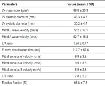
Table 2 - Baseline echocardiographic characteristics
Parameters Values (mean ± SD)
LV mass index (g/m2) 89.8 ± 25.3
LV diastolic diameter (mm) 48.3 ± 4.7 LV systolic diameter (mm) 30.2 ± 4.7 Mitral E wave velocity (cm/s) 72.2 ± 17.1 Mitral A wave velocity (cm/s) 62.7 ± 18.3
E/A ratio 1.24 ± 0.47
E wave deceleration time (ms) 212.7 ± 57.6 Mitral annulus e’ velocity (cm/s) 9.9 ± 3.9 Mitral annulus a’ velocity (cm/s) 9.9 ± 3.8 Mitral annulus S velocity (cm/s) 8.9 ± 2.8
E/e’ ratio 7.8 ± 2.9
Determinants of functional exercise capacity on exercise test
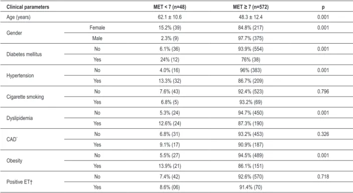
Univariate analysis of the exercise capacity in MET was carried out in relation to clinical and echocardiographic parameters. We observed that the clinical parameters that correlated with a low functional capacity (MET < 7) were female gender, age, presence of diabetes mellitus (DM), presence of hypertension, and obesity (Table 3). In relation to the echocardiographic parameters, we observed that the LV mass index, A wave velocity of the mitral flow velocity, E/A ratio of the mitral flow, e’ wave velocity of tissue Doppler of the mitral annulus, S wave velocity of tissue Doppler of the mitral annulus, and the E/e’ ratio were associated with MET < 7 (Table 4).
The clinical and echocardiographic parameters which were independently associated with a low exercise capacity in the adjusted logistic regression model were age, female gender, and A wave velocity of the mitral flow (Table 5).
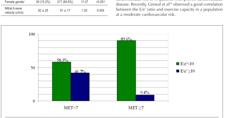
The comparative analysis of the MET < 7 and MET > 7 groups in relation to the presence of increased filling pressures (Graph 1) showed that 41.6% of the individuals with low functional capacity had a E/e’ ratio > 10, whereas only 9.4% of the individuals had a good functional capacity. The same was observed in relation to the presence of diastolic dysfunction: 76.6% of the patients with low functional capacity had some degree of diastolic dysfunction in comparison to 34.1% of the patients with a better functional capacity (Graph 2).
Discussion
The novel and important finding of the present study is that even in patients at low risk of CAD there is a significant correlation between diastolic function and exercise
capacity. Another significant finding corroborating other studies1,10,21 is that clinical parameters such as advanced
age and female gender are independent predictors of a low functional capacity.
Undoubtedly, the presence of diastolic dysfunction has a physiological ground on the decreased exercise capacity since, during exercise, the maximal cardiac output is dependent
Table 3 - Clinical parameters in the univariate analysis
Clinical parameters MET < 7 (n=48) MET ≥ 7 (n=572) p
Age (years) 62.1 ± 10.6 48.3 ± 12.4 0.001
Gender Female 15.2% (39) 84.8% (217) 0.001
Male 2.3% (9) 97.7% (375)
Diabetes mellitus No 6.1% (36) 93.9% (554) 0.001
Yes 24% (12) 76% (38)
Hypertension No 4.0% (16) 96% (383) 0.001
Yes 13.3% (32) 86.7% (209)
Cigarette smoking No 7.6% (43) 92.4% (523) 0.796
Yes 6.8% (5) 93.2% (69)
Dyslipidemia No 5.3% (24) 94.7% (450) 0.001
Yes 12.6% (24) 87.3% (190)
CAD* No 6.8% (31) 93.2% (453) 0.326
Yes 9.1% (17) 90.9% (187)
Obesity No 5.5% (27) 94.5% (489) 0.001
Yes 13.9% (21) 86.1% (151)
Positive ET† No 7.4% (42) 92.6% (570) 0.718
Yes 8.6% (06) 91.4% (70)
*CAD - coronary artery disease; †ET - exercise test.
Table 4 - Echocardiographic parameters in the univariate analysis
Echocardiographic parameters
MET < 7 (n=48)
MET ≥ 7
(n=572) p
LV mass index (g/m2) 101 ± 34 88 ± 25 0.011
LV diastolic diameter
(mm) 49 ± 6 48 ± 4 0.384 LV systolic diameter (mm) 32 ± 7 30 ± 4 0.081 Mitral E wave velocity
(cm/s) 73 ± 16 72 ± 17 0.757 Mitral A wave velocity
(cm/s) 82 ± 25 61 ± 17 <0.001
E/A ratio 0.96 ± 0.4 1.27 ± 0.5 <0.001
Deceleration time 217 ± 43 212 ± 59 0.330
Mitral annulus e’ velocity
(cm/s) 7.4 ± 2 10.2 ± 3 <0.001 Mitral annulus a’ velocity
(cm/s) 10.2 ± 3 9.8 ± 2 0.560 Mitral annulus S velocity
Table 5 - Clinical and echocardiographic determinants of low exercise capacity (logistic regression)
Independent parameters
MET < 7 (n=48)
MET ≥ 7
(n=592) OR P
Age (years) 62±11 48 ± 2 1.09 <0.001 Female gender 39 (15.2%) 217 (84.8%) 11.37 <0.001
Mitral A wave
velocity (cm/s) 82 ± 25 61 ± 17 1.03 0.004
Graph 1 - Relationship between increased left ventricular illing pressure and functional capacity.
Graph 2 - Relationship between left ventricular diastolic function and functional capacity. DF - diastolic function.
filling rates are achieved and become inadequate to supply the cardiac output required during exercise, with a subsequent increase in filling pressures and decrease in the maximal capacity22. The more abnormal the baseline diastolic function,
the lower the exercise capacity.
Several studies have demonstrated a good correlation between tissue Doppler of the mitral annulus and functional exercise capacity23,24, probably due to the association of the
E/e’ ratio with left ventricular filling pressures19,25. However,
most of these studies were conducted in populations at high cardiovascular risk or in patients with proven cardiovascular disease. Recently, Grewal et al10observed a good correlation
between the E/e’ ratio and exercise capacity in a population at a moderate cardiovascular risk.
Our study population was characterized as at a low cardiovascular risk; 62% of the individuals had a normal diastolic function, and 60% showed a low probability of CAD. Nonetheless, we could find correlations between diastole and exercise capacity.
In our protocol model, the number of patients with normal diastolic function was high, and the fact that the predominant diastolic dysfunction was relaxation abnormality (30% of the patients) may explain why the low functional capacity, but not the E/e’ ratio, is associated with increased A wave velocity in conventional mitral Doppler. A wave velocity increases in patients with diastolic dysfunction with relaxation abnormality and becomes a significant parameter in a population with 30% of the patients classified in this degree of diastolic dysfunction.
Firstenberg et al’s study26 showed absence of correlation
between E/e’ ratio and left ventricular filling pressures in individuals without cardiovascular diseases, and this corroborates the findings of the present protocol, in which most of the patients did not present with cardiovascular disease (60%). The physiological basis for this lack of correlation may be reinforced by recent evidences that suggest that the early diastolic velocity of the septal annulus (e’) is significantly influenced by the preload in normal ventricles, but this does not occur in ventricles with abnormal diastolic function, especially abnormalities in ventricular relaxation27.
Advanced age was another independent factor related to low functional capacity. Decrease in functional capacity is evidenced as from 50 years of age, and becomes more evident after 75 years of age in both genders28. In our study,
the mean age of the individuals in the MET < 7 group was 62.1 years, thus corroborating the findings previously described. Multiple factors are related to this decreased functional capacity, such as decrease in joint motion, muscle mass, strength, coordination, and endurance, as well as the presence of chronic diseases28,29.
Another clinical variable related to low functional capacity in our study was the female gender. The factors that explain this difference between genders are a reduced capacity of oxygen transportation, smaller muscle mass, higher percentage of fat, and lower cardiorespiratory fitness observed in women30.
The major limitations of this study are its retrospective design; the use of only some parameters in the analysis of the diastolic function on echocardiogram; indirect assessment of physical capacity by exercise test instead of cardiopulmonary exercise test; and the fact that the interval between tests was 30 days at most. We should also mention that laboratory data and pulmonary functional capacity data were not available, since innumerous individuals were being followed-up in other services. However, the population studied is at a low risk for CAD and is representative of patients who undergo frequent assessments of the cardiovascular risk, which makes the parameters found useful in the clinical practice.
Conclusion
Diastolic dysfunction as determined by echocardiography, female gender, and age are associated with a lower exercise capacity in a population at a low risk for CAD with preserved left ventricular function.
Potential Conflict of Interest
No potential conflict of interest relevant to this article was reported.
Sources of Funding
There were no external funding sources for this study.
Study Association
This study is not associated with any post-graduation program.
References
1. Woo JS, Derleth C, Stratton JR, Levy WC. The influence of age, gender, and training on exercise efficiency. J Am Coll Cardiol. 2006; 47 (5): 1049-57.
2. Wong CY, O’Moore-Sullivan T, Fang ZY, Haluska B, Leano R, Marwick TH. Myocardial and vascular dysfunction and exercise capacity in the metabolic syndrome. Am J Cardiol. 2005; 96 (12): 1686-91.
3. Shubair MM, Kodis J, McKelvie RS, Arthur HM, Sharma AM. Metabolic profile and exercise capacity outcomes: their relationship to overweight and obesity in a Canadian cardiac rehabilitation setting. J Cardiopulm Rehabil. 2004; 24 (6): 405-13.
4. Myers J, Prakash M, Froelicher V, Do D, Partington S, Atwood JE. Exercise capacity and mortality among men referred for exercise testing. N Engl J Med. 2002; 346 (11): 793-801.
5. Blair SN, Kampert JB, Kohl HW, 3rd, Barlow CE, Macera CA, Paffenbarger RS, et al. Influences of cardiorespiratory fitness and other precursors on cardiovascular disease and all-cause mortality in men and women. JAMA. 1996; 276 (3): 205-10.
6. Kavanagh T, Mertens DJ, Hamm LF, Beyene J, Kennedy J, Corey P, et al. Prediction of long-term prognosis in 12 169 men referred for cardiac rehabilitation. Circulation. 2002; 106 (6): 666-71.
7. Mancini D, LeJemtel T, Aaronson K. Peak VO(2): a simple yet enduring standard. Circulation. 2000; 101 (10): 1080-2.
8. Skaluba SJ, Litwin SE. Mechanisms of exercise intolerance: insights from tissue Doppler imaging. Circulation. 2004; 109 (8): 972-7.
9. Kitzman DW, Higginbotham MB, Cobb FR, Sheikh KH, Sullivan MJ. Exercise intolerance in patients with heart failure and preserved left ventricular systolic function: failure of the Frank-Starling mechanism. J Am Coll Cardiol. 1991; 17 (5): 1065-72.
10. Grewal J, McCully RB, Kane GC, Lam C, Pellikka PA. Left ventricular function and exercise capacity. JAMA. 2009; 301 (3): 286-94.
11. Okura H, Inoue H, Tomon M, Nishiyama S, Yoshukawa T, Yoshida K, et al. Impact of Doppler-derived left ventricular diastolic performance on exercise capacity in normal individuals. Am Heart J. 2000; 139 (4): 716-22.
12. Nagueh SF, Middleton KJ, Kopelen HA, Zoghbi WA, Quinones MA. Doppler tissue imaging: a noninvasive technique for evaluation of left ventricular relaxation and estimation of filling pressures. J Am Coll Cardiol. 1997; 30 (6): 1527-33.
middle-aged and elderly adults: the Strong Heart Study. Circulation. 2002; 105 (18): 1928-33.
14. Myers J, Buchanan N, Smith D, Neutel J, Bowes E, Walsh D, et al. Individualized ramp treadmill: observations on a new protocol. Chest. 1992; 101 (5 Suppl): 236S-41S.
15. Franklin B, Whaley M, Howley E. Guidelines for exercise testing and prescription. 6th ed. Baltimore :Lippincott Williams & Wilkins; 2000.
16. Quinones MA, Otto CM, Stoddard M, Waggoner A, Zoghbi WA. Recommendations for quantification of Doppler echocardiography: a report from the Doppler Quantification Task Force of the Nomenclature and Standards Committee of the American Society of Echocardiography. J Am Soc Echocardiogr. 2002; 15 (2): 167-84.
17. Burgess MI, Jenkins C, Sharman JE, Marwick TH. Diastolic stress echocardiography: hemodynamic validation and clinical significance of estimation of ventricular filling pressure with exercise. J Am Coll Cardiol. 2006; 47 (9): 1891-900.
18. Lester SJ, Tajik AJ, Nishimura RA, Oh JK, Khandheria BK, Seward JB. Unlocking the mysteries of diastolic function: deciphering the Rosetta Stone 10 years later. J Am Coll Cardiol. 2008; 51 (7): 679-89.
19. Ommen SR, Nishimura RA, Appleton CP, Miller FA, Oh JK, Redfield MM, et al. Clinical utility of Doppler echocardiography and tissue Doppler imaging in the estimation of left ventricular filling pressures: a comparative simultaneous Doppler-catheterization study. Circulation. 2000; 102 (15): 1788-94.
20. Morise AP, Haddad WJ, Beckner D. Development and validation of a clinical score to estimate the probability of coronary artery disease in men and women presenting with suspected coronary disease. Am J Med. 1997; 102 (4): 350-6.
21. Weiss EP, Spina RJ, Holloszy JO, Ehsani AA. Gender differences in the decline in aerobic capacity and its physiological determinants during the later decades of life. J Appl Physiol. 2006; 101 (3): 938-44.
22. Oldershaw PJ, Dawkins KD, Ward DE, Gibson DG. Diastolic mechanisms of impaired exercise tolerance in aortic valve disease. Br Heart J. 1983; 49 (6): 568-73.
23. Kim HK, Kim YJ, Cho YS, Solin DW, Lee MM, Park YB, et al. Determinants of exercise capacity in hypertensive patients: new insights from tissue Doppler echocardiography. Am J Hypertens. 2003; 16 (7): 564-9.
24. Matsumura Y, Elliott PM, Virdee MS, Sorajja P, Doi Y, McKenna WJ. Left ventricular diastolic function assessed using Doppler tissue imaging in patients with hypertrophic cardiomyopathy: relation to symptoms and exercise capacity. Heart. 2002; 87 (3): 247-51.
25. Nagueh SF, Lakkis NM, Middleton KJ, Spencer WH 3rd, Zoghbi WA, Quinones MA. Doppler estimation of left ventricular filling pressures in patients with hypertrophic cardiomyopathy. Circulation. 1999; 99 (2): 254-61.
26. Firstenberg MS, Levine BD, Garcia MJ, Greenberge NL, Lardon L, Morehead AJ, et al. Relationship of echocardiographic indices to pulmonary capillary wedge pressures in healthy volunteers. J Am Coll Cardiol. 2000; 36 (5): 1664-9.
27. Takatsuji H, Mikami T, Urasawa K, Teranishi J, Onozuka H, Takagi C, et al. A new approach for evaluation of left ventricular diastolic function: spatial and temporal analysis of left ventricular filling flow propagation by color M-mode Doppler echocardiography. J Am Coll Cardiol. 1996; 27 (2): 365-71.
28. Wright VJ, Perricelli BC. Age-related rates of decline in performance among elite senior athletes. Am J Sports Med. 2008; 36 (3): 443-50.
29. Baker AB, Tang YQ, Turner MJ. Percentage decline in masters superathlete track and field performance with aging. Exp Aging Res. 2003; 29 (1): 47-65.