C
a s eR
e p o Rt3 3 0 Arq Bras Oftalmol. 2017;80(5):330-1 http://dx.doi.org/10.5935/0004-2749.20170081
ABSTRACT
A 37-year-old female presented with severe apraxia of lid opening (ALO) affec-ting the right upper lid associated with Becker congenital myotonia (MC). The patient had a history of right upper lid ptosis for 25 years that was exacerbated over the previous month with severe incapacity to open her right eye. No other associated neurological or ophthalmic symptoms were observed. The patient was treated with botulinum toxin (BoNT-A) injection into the pretarsal and lateral canthus region of the orbicularis oculi of the affected eyelid. Treatment with BoNT-A is an effective method of managing ALO in Becker MC. This is the first case of unilateral ALO in the course of Becker MC that was successfully treated with injections of botulinum toxin.
Keywords: Myotonia congenita; Botulinum toxins, type A; Apraxias; Oculomotor mus cles; Humans; Case reports
RESUMO
Trata-se de uma mulher de 37 anos apresentando grave apraxia de abertura da pál-pebra (AAP) superior direita associada com miotomia congênita de Becker (MC). A paciente há 25 anos apresentava ptose palpebral a direita e há um mês desenvolveu incapacidade de abertura do olho direito. Não havia associação com outro sintoma neurológico ou oftalmológico. A paciente recebeu injeção de botulinum toxin (BoNT-A) no músculo orbicular a direita, na região pretarsal e no canto lateral. A BoNT-A foi efetiva para o tratamento da AAP associada com miotomia congênita de Becker.
Descritores: Miotonia congênita; Toxinas botulínicas tipo A; Apraxias; Músculos ocu-lomotores; Humanos; Relatos de casos
INTRODUCTION
There are two types of myotonia congenita (MC; also congenital myotonia): Thomsen and Becker. Thomsen is autosomal dominant, with onset of symptoms in early childhood. Becker is autosomal recessive and is characterized by muscle stiffness present since childhood or with onset in the third or fourth decade of life. All stria-ted muscle groups may be involved(1).
Apraxia of lid opening (ALO) is the inability to open the eyes after voluntary closure of the eyelids. ALO can interfere with normal lid opening, which results in significant vision interference and adversely affects activities of daily living. The condition has been described as a bilateral phenomenon related to multiple causes(2-4). We present a case of unilateral Becker MC associated with unilateral ALO that was successfully treated with botulinum toxin (BoNT-A).
CASE REPORT
A 37-year-old Caucasian female presented with a 25-year history of unilateral right upper lid ptosis diagnosed by a neurologist as Becker MC. The patient’s older brother had MC, which was confirmed by chloride voltage-gated channel 1 positivity. The ptosis was worse in the mornings and varied over the day. The condition was controlled, had remained stable for the previous 25 years, and the patient had an acceptable quality of life. Three weeks prior to presentation, she developed progressive worsening of her right upper lid ptosis. The patient was taking cyproterone acetate, ethinylestradiol (Diane; Bayer
Hispania S.L., Barcelona, Spain) to treat a deficiency of coagulation factor VII, diazepam 5 mg (Laboratorios Normon, Madrid, Spain) and sertralina 100 mg (Laboratorios Alter S.A., Madrid, Spain).
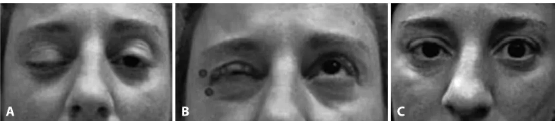
On examination, best corrected visual acuity was 20/20 in both eyes. The palpebral fissure opening was 1 mm for the right eye (OD) and 9 mm for the left eye (OS). Full extraocular motility and pupils were normal. The margin reflex distance OD was -4 mm and 3 mm OS, and levator function was 12 OD and 15 mm OS. The right orbicularis muscle maintained contraction in the right upper and lower eyelid, and voluntary opening of the OD was impaired throughout the day. The closure function of the orbicularis was normal. The patient stated that the condition disrupted activities of daily living, such as driving, going out alone, and reading on the computer, all of which decreased her quality of life (Figure 1 A).
Neurological examination indicated abnormal movements that affected the upper lid unilaterally, and all other facial movements and sensitivity were normal with no aberrant regeneration and no blepharospasm. Interestingly OS was normal. Systemically, there was mild generalized muscle hypertrophy, and phonation and swallowing were normal.
The patient was treated with botulinum neurotoxin type-A (BoNT-A; Botox®; Allergan plc., Dublin, Ireland). One hundred units of dry Botox® were diluted with 2 mL of non-preserved saline solution. We used a total of 12 units (IU). Two IUs were delivered per loci at two points of the pretarsal orbicularis in the upper right lid and 4 IU per loci were delivered subcutaneously at two points in the lateral canthus (Figure 1 B).
Botulinum toxin for treating unilateral apraxia of eyelid opening in a patient with
congenital myotonia
Tratamento da apraxia de abertura palpebral unilateral em portador de miotonia congênita com
botulinum toxin
EstrElla FErnándEz1, Marta latasiEwicz1, laura PElEgrin1, ManuEl roMEra2, silvana schEllini3,4, alicia galindo-FErrEiro5
Submitted for publication: March 27, 2017 Accepted for publication: May 18, 2017
1 Hospital Clínic de Barcelona, Institut Clínic d’Oftalmologia, Barcelona, Spain. 2 Department of Ophthalmology, Institut Condal Oftalmologia, Barcelona, Spain.
3 Department of Ophthalmology Faculdade de Medicina de Botucatu - UNESP, São Paulo, Brasil. 4 Oculoplastics and Orbit Division, King Khaled Eye Specialist Hospital, Riyadh, Saudi Arabia. 5 Department of Ophthalmology, Rio Hortega University Hospital, Dulzaina St, Valladolid, Spain.
Funding: No specific financial support was available for this study.
Disclosure of potential conflicts of interest: None of the authors have any potential conflict of interest to disclose.
Fe r n á n d e z e, e ta l.
3 3 1 Arq Bras Oftalmol. 2017;80(5):330-1 At the 1-week and 5-month follow-up, there was a sustained sa
tis-factory palpebral aperture opening of 7 mm and 9 mm, respectively. There were no complications during the 5 months of follow-up. The patient reported an improvement in activities of daily living, such as driving and working on the computer, and a greater visual field. At the 6-month follow-up, repeat BoNT-A injections were required and gave similar outcomes as those of the first treatment. Currently, this patient is seen at follow-up examinations every 5 months, and injections are delivered as required. To date, the patient has been followed for 6 years with good outcomes and no adverse events.
DISCUSSION
Treatment for ALO with BoNT-A was used to decrease the acti-vity of the orbicularis oculi to improve lid opening. Although there is a greater preponderance of males who are affected by MC, our patient was female(5). Onset of variable upper lid ptosis OD occurred when she was 12 years old and was concurrent with onset of puberty, which was diagnosed as Becker MC. Symptoms can be augmented during puberty, pregnancy, menstruation, hypothyroidism, and stress or in cold environments(5).
Additionally, individuals with Becker MC experience bilateral muscle stiffness, which can be observed in almost every facial mus-cle, the tongue, and extraocular muscles. The patient in this case had isolated abnormal movements that only affected the upper lid unilaterally. Patients with MC typically present other signs, inclu-ding variable muscle hypertrophy, which is usually expressed in the neck and upper limb region, or generalized hypertrophy as in our case(5). Our patient complained of daily fluctuation of ptosis, which was worse in the morning and varied throughout the day, over 25 years. At her first examination at our facility, she reported a recent and sudden worsening of ptosis and an inability to open her lids properly. She was diagnosed as having ALO associated with Becker MC of the same (right) upper lid.
ALO usually presents in patients older than our patient (37 years old)(5). We are uncertain about the cause of ALO that is associated with Becker MC in a 37-year-old patient, as in our case. Isolated
ALO may also occur, as observed in our patient who had no other abnormality of movement. ALO usually occurs more commonly in association with essential blepharospasm and extrapyramidal diseases, such as Parkinson’s disease and progressive supranuclear palsy(5,6). Some individuals with MC report painful muscular contrac-tions, and spora dic muscle weakness.(5) However, these symptoms were absent in our patient, who was under systemic treatment to decrease muscle contraction. However, the apraxia did not improve. Hence, we elected to treat ALO with BoNT-A. A similar case of MC, ALO, and bone dysplasia treated with BoNT-A that resulted in a good outcome has been reported(7).
In our case, the first dose of BoNT-A was empiric. With this treatment, the outcome was successful and the patient returned to her daily activities. In similar cases, general measures, such as adjusting daily activity and lifestyle can be suggested.
In conclusion, we experienced a rare case of Becker MC associa-ted with ALO. This case was unique because of the patient’s relatively young age and the unilaterality of presentation. We believe that treatment with BoNT-A can be useful.
REFERENCES
1. Duno M, Colding-Jorgensen E. Myotonia congenita. In: Pagon RA, Adam MP, Ardinger HH, Wallace SE, Amamiya A, Bean LJ, et al., editors. GeneReviews(R). Seattle (WA): Uni-versity of Washington;1993.
2. Piccione F, Mancini E, Tonin P, Bizzarini M. Botulinum toxin treatment of apraxia of eyelid opening in progressive supranuclear palsy: report of two cases. Arch Phys Med Rehabil. 1997;78(5):525-9.
3. Jankovic J. Apraxia of lid opening. Mov Disord. 1995;10(5):686-7.
4. Cherian V, Foroozan R. Benign unilateral apraxia of eyelid opening. Ophthalmology. 2010;117(6):1265-8.
5. Morales F, Cuenca P, del Valle G, Vásquez M, Brian R, Sitytenfeld M, Johnson K, et al. Clinical and molecular diagnosis of a Costa Rican family with autosomal recessive myo tonia congenita (Becker disease) carrying a new mutation in the CLCN1 gene. Rev Biol Trop. 2008;56(1):1-11.
6. Tozlovanu V, Forget R, Iancu A, Boghen D. Prolonged orbicularis oculi activity: a major factor in apraxia of lid opening. Neurology. 2001;57(6):1013-8.
7. Morrison DA, Mellington FB, Hamada S, Moore AT. Schwartz-Jampel syndrome: surgi-cal management of the myotonia-induced blepharospasm and acquired ptosis after failure with botulinum toxin A injections. Ophthal Plast Reconstr Surg. 2006;22(1):57-9.
A B C