COMPUTED TOMOGRAPHY IN THE ANALYSIS OF CALCIFICATION
PATTERNS IN PEDIATRIC BONE TUMORS OF THE HIP: A NEW
APPROACH*
Gabriel Antônio de Oliveira1
, Henrique Zambenedetti Werlang2
, Pedro Martins Bergoli2 , Madalena Frechiani3
, Fernão Oliveira4
OBJECTIVE: In the pediatric group, the radiological diagnosis of bone tumors of the hip is difficult and pre-sents some peculiarities, but reviewed literature does not approach this specific problem. The objective of the present study was to investigate the existence of reliable radiological patterns for the differential diag-nosis of these tumors. MATERIALS AND METHODS: Radiological findings of bone tumors of the hip in ten patients in the age range between 8 and 19 years have been reviewed. RESULTS: Bone reaction (sclerosis or lysis), periosteal reaction (lamellar with single or multiple layers, or radial), tumor extent in the bone and level of soft tissues invasion have presented low specificity. Soft tissue calcifications, when considered as a whole, were non-specific. However, when those calcifications with varied shapes and sizes, nearby the affected bone (pattern I) were separated from those, thin and amorphous, away from the bone (pattern II), we have observed that the pattern I was totally non-specific, and the pattern II was found in the three cases of osteosarcoma (100%) and in only one case of Ewing’s sarcoma (16.6%). CONCLUSION: In the present study, pattern II calcifications have shown a 100% sensitivity and 90% specificity for osteosarcoma. How-ever, their importance may be not limited to the radiological diagnosis. Pattern II calcifications indicate prob-ably ideal sites for biopsy.
Keywords: Bone tumors; Ilium; Hip; Pelvis; Pediatrics; Computed tomography.
Tomografia computadorizada na análise dos padrões de calcificações nos tumores ósseos da bacia em pediatria: nova aboardagem.
OBJETIVO: No grupo pediátrico, o diagnóstico radiológico dos tumores dos ossos ilíacos, ísquios e púbis apresenta dificuldades e peculiaridades próprias, mas a literatura revisada não trata especificamente desse tema. Este trabalho pretende investigar a existência de padrões radiológicos confiáveis para o diagnóstico diferencial desses tumores. MATERIAIS E MÉTODOS: Foram revistos os achados radiológicos de tumores dos ossos do quadril em dez pacientes com idades entre 8 e 19 anos. RESULTADOS: Reação óssea (escle-rose ou lise), reação periosteal (lamelar em camada única, múltiplas camadas ou radial), extensão do tumor no osso e grau de invasão das partes moles revelaram baixa especificidade. As calcificações nas partes moles, consideradas em conjunto, foram inespecíficas. Contudo, separando as próximas do osso comprometido, que apresentam formas e tamanhos variados — padrão I —, daquelas afastadas do osso, finas e amorfas — padrão II —, observamos que o padrão I foi totalmente inespecífico e o padrão II foi identificado nos três casos de osteossarcoma (100%) e em apenas um dos casos de Ewing (16,6%). CONCLUSÃO: Neste ma-terial, as calcificações padrão II revelaram sensibilidade de 100% e especificidade de 90% para osteossar-coma. Contudo, sua importância pode não se limitar ao diagnóstico radiológico. As calcificações padrão II indicam, provavelmente, os sítios ideais para biópsia.
Unitermos: Tumores ósseos; Ilíaco; Quadril; Pediatria; Tomografia computadorizada. Abstract
Resumo
* Study developed at Service of Diagnostic Imaging – Hospi-tal Infantil Nossa Senhora da Glória, Vitória, ES, Brazil.
1. MD, Radiologist at Hospital Infantil Nossa Senhora da Glória, Preceptor in Pediatric Radiology – Medical Residence of Diag-nostic Imaging Center at Hospital Universitário Cassiano Antônio de Morais/Hospital Infantil Nossa Senhora da Glória.
2. MD, Radiologists, ex-Residents at Diagnostic Imaging Center – Hospital Universitário Cassiano Antônio de Morais/Hospital Infantil Nossa Senhora da Glória.
3. MD, Pediatric Oncologist at Hospital Infantil Nossa Senhora da Glória.
4. MD, Radiologist at Hospital Infantil Nossa Senhora da Glória.
Mailing address: Dr. Gabriel de Oliveira. Rua Sagrado Coração de Maria, 220, Praia do Canto. Vitória, ES, Brazil 29055-770. E-mail: [email protected]
Received September 8, 2005. Accepted after revision March 29, 2006.
INTRODUCTION
In long bones, orthogonal plain films are very useful in the formulation of diag-nostic hypotheses. Different lesions tend towards presenting higher incidence in epi-physes, metaphyses or diaphyses(1).
Os-teosarcomas, for example, use to be meta-physeal; the Ewing’s sarcoma, diaphyseal or metadiaphyseal. In the hip bones (ilium, ischium and pubis), on the contrary, or-thogonal incidences are not feasible and the
In the reviewed pediatrics literature, we have not found any specific study on the radiological semiology of bone tumors of the hip.
MATERIALS AND METHODS
We have retrospectively analyzed the dossiers of ten patients with histologically confirmed bone tumors of the hip docu-mented by means of plain films and CT. The CT studies have been analyzed in the non-contrast phase to avoid the confusion between tumor vessels and calcifications. Magnetic resonance imaging (MRI) is poorly appropriate for diagnosis, and has not been presented in this study, despite its fundamental role in the staging of this kind of lesions. Post-chemo-radiotherapy im-ages have not been utilized, as well as post-contrast images. In the first case, because of a significant calcification increase in the soft parts of the tumor, probably resulting from tissular necrosis. In the second case, because the tissular, neovascular enhance-ment could be confused with tumor calci-fications.
The sacrum and coccyx, although in-cluded in the bone structure of the hip, have been excluded from this study for present-ing peculiar characteristics, besides other in common with vertebrae.
The casuistic has been limited to the first decades of life because of the several diseases affecting these bones in the other age ranges. Six cases had histological di-agnosis for Ewing’s sarcoma, three for os-teosarcoma, and one for chondrosarcoma. Ages ranged between 8 and 19 years. Six patients were male and four, female.
Other criteria employed in the analysis are shown in Table 1.
RESULTS
All of the patients presented with pain as their initial symptom, reported as re-stricted to the hip in seven patients; in the hip irradiating to the coxa in one; and in the coxa and/or knee in two. All of them pre-sented with a palpable mass at diagnosis. The lesion involved less than 25% of the bone in two cases of Ewing’s sarcoma, and in one case of chondrosarcoma. Be-tween 25% and 50% in one case of Ewing’s
sarcoma, and in one of osteosarcoma, and more than 50% in three cases of Ewing’s sarcoma, and in two of osteosarcomas.
All of the three cases of osteosarcomas had predominantly sclerotic bone lesions (cases 7, 8 and 9). In the cases of Ewing’s sarcoma, there was a predominance of scle-rotic lesions in three (cases 1, 4 and 6), lytic in two (cases 2 and 5) and a balance be-tween sclerosis and lysis in one (case 3). In the chondrosarcoma, there was a lytic le-sion (case 10). Lele-sions presented with a
permeative pattern in all of the cases, with ill-defined limits between the normal and affected areas.
Cortical involvement had occurred in all of the cases.
Periosteal reaction occurred in the three osteosarcomas, in one of them associated with single lamellar reaction (case 9). In the Ewing’s sarcomas, two have not presented any periosteal reaction, two presented with single lamellar reaction (cases 1 and 2), and two, mixed reaction (cases 3 and 6). In the
Table 1 Criteria employed in tumors evaluation.
Clinical presentation
Affected bones Bone lesion size*
Predominant osseous reaction Cortical involvement Type of periosteal reaction Extra-osseous tumor mass†
Pattern I calcifications‡
Pattern II calcifications§
Pattern III calcifications¶
Contiguity invasion** Metastases at diagnosis
Ilium; isquium; pubis < 25%; 25–50%; > 50%
Lytic; sclerotic; mixed (balanced lytic + sclerotic) Absent; present
Sunburst; single lamellar; multiple lamellar Absent; present Absent; present Absent; present Absent; present Absent; present Absent; present
* Considered in relation to the percentage of bone affected. If more than one bone (ilium, ischium or pubis) is involved, the size of the lesion in the largest bone will be considered.
† Considered in relation to the highest thickness of the affected bone.
‡ With different sizes and shapes, localized at a distance equal or lower than the measure of the highest thickness
of the contralateral, normal bone in the same cut plane (Case 3, Figure C).
§ When thin and amorphous, localized at a distance higher than the highest thickness of the contralateral normal
bone, in the same cut plane.
¶ When rounded, defined, usually found in calcified chondroid matrices.
** In the absence of a cartilage separating the ilium, ischium and pubis, the involvement of more than one of these bones will not be considered as a contiguity invasion.
Figure 4. Case 4. Ewing’s sar-coma. Fourteen-year-old girl. Left-sided iliac predominantly sclerotic lesion; on the detail cortical rupture is observed.
Figure 3. Case 3. Ewing’s sarcoma. Thirteen-year-old boy. A: Left-sided iliac permeative, mixed pattern (sclerosis and lysis) lesion presenting sunburst periosteal reaction (arrows); some gross calcifications maybe representing osseous fragments. B: Periosteal, lamellar reaction (multiple layers)(arrow)C:
Another CT slice showing calcifications in soft parts at a distance lower than the greatest diameter of the contralateral bone taken as a reference.
Figure 5. Case 5. Ewing’s sar-coma. Male, 16-year-old pa-tient. Right-sided pubic, pre-dominantly lytic lesion.
chondrosarcoma, the reaction was of lamel-lar nature.
An extra-osseous mass was demon-strated in eight cases whose CT and/or MRI were available, always occurring from both faces and exceeding the major transverse axis of the bone.
Calcifications in soft parts were evalu-ated every time CT was available for analy-sis; pattern I calcifications were found in all of the cases (Ewing’s sarcoma, osteosa-rcoma, chondrosarcoma); pattern II, in os-teosarcomas and in one Ewing’s sarcoma (case 6). The majority of calcifications near the bone were pattern I; and the distant ones were pattern II. Pattern III calcifications have not been found.
Contiguity invasion into the sacrum has occurred in only one case of primary iliac osteosarcoma (case 8).
Figure 4 Figure 5
Figure 6.Case 6. Ewing’s sarcoma. Twelve-year-old boy. A: X-ray evidencing extensive sclerosis in left ilium, associated with soft tissue mass dislocating the bladder. The detail shows multiple lamellar reaction. B: Non-contrast en-hanced CT showing significantly extensive extra-osseous tumor and a small focus of amorphous calcification (arrow). C: Contrast-enhanced CT and os-seous window demonstrating periosteal reaction with sunburst and lamellar components. An increase in the bone diameter due to periosteal apposition is observed. (Case assigned by Dr. Mário Flores, from Instituto da Criança – Hospital das Clínicas - Universidade de São Paulo).
Metastasis have been found at diagno-sis in only one case of Ewing’s sarcoma (case 6, pulmonary metastasis).
DISCUSSION
Iliac involvement occurs in 18% of Ewing’s sarcoma, and in 12% of osteosar-comas. Chondrosarcomas, most frequently, affect this bone (60%), but they are rare in
the childhood and adolescence(2).
There-fore, the casuistic presented in this study may be considered as representative.
Seven patients presented with a severe bone involvement by the tumor, most prob-ably related to delay in diagnosis. Small alterations on the first x-ray of the hip had been neglected in case 1. In case 6, the patient reported pain in the knee, so the
initial imaging investigation — including a MRI — was restricted to this site. Al-though it is a well known fact, it will never be enough to remind that pain in the coxa and/or knee may be related to a hip bone lesion. Therefore, in cases where knee or coxa x-ray images are normal, the necessity of extending the radiological investigation to the hip must be considered.
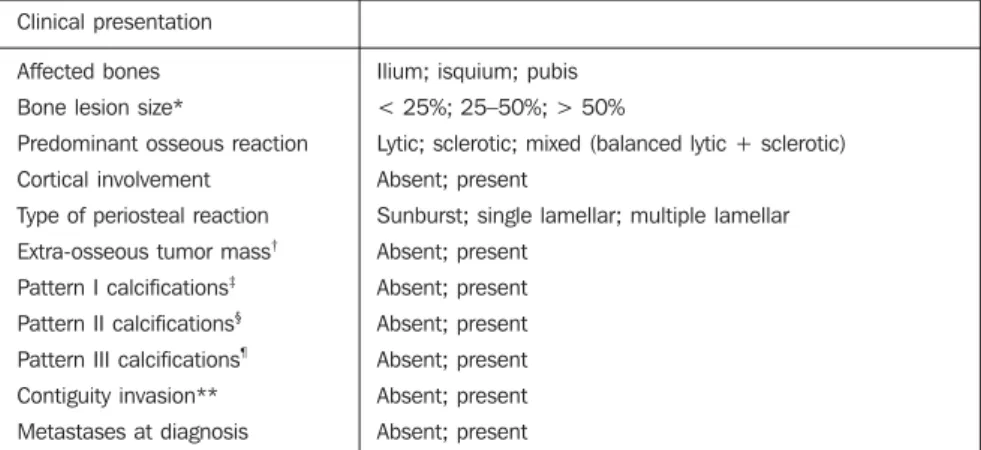
Figure 7.Case 7. Osteosar-coma. Male, 18-year-old pa-tient. A: Topogram showing sclerosis affecting the whole left ilium. B: CT demonstrat-ing, besides sclerotic bone lesion, mild, amorphous cal-cifications distant from the ilium (arrows). (Case assigned by Dr. Reinaldo B. Salgado, from Hospital São Lucas, Vitória, ES).
Other factors may be taken into consid-eration for the existence of so extensive le-sions. Differently from the long bones, in the ilium, ischium and pubis, the barriers constituted by conjugation cartilages and yellow bone marrow — hypovascularized — fat are absent. This absence makes the tumor cells dissemination easier.
Sunburst or lamellated periosteal reac-tions occurred in all the types of tumors, al-though the first one has predominated in osteosarcomas. Cortical involvement and extra-osseous tumor mass have been present in all of the cases.
Tumor cells have the capacity to stimu-late osteoblasts to produce sclerosis, and osteoclasts, lysis. Mixed lesions occur when these two functions coexist. In
osteo-genic sarcomas, additionally to this stimu-lus, tumor cells also produce osteoid ma-trix (an intercellular substance produced by normal or tumor osteoblasts). Only miner-alized matrices can be detected in images(1).
Also it is important to note that the amount of osteoid produced by osteosarcomas de-pends on the predominant differentiation of tumor cells: osteoblastic, chondroblastic and fibroblastic. However, it is impossible to differentiate the reactional sclerosis from that resulting from the production of os-teoid matrix by the tumor osteoblasts.
Predominance of sclerosis has occurred in all of the cases of osteosarcoma, but also in half of cases of Ewing´s sarcoma. Pre-dominance of osteolysis has only occurred in Ewing’s sarcomas, but it may also occur
in osteosarcomas, mainly the telangiectatic ones, and in osteomyelitis.
With regard to calcification in soft parts, they may occur in areas of tissular necro-sis, tumor-induced bone metaplasia, os-seous fragments dislocated by the tumor, periosteal reaction, or resulting from cal-cium deposition on the tumor matrix (os-teoid or chondroid). These calcifications present different patterns according Table 1. Pattern I calcifications were totally non-specific, probably because of the impossi-bility of distinguishing calcifications re-sulting from the osteoid matrix (in osteosar-comas) from those resulting from peri-osteal reaction and dislocated osseous frag-ments. Pattern II calcifications have been evidenced in all the osteogenic sarcomas
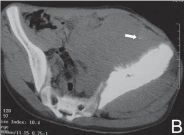
Figure 8.Case 8. Osteosarcoma. Female, 19-year-old patient. A: X-ray demonstrating sclerosis in the whole left ilium and mild soft tissue calcifications, adjacent to the iliac crest. B,C: CT showing mild, amorphous calcifications, some of them distant from the bone (white arrows), and lytic lesion on the left wing of the sacrum, characterizing invasion (black arrow).
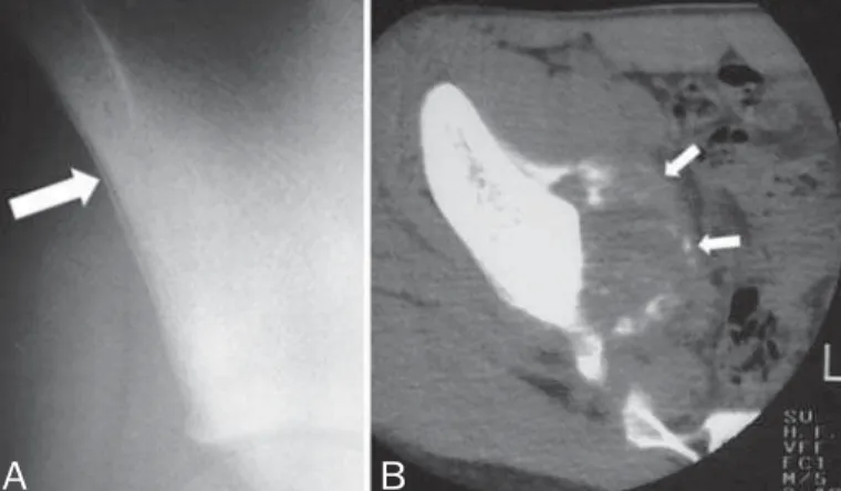
Figure 9.Case 9. Osteosarcoma. Fourteen-year-old boy. A: X-ray demonstrating sclerosis and periosteal, single lamellar reaction on right ilium (arrow). B: CT showing amorphous, cloudlike calcifications, some of them very distant from the bone (arrows).
Figure 10.Case 10. Chondrosarcoma. Thirteen-year-old girl. A: Peri-osteal, single lamellar reaction (arrow). B: Cortical rupture (arrow) and extra-osseous extension.
and in one of the Ewing’s sarcomas (case 6). In osteosarcomas, presumably, they have resulted from the tumor osteoid ma-trix calcification (ossification). Since Ewing’s sarcomas do not produce osteoid matrix, when present, these calcifications could result from necrosis, or, eventually, from tumor-induced bone metaplasia(3).
Resnick says that 9% of Ewing’s sarcomas calcify, but he neither specify which bones are affected nor the types of calcifications. The same author reports two cases of Ewing’s sarcoma of the ilium with calcifi-cations in soft parts. In this situation, a mistaken radiological diagnosis of osteosa-rcoma may be induced(4).
Despite the recent developments of his-topathology, immunohistochemistry, cyto-genetics and electronic microscopy, the di-agnosis of Ewing’s sarcoma remains as a challenge, and it is difficult to differenti-ate them from other tumors, especially small cell osteosarcomas(1,3,5–15),
neuroec-todermal tumors, mainly the primitive ones, and lymphomas(1,4). It is known that
bone tumors may present different histo-logical aspects, depending on the biopsy area. On CT, areas with pattern II calcifi-cations suggest the presence of osteoid matrix. In a tumor, provided the likelihood
of an induced bone metaplasia is remote, the presence of osteoid matrix is usually considered as an indication of osteosar-coma. However, the likelihood of such cal-cifications being a result of tissular necro-sis should not be disregarded.
Therefore, in the hypothesis of the bi-opsy does not reach areas with pattern II calcifications, there is a possibility of the osteoid tissue - existing only in these areas - not being included in the biopsy slide. The result would be a diagnostic mistake. CT-guided biopsy aimed at areas with pattern II calcifications could avoid this mistake.
In summary, in iliac bone tumors, soft parts calcifications seem to be of signifi-cance for the presumed radiological diag-nosis, and even more for biopsy guidance. A specific protocol adopting this approach could be employed in prospective studies, especially the multicentric ones.
REFERENCES
1. Baunin C, Rubie H, Sales de Gauzy J. Sarcoma d’Ewing. Ecycl Méd Chir Radiodiagnostic Appa-reil Locomoteur 2001;31:520-A-50,9p. 2. Schajowicz F. Neoplasias ósseas e lesões
pseu-dotumorais. 2ª ed. Rio de Janeiro: Revinter, 2000. 3. Schajowicz F. Current trends in the diagnosis and treatment of malignant bone tumors. Clin Orthop Relat Res 1983;180:220–252.
4. Resnick D. Tumor and tumor like lesions of bone:
image and pathology of specific lesions. In: Res-nick D, editor. Diagnosis of bone and joint disor-ders. 4th ed. Philadelphia: WB Saunders, 2002; 3763–4128.
5. Sim FH, Unni KK, Beabout JW, et al.
Osteosar-coma with small cell simulating Ewing’s tumors. J Bone Joint Surg [Am] 1979;61:207–215.
6. Martin SE, Dwyer A, Kissane JM, et al.
Small-cell osteosarcoma. Cancer 1982;50:990–996.
7. Roessner A, Immenkamp M, Hiddemann W, et al.
Case report 331. Small cell osteosarcoma of the tibia with diffuse metastatic disease. Skeletal Radiol 1985;14:216–225.
8. Edeiken J, Raymond AK, Ayala AG, et al. Small-cell osteosarcoma. Skeletal Radiol 1987;16:621– 628.
9. Ayala AG, Ro JY, Raymond AK, et al. A clinico-pathologic study of 27 cases. Cancer 1989;64: 2162–2173.
10. Kyriakos M, Gilula LA, Becich MJ, et al. Intra-cortical small cell osteosarcoma. Clin Orthop 1992;279:269–280.
11. Sanjay B, Raj GA, Vishwakarma G. A small cell osteosarcoma with multiple skeletal metastasis. Arch Orthop Trauma Surg 1988;107:58–60. 12. Ayala AG, Ro JY, Papadopoulos NK, et al. Small
cell osteosarcoma. Cancer Treat Res 1993;62: 139–149.
13. Mawad JK, MacKay B, Raymond AK, et al.
Elec-tron microscopy in the diagnosis of small round cell tumors of bone. Ultrastruc Pathol 1994;18: 263–268.
14. Nakajima H, Sim FH, Bond JR, et al. Small cell osteosarcoma of bone: review of 72 cases. Can-cer 1997;79:2095–2106.
15. Mulligan ME, Lewis DR Jr, Resnick CS, et al.