ABSTRACT
Prevalence and m orphom et ric analysis of t
hree-URRWHG PDQGLEXODU ¿UVW PRODUV LQ D %UD]LOLDQ
subpopulat ion
Clarissa Teles RODRIGUES1, Christiano de OLIVEIRA-SANTOS2, Norberti BERNARDINELI1, Marco Antonio Hungaro DUARTE1, Clovis Monteiro BRAMANTE1, Paloma Gagliardi MINOTTI-BONFANTE1, Ronald ORDINOLA-ZAPATA1
1- Universidade de São Paulo, Faculdade de Odontologia de Bauru, Departamento de Dentística, Endodontia e Materiais Odontológicos, Bauru, SP, Brasil. 2- Universidade de São Paulo, Faculdade de Odontologia de Ribeirão Preto, Departamento de Estomatologia, Saúde Coletiva e Odontologia Legal, Ribeirão Preto, SP, Brasil.
Corresponding address: Clarissa Teles Rodrigues - Faculdade de Odontologia de Bauru - USP - Al. Octávio Pinheiro Brisolla, 9-75 - 17012-901 - Bauru-SP - Brazil - Phone: +551432358344 - e-mail: [email protected]
6XEPLWWHG2FWREHU0RGL¿FDWLRQ0DUFK$FFHSWHG0D\
T
he know ledge of t he int ernal anat om y of t hree- root ed m andibular m olars m ay help clinicians t o diagnose and plan t he root canal t reat m ent in order t o provide adequat e t herapy w hen t his variat ion is present . Obj ect ives: To det erm ine t he prevalence of t hree-root ed m andibular m olars in a Brazilian populat ion using cone beam com put ed t om ography&%&7DQGWRDQDO\]HWKHDQDWRP\RIPDQGLEXODU¿UVWPRODUVZLWKWKUHHURRWVWKURXJK
m icro- CT. Mat erial and Met hods: CBCT im ages of 116 pat ient s w ere review ed t o det erm ine
WKH SUHYDOHQFH RI WKUHHURRWHG ¿UVW PDQGLEXODU PRODUV LQ D %UD]LOLDQ VXESRSXODWLRQ )XUWKHUPRUHZLWKWKHXVHRIPLFUR&7H[WUDFWHGWKUHHURRWHGPDQGLEXODU¿UVWPRODUV ZHUHVFDQQHGDQGUHFRQVWUXFWHGWRDVVHVVURRWOHQJWKGLVWDQFHEHWZHHQFDQDORUL¿FHV DSLFDO GLDPHWHU 9HUWXFFL¶V FODVVL¿FDWLRQ SUHVHQFH RI DSLFDO GHOWD QXPEHU RI IRUDPLQD DQG IXUFDWLRQV ODWHUDO DQG DFFHVVRU\ FDQDOV 7KH GLVWDQFH EHWZHHQ WKH RUL¿FH RQ WKH SXOSFKDPEHUÀRRUDQGWKHEHJLQQLQJRIWKHFXUYDWXUHDQGWKHDQJOHRIFDQDOFXUYDWXUH
w ere analyzed in t he dist olingual root . Dat a w ere com pared using t he Kruskal–Wallis t est
Į 5HVXOWV7KHSUHYDOHQFHRIWKUHHURRWHGPDQGLEXODU¿UVWPRODUVZDVRI
Mesial root s show ed com plex dist ribut ion of t he root canal syst em in com parison t o t he dist al root s. The m edian of m aj or diam et ers of m esiobuccal, m esiolingual and single m esial canals w ere: 0.34, 0.41 and 0.60 m m , respect ively. The higher values of m aj or diam et ers were found in t he dist obuccal canals ( 0.56 m m ) and t he lower diam et ers in t he dist olingual
FDQDOVPP7KHORZHVWRUL¿FHGLVWDQFHZDVIRXQGEHWZHHQWKHPHVLDOFDQDOV0%
ML) and t he highest dist ance bet w een t he dist al root canals ( DB- DL) . Alm ost all dist al root s had one root canal and one apical foram en w it h few accessory canals. Conclusions: Dist olingual root generally has short lengt h, severe curvat ure and a single root canal w it h low apical diam et er.
Ke y w or d s: Anat om y. Cone- beam com put ed t om ography. Root canal t herapy. X- ray m icrot om ography.
I N TROD UCTI ON
The underst anding of t he num ber of canals of hum an t eet h, t heir t ransverse sect ion and possible var iat ion s, is of u t m ost im por t an ce t o ach iev e t he decont am inat ion goals of endodont ic t herapy because necrot ic t issue in unt reat ed root canals can lead t o persist ent chronic apical periodont it is4.
Despit e t he m any anat om ical variat ions of t he root
FDQDO V\VWHP RI WKH PDQGLEXODU ¿UVW PRODU WKH H[WHUQDO DQDWRP\ W\SLFDOO\ KDV WZR ZHOOGH¿QHG
denom inat ed radix param olaris3.
As w it h ot her anat om ical var iat ions including C- sh ap ed m an d ib u lar secon d m olar s1 6, it h as been show n t hat t he incidence of a t hird root in
WKH PDQGLEXODU ¿UVW PRODU LV FORVHO\ UHODWHG WR
et hnicit y8. This var iabilit y has higher pr evalence
LQ VSHFL¿F SRSXODWLRQV HJ 0RQJRORLG 1DWLYH
Am erican, Eskim o and Chinese, for w hich it can be
FRQVLGHUHGDQRUPDO¿QGLQJ6,11,25.
Cu r r en t ly, im ag es of con e b eam com p u t ed t om ography ( CBCT) have been used t o st udy t he prevalence of t hree- root ed m andibular m olars in several populat ions23,25. I n addit ion t o t he CBCT
m et h od , m icr com p u t ed t om og r ap h y ( m icr o-CT) h a s a l so b e e n u se d t o d e scr i b e se v e r a l m orphom et ric aspect s of t hree- root ed m andibular m olar s in clu d in g p u lp ch am b er, cu r v at u r e an d m orphom et ric analysis.
A p r ev iou s p r ev alen ce st u d y in a Br azilian
SRSXODWLRQXVLQJ&%&7GLGQRW¿QGDQ\WKUHHURRWHG PDQGLEXODU ¿UVW PRODUV20. Unt il now, few st udies
have addr essed t heir pr evalence and com par ed qualit at ive and quant it at ive dat a of t he root canal
syst em s of t his anat om ical variat ion11,22. The purpose
of t his st udy was t o invest igat e t he prevalence of
WKUHHURRWHGSHUPDQHQWPDQGLEXODU¿UVWPRODUVLQD
Brazilian populat ion using CBCT im ages of pat ient s w ho had under gone CBCT scanning for im plant or t hird m olar surgery t reat m ent planning and t o analyze in vit ro t he m orphom et ric aspect s of t he
LQWHUQDODQDWRP\RIWKUHHURRWHGPDQGLEXODU¿UVW
m olars t hrough m icro- CT.
M ATERI AL AN D M ETH OD S
CBCT a n a lysis
Con e b eam com p u t ed t om og r ap h y ( CBCT) im ages of m andibular m olars w ere collect ed from 116 pat ient s who had undergone CBCT scanning for im plant or t hird m olar surgery t reat m ent planning. The inclusion crit eria w ere im ages displaying fully m at u r ed an d er u p t ed m an d ib u lar f ir st m olar s
ELODWHUDOO\ZLWKRXWURRWFDQDO¿OOLQJVSRVWVRUFURZQ
r est orat ions. The exclusion cr it er ia w er e im ages lacking t echnical qualit y or absence of one of t he t eet h t o be evaluat ed. All t he CBCT im ages w ere acquir ed using an i- CAT CBCT dev ice ( I m aging
6FLHQFHV,QWHUQDWLRQDO,QF+DW¿HOG3$86$7KH
scanner was operat ed at 120 kVp, 8 m A and a voxel size of 0.25 m m . Sagit t al, coronal and axial im ages w er e analy zed w it h t he use of t he i- CAT Vision soft ware by an experienced Oral and Maxillofacial radiologist in or der t o det er m ine t he num ber of
URRWVLQWKHPDQGLEXODU¿UVWPRODUV
M icr oCT a n a lysis
Th is st u d y w as ap p r ov ed accor d in g t o t h e gu idelin es of t h e local Hu m an Resear ch Et h ics
Com m it t ee. For t he in vit ro analysis, 55 m andibular
¿UVW PRODUV ZLWK WKUHH URRWV ZHUH VHOHFWHG IURP
a pool of ex t ract ed t eet h. Pat ient s’ gender and age w ere unknow n. All t eet h w ere scanned w it h a m icr o CT sy st em ( Sk y Scan 1 1 7 4 v 2 ; Br u k er -m icroCT, Kont ich, Belgiu-m ) using 50 kV, 800 -m A,
DQLVRWURSLFUHVROXWLRQRINjPDURWDWLRQVWHSRI
0.8 degrees and 360- degree rot at ion. Radiographs of each specim en were reconst ruct ed wit h dedicat ed soft ware ( NRecon v.1.6.3; Bruker- m icroCT, Kont ich, Belgium ) providing axial cross- sect ions of t he inner st ruct ure of t he t eet h.
7ULGLPHQVLRQDOFODVVL¿FDWLRQ
Three- dim ensional m odels w ere reconst ruct ed aft er binarizat ion of t he source im ages, export ed
DV 3* ¿OHV XVLQJ WKH &7$Q VRIWZDUH %UXNHU
m icr oCT, Kont ich, Belgium ) . Dat aView er v. 1. 4. 4 ( SkyScan, Kont ich, Belgium ) and CTVol soft wares ( Br u k er - m icr oCT, Kon t ich , Belgiu m ) w er e u sed for visualizat ion and qualit at ive evaluat ion of t he
VSHFLPHQVDFFRUGLQJWR9HUWXFFL¶VFODVVL¿FDWLRQ24. Th e p r esen ce o f ap i cal d el t a, t h e n u m b er o f foram ina and furcat ions, t he lat eral and accessory can al s w er e al so r eco r d ed . Fu r t h er m o r e, t h e lengt h of all root s was m easured from t he j unct ion of t he cem ent oenam el w it h t he apex13 using t he Dat aView er soft ware.
M orphom et ric a na lysis of t he cross- sect ions of t h e r oot ca n a l
Two- dim ensional cross- sect ions w ere select ed from 1 t o 3 m m apical level for quant it at ive analysis of ar ea, per im et er, r oundness, m aj or diam et er, m inor diam et er and aspect rat io using t he CTAn soft ware. Area and perim et er were calculat ed using t he Prat t algorit hm . The cross- sect ional appearance ( i.e. round or m ore ribbon- shaped) was expressed as roundness. The value of roundness ranges from 0–1, w it h 1 m eaning a perfect circle10. Maj or and
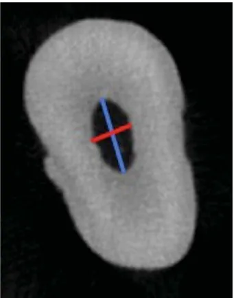
PLQRUGLDPHWHUVZHUHGH¿QHGDVVKRZQLQ)LJXUH
1. The aspect rat io is a quant it at ive index t hat also helps t o describe t he shape of t he root canal. I t is
GH¿QHGDVWKHUDWLREHWZHHQPDMRUGLDPHWHUDQG
m inor diam et er, i.e., t he closer t he values are t o 1
WKHOHVVÀDWWHQHGWKHFDQDOVDUH
0RUSKRORJ\RIWKHSXOSÀRRU
7KHLQWHURUL¿FHGLVWDQFHVRQWKHSXOSÀRRUZHUH
m easured using t he Dat aView er soft ware. A line
ZDVGUDZQEHWZHHQWKHFHQWHUVRIWKHRUL¿FHVDQG
t he dist ances w ere m easured using t he geom et ric m easurem ent m odule.
Cu r va t u r e of t h e dist olin gu a l r oot
of Healt h, Bet hesda, MD, USA) as descr ibed by Schneider18 ZLWKPRGL¿FDWLRQVE\Gu, et al.13
( 2011),WZDVFODVVL¿HGLQWRWKUHHJURXSVVWUDLJKW
( 10 degrees or less) , m oderat e ( 10 t o 20 degrees) or severe ( 20 degrees or m ore) . Ot her anat om ical landm arks m easured included t he dist ance bet ween
SXOSFKDPEHUÀRRUDQGEHJLQQLQJRIWKHFXUYDWXUH
and from t his point t o t he apical foram en.
St a t ist ica l a n a lysis
The result s of t he 2D analysis, t he angles and t he dist ances bet w een t he anat om ic landm ar k s w ere described as having m edian, m inim um and
PD[LPXPYDOXHV7KHDQDO\VLVRIWKHLQWHURUL¿FH GLVWDQFHVRQWKHSXOSÀRRUDQGWKHm orphom et ric analysis of t he cross- sect ions of t he root canal did not show norm al dist ribut ions t hus nonparam et ric t est s w ere used. Dat a was st at ist ically com pared using Kruskal–Wallis post - hoc Dunn t est , w it h t he
VLJQL¿FDQFH OHYHO VHW DW S XVLQJ *UDSK3DG
Prism 5 ( GraphPad Soft ware I nc, La Jolla, CA, USA) .
7KH FDWHJRUL]DWLRQ XVLQJ 9HUWXFFL¶V FODVVL¿FDWLRQ
was present ed descript ively.
RESULTS
CBCT a n a lysis
$ WRWDO RI PDQGLEXODU ¿UVW PRODUV IURP
a sam ple of 116 pat ient s w er e analy zed. Thr ee pat ient s ( 2 w om en and 1 m an) had t hree- root ed f ir st m olar s ( 2 . 5 8 % ) . On e b ilat er al case w as obser v ed, t h er ef or e, a t ot al of 4 t h r ee- r oot ed
PDQGLEXODU ¿UVW PRODUV ZHUH IRXQG DOO RI ZKLFK
had dist olingual root ( radix ent om olaris) ( Figure 2) . The radix param olaris anat om ical variat ion was not
SUHVHQWLQWKHPDQGLEXODU¿UVWPRODUV
M i c r o - CT q u a l i t a t i v e a n d q u a n t i t a t i v e a n a lysis
I n dist olingual root s, t he m ost prevalent canal
FRQ¿JXUDWLRQZDV9HUWXFFLW\SH,)LJXUH
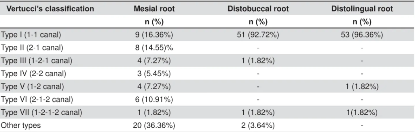
3) . Only one case had t ype V anat om y and anot her, t y pe VI I . Ty pe I anat om y was also found m or e frequent ly in dist obuccal root s ( 92.72% ) follow ed by t ype I I I ( one t oot h) , t ype VI I ( one t oot h) . The m esial root show ed a m ore com plex dist ribut ion of t he root canals: Vert ucci t ype I in 16.36% , t ype I I in 14.55% , t ype I I I in 7.27% , t ype I V in 5.45% , t ype V in 7.27% , t ype VI in 10.91% and t ype VI I in 1.82% ( Table 1) . Root lengt h, presence of apical
Figure 1- MicroCT cross-section demonstrating the major diameter (blue line) and minor diameter (red line). The major diameter was determined by drawing a line between the two most distant pixels of the root canal ZDOOV7KHPLQRUGLDPHWHUZDVGH¿QHGDVWKHORQJHVWOLQH drawn perpendicular to that of the major diameter11
delt a, lat eral and accessory canals, and num ber of foram ina, are show n in Table 2. A furcat ion canal was observed in only one t oot h.
At 1 m m apical level, t he lowest area values were found in m esiobuccal, m esiolingual and dist olingual root canals ( p> 0.05) . The highest values for area
Figure 3- 'UHFRQVWUXFWLRQVRIWKUHHURRWHGPDQGLEXODU¿UVWPRODUVREVHUYHGIURPWKHEXFFDOYLHZ$'7KHVKRUWOHQJWK
of the distolingual root is observed. The mesial view of these molars (E-H) shows a more complex anatomical root canal system of the mesial root, compared to the distobuccal and distolingual roots and the presence of severe curvatures in the distolingual root
9HUWXFFL¶VFODVVL¿FDWLRQ Mesial root Distobuccal root Distolingual root
Q Q Q
Type I (1-1 canal) 9 (16.36%) 51 (92.72%) 53 (96.36%)
Type II (2-1 canal) 8 (14.55)% -
-Type III (1-2-1 canal) 4 (7.27%) 1 (1.82%)
-Type IV (2-2 canal) 3 (5.45%) -
-Type V (1-2 canal) 4 (7.27%) - 1 (1.82%)
Type VI (2-1-2 canal) 6 (10.91%) -
-Type VII (1-2-1-2 canal) 1 (1.82%) 1 (1.82%) 1(1.82%)
Other types 20 (36.36%) 2 (3.64%)
-Table 1- 'LVWULEXWLRQRI9HUWXFFLV&ODVVL¿FDWLRQW\SHVEHWZHHQPHVLDOGLVWREXFFDODQGGLVWROLQJXDOURRWV
Column1 Mesial Distobuccal Distolingual
Root length (mm) 14.02 (10.41-17.50)
12.58 (8.51-15.40)
11.55 (7.84-16.11)
Apical delta 8 7 6
Lateral canals 4 3 1
Accessory canals 1 3 2
1 foramen 32.7% 84% 80%
2 foramina 50% 14% 20%
3 foramina 17.3% 2% 0
an d p er im et er p ar am et er s w er e f ou n d in t h e dist obuccal and single m esial canals ( Table 3) .
Dist olingual canals show ed higher r oundness values and low er aspect rat io values in com parison t o t he ot her root canals evaluat ed ( p< 0.05) . The m edian of m aj or diam et er s of m esiobuccal and m esiolin gu al an d sin gle m esial can als w er e as follows: 0.34, 0.41 and 0.60 m m , respect ively. The highest values of m aj or diam et ers w ere found in t he dist obuccal canals ( 0.56 m m ) and t he low est values in t he dist olingual canals ( 0.29 m m ) . Ot her values corresponding t o 2 and 3 m m apical levels are show n in Table 3.
Al l t h e d i st o l i n g u al r o o t s ex h i b i t ed sev er e
FXUYDWXUHV)LJXUH$7KHVKRUWHVWRUL¿FH
dist ance was found bet ween t he m esial canals ( MB-ML) and t he longest dist ance bet w een t he dist al root canals ( DB- DL) ( p< 0.05) Fig. 4B.
D I SCUSSI ON
7KHLGHQWL¿FDWLRQRIDGGLWLRQDOFDQDOV\VWHPVLQ
t he m esial and dist al root of m andibular m olars is considered im port ant for successful disinfect ion and
¿OOLQJRIWKHURRWFDQDOV\VWHPDQGFRQVHTXHQWO\
for t he long- t erm prognosis of t he endodont ically t reat ed t oot h. The presence of a t hird root , usually a dist olingual root , is t he m ost com m on anat om ical variat ion in m andibular m olars7.
I t seem s plausible t hat et hnical back gr ound
MB ML DB DL M single
Median (Minimum-Maximum)
Median (Minimum-Maximum)
Median (Minimum-Maximum)
Median (Minimum-Maximum)
Median (Minimum-Maximum)
1 mm apical
Area (mm2) 0.05 (0.01-0.80)a 0.07 (0.01-0.79)a 0.13 (0.04-0.72)b 0.04 (0.01-0.54)a 0.13 (0.05-1.35)b
Perimeter (mm) 0.95 (0.46-5.63)a 1.16 (0.34-5.56)a 1.48 (0.83-3.82)b 0.78 (0.30-3.23)a 1.61 (0.24-5.34)b
Roundness 0.69 (0.16-0.85)ab 0.59 (0.17-0.78)a 0.56 (0.22-0.80)a 0.69 (0.34-0.91)b 0.47 (0.22-0.80)a
Major diameter (mm)
0.34 (0.16-2.50)ab 0.41 (0.12-2.40)b 0.56 (0.29-1.51)b 0.29 (0.11-1.22)a 0.60 (0.15-1.88)b
Minor diameter (mm)
0.23 (0.05-0.82)a 0.23 (0.03-0.54)a 0.35 (0.15-0.74)b 0.19 (0.07-0.59)a 0.33 (0.15-3.78)b
Aspect Ratio 1.36 (0.92-5.76)ab 1.55 (1.06-4.62)ab 1.55 (0.0-3.53)ab 1.36 (0.97-3.18)a 2.05 (1.23-4.17)b
2 mm apical
Area (mm2) 0.11 (0.03-0.85)a 0.10 (0.02-0.98)ab 0.17 (0.05-0.68)ab 0.06 (0.01-0.75)a 0.19 (0.08-0.86)b
Perimeter (mm) 1.44 (0.72-6.62)b 1.45 (0.61-7.38)bc 1.61 (0.84-4.29)bc 0.92 (0.26-3.47)a 2.25 (1.18-4.57)c
Roundness 0.52 (0.08-0.85)ab 0.39 (0.08-0.80)a 0.59 (0.16-0.91)b 0.72 (0.13-0.87)c 0.30 (0.10-0.55)a
Major diameter (mm)
0.50 (0.24-2.92)b 0.57 (0.23-3.24)b 0.58 (0.27-1.82)bc 0.33 (0.09-1.29)a 0.99 (0.44-1.83)c
Minor diameter (mm)
0.27 (0.13-0.86)a 0.25 (0.13-0.63)a 0.37 (0.15-0.90)b 0.25 (0.03-0.74)ab 0.33 (0.19-0.96)ab
Aspect Ratio 1.84 (0.92-9.58)bc 2.21 (1.13-9.58)c 1.59 (0.99-5.75)ab 1.31 (0.91-4.34)a 3.03 (1.47-7.21)c
3 mm apical
Area (mm2) 0.17 (0.04-1.25)b 0.16 (0.03-1.30)b 0.25 (0.07-0.80)bc 0.07 (0.01-0.92)a 0.27 (0.17-1.45)c
Perimeter (mm) 1.72 (0.78-8.02)b 2.02 (0.72-7.49)b 2.18 (1.04-5.36)bc 1.05 (0.35-3.70)a 3.49 (1.95-7.27)c
Roundness 0.37 (0.06-0.80)ab 0.44 (0.07-0.76)a 0.60 (0.14-0.87)b 0.75 (0.39-0.89)c 0.20 (0.05-0.43)a
Major diameter (mm)
0.68 (0.26-3.57)b 0.69 (0.29-3.29)b 0.79 (0.34-1.71)b 0.36 (0.12-1.31)a 1.54 (0.82-2.97)c
Minor diameter (mm)
0.33 (0.10-0.86)a 0.31 (0.15-0.66)a 0.46 (0.19-0.94)b 0.31 (0.11-0.90)a 0.31 (0.19-0.74)ab
Aspect Ratio 2.29 (1.13-9.71)c 2.25 (1.15-9.71)c 1.55 (1.04-4.54)b 1.26 (0.92-2.51)a 4.77 (2.21-10.06)c
Different superescript letters (a, b and c) in the same row indicate statistical difference between the groups (p<0.05).
LV D PDMRU IDFWRU WKDW LQÀXHQFHV WKH SUHYDOHQFH
o f a d i s t o l i n g u a l r o o t , a s p r e v i o u s s t u d i e s h av e d em on st r at ed h ig h er p r ev alen ce am on g individuals of Asian origin, varying from 24.5% t o 33.3%6,21,23,25,26. The prevalence found in our st udy is sim ilar t o what was report ed by Shem esh, et al.19 ( 2015) , in an I sraeli populat ion w it h CBCT.
Fer r az & Pecor a9 ( 1 9 9 2 ) obser v ed a sim ilar prevalence of t hree- root ed m andibular m olars in a Brazilian populat ion ( 2.8% for Black origin and 4 . 2 % for Cau casian ) . How ev er, t h is st u dy w as carried out w it h periapical radiographs. I n a CBCT st udy w it h a Brazilian populat ion, Silva, et al.20
GLGQRW¿QGDQ\WKUHHURRWHG¿UVWPRODUV
To iden t ify an addit ion al r oot in m an dibu lar m olars, changes in t he horizont al angulat ion during X- ray exposure m ay be useful, in order t o at t em pt t o overcom e t he lim it at ions of radiographs, such as super im posit ions by sur r ounding st r uct ur es. How ever, t he addit ion of m ult iple radiographs does
QRW HQVXUH WKH LGHQWL¿FDWLRQ RI WKLV DQDWRPLFDO
variabilit y1,22. Due t o t his fact , alt hough CBCT should not be used as a rout ine procedure in endodont ics, it m ay be indicat ed in t he assessm ent and t reat m ent o f co m p l e x e n d o d o n t i c co n d i t i o n s2, b e ca u se t his t echnique pr ov ides a bet t er v isualizat ion of anat om ical variat ions in t he num ber of root s and
root canals1,22. According t o Abella, et al.1 ( 2011) , when an addit ional root is det ect ed before root canal t reat m ent , t he clinician can plan t he procedures bet t er, such as enlarging t he opening access cavit y
LQRUGHUWRORFDWHDOOWKHRUL¿FHVRIWKHFDQDOV
I n t he present st udy, 55 t hree- root ed m andibular f ir st m olar s w er e ev alu at ed t h r ou g h m icr o- CT analysis, a num ber t hat can be considered superior t o pr ev ious anat om ical st udies w hich addr essed t he m orphom et ric aspect s of t his variat ion t hrough m icro- CT11- 13,22.
Alm ost all dist olingual and dist obuccal r oot s had one root canal ( Vert ucci t ype I ) and one apical foram en. The dist obuccal and dist olingual r oot s had a single canal in 92.72% and 96.36% of t he cases, respect ively, w hich cont rast s w it h t he low er prevalence of single canals of dist al root s of t w
o-URRWHGPDQGLEXODU¿UVWPRODUV10,14. On t he
cont rary, t he m esial root show ed a m ore com plex d ist r ib u t ion of t h e in t er n al an at om y w it h t h e presence of t w o foram ina being t he m ost com m on
¿QGLQJ$PRQJPHVLDOURRWVVLQJOHFDQDOV
w ere m arkedly less com m on ( 16.36% ) and a large
YDULDELOLW\LQ9HUWXFFL¶VFODVVL¿FDWLRQZDVREVHUYHG
I n t his st udy, t he dist al root s result s are sim ilar t o t he cat egories found by Gu, et al.11 ( 2010) and
Wang, et al.25 ( 2010) .On t he ot her hand, m esial Figure 4-$7KHPHGLDQDQGUDQJHYDOXHVRIWKHGLVWDQFHEHWZHHQWKHSXOSFKDPEHUÀRRUDQGWKHEHJLQQLQJRIWKH
root s showed a different dist ribut ion which cont rast s w it h t he w ork of t hese aut hors. The descript ion of t he m esial and dist al root s of m andibular m olars
DFFRUGLQJWR9HUWXFFL¶VFODVVL¿FDWLRQDQGPLFUR&7
im aging has been rest rict ed t o only one previous st udy, w hich observed 81.8% of dist al root s w it h a single canal15. One can t hus speculat e t hat t he
com plex anat om y of dist al root s is less frequent w hen an ext ra dist olingual root is present .
Gu, et al.11 ( 2010) found out t hat accessory and
lat eral canals rarely occurred in dist olingual root s. Sim ilarly, in t his st udy, lat eral and accessory canals were found in only one and t wo cases, respect ively. How ever, such anat om ical variat ions are also not com m on in m esial and dist obuccal root s.
The analy sis of m or phom et r ic dat a at 1 m m level dem onst rat ed low er m edian values of apical diam et er in t h e dist olin gu al can als ( 0 . 3 0 m m ) co m p a r e d t o d i st o b u cca l ca n a l s ( 0 . 5 6 m m ) .
I n a d d i t i o n , di st o l i n g u a l ca n a l s a r e r o u n d e r in sh ap e, w h er eas t h e m esial an d d ist ob u ccal
FDQDOV DUH VLJQL¿FDQWO\ PRUH RYDO VKDSHG ZKLFK
is in agreem ent w it h t he st udies by Gu, et al.13
( 2011) and Souza- Flam ini, et al.22 ( 2014) . I n t his invest igat ion, t he m aj or apical diam et er values were sim ilar t o t he ones previously report ed by Harris, et al.15 ( 2013) .
Accor ding t o t he pr esent st udy, t he dist ance o f t h e o r i f i ce s o n t h e p u l p a l f l o o r su g g e st s t hat t he endodont ic access should be enlar ged fr om a t r iangular t o a t rapezoidal opening w it h an ex t en sion t o t h e d ist olin g u al ar ea t o h elp locat ing t he DL canal3,7. The use of an operat ive
m icroscope can be useful in det ect ing a t hird root in a m andibular m olar. Once t he addit ional root is
LGHQWL¿HGLQWKHSUHRSHUDWLYHGLDJQRVLVUDGLRJUDSK
or CBCT, t he dist ance bet w een t he m esiobuccal and m esiolingual root s can be used as a guide for
¿QGLQJWKHGLVWROLQJXDOURRWFDQDOVLQFHWKHGLVWDQFH
bet w een t he dist obuccal and dist olingual root s is usually bet w een 0.5 and 1 m m longer t han t he m esiobuccal t o m esiolingual dist ance11.
S t u d i e s h a v e s h o w n t h a t t h e a d d i t i o n a l dist olin gu al r oot is gen er ally sm aller t h an t h e m esial and dist obuccal root s5,13,22 and has severe cu r v at u r e1 2. I n a p r ev i o u s st u d y1 3, t h e m ean lengt h of t he dist olingual root was 10.65 m m wit h a curvat ure of 32 degrees, w hich is sim ilar t o t he result s of t he present st udy. Anot her st udy in a Brazilian populat ion show ed a low er m ean lengt h i.e. 7.65 m m22. The variat ion can be explained by t he difference in t he num ber of sam ples st udied, since Souza- Flam ini, et al.22 ( 2014) used only 19 t eet h.
Clinicians need t o know about t he short lengt h and severe curvat ure of dist olingual root s because it can increase t he risk of accident s such as inst rum ent separat ion or ledge form at ion. I t is known t hat cyclic
fat igue decreases w it h an increase in t he angle of curvat ure17. Thus, decreasing t aper conicit y, using
VPDOOHUDSLFDOGLDPHWHUVQLFNHOWLWDQLXP¿OHVDQG SUHÀDULQJRIWKHFHUYLFDOWKLUGDUHLQGLFDWHGLQRUGHU
t o avoid accident s.
Considering t he prevalence and charact erist ics of t he dist olingual root , clinicians should be able t o diagnose and develop skills t o provide adequat e root canal t reat m ent when t his variat ion is present .
CON CLUSI ON
Th e p r ev alen ce of t h r ee- r oot ed m an d ib u lar m olars in a Brazilian subpopulat ion was of 2.58% . Dist olingual root s had short lengt h, severe curvat ure and a low apical diam et er in com par ison t o t he dist obuccal and m esial root s. Single canals w ere highly prevalent in bot h dist al root s in com parison t o t he m esial root w hich show ed a m ore com plex anat om ical dist ribut ion.
ACKN OW LED GEM EN TS
This work was supported by FAPESP (2013/ 03695-0 and 203695-0103695-0/ 1603695-072- 2) .
REFEREN CES
1- Abella F, Mercade M, Duran- Sindreu F, Roig M. Managing severe curvat ure of radix ent om olaris: t hree- dim ensional analysis w it h cone beam com put ed t om ography. I nt Endod J. 2011; 44: 876- 85. 2- Am erican Associat ion of Endodont ist s, Am erican Academ y of Oral and Maxillofacial Radiology. Use of cone- beam com put ed t om ogr aphy in en dodon t ics. Join t Posit ion St at em en t of t h e Am erican Associat ion of Endodont ist s and t he Am erican Academ y of Oral and Maxillofacial Radiology. Oral Surg Oral Med Oral Pat hol Oral Radiol Endod. 2011; 111: 234- 7.
3- Calberson FL, De Moor RJ, Deroose CA. The radix ent om olaris an d par am olar is: clin ical appr oach in en dodon t ics. J En dod. 2007; 33: 58- 63.
4 - Can t at or e G, Ber u t t i E, Cast ellu cci A. Missed an at om y : frequency and clinical im pact . Endod Topics. 2006; 15: 3- 31. 5- Chen YC, Lee YY, Pai SF, Yang SF. The m orphologic charact erist ics
RIWKHGLVWROLQJXDOURRWVRIPDQGLEXODU¿UVWPRODUVLQD7DLZDQHVH
populat ion. J Endod. 2009; 35: 643- 5.
6- Curzon ME, Curzon JA. Three- root ed m andibular m olars in t he Keewat in Eskim o. J Can Dent Assoc. 1971; 37: 71- 2.
7- De Moor RJ, Deroose CA, Calberson FL. The radix ent om olaris
LQPDQGLEXODU¿UVWPRODUVDQHQGRGRQWLFFKDOOHQJH,QW(QGRG
J. 2004; 37: 789- 99.
8- De Pablo OV, Est evez R, Péix Sánchez M, Heilborn C, Cohenca N.
5RRWDQDWRP\DQGFDEDOFRQ¿JXUDWLRQRIWKHSHUPDQHQWPDQGLEXODU ¿UVWPRODUDV\VWHPDWLFUHYLHZ-(QGRG
9 - Fer raz JA, Pécora JD. Thr ee- r oot ed m andibular m olar s in pat ient s of Mongolian, Caucasian and Negro origin. Braz Dent J. 1992; 3: 113–7.
10- Filpo- Perez C, Bram ant e CM, Villas- Boas MH, Húngaro Duart e MA, Versiani MA, Ordinola- Zapat a R. Micro- com put ed t om ographic an aly sis of t h e r oot can al m or ph ology of t h e dist al r oot of
PDQGLEXODU¿UVWPRODU-(QGRG
1 1 - Gu Y, Lu Q, Wan g H, Din g Y, Wan g P, Ni L. Root can al
12- Gu Y, Lu Q, Wang P, Ni L. Root canal m orphology of perm anent
WKUHHURRWHGPDQGLEXODU¿UVWPRODUV3DUW,,±PHDVXUHPHQWRI
root canal curvat ures. J Endod. 2010; 36: 1341- 6.
13- Gu Y, Zhou P, Ding Y, Wang P, Ni L. Root canal m orphology
RISHUPDQHQWWKUHHURRWHGPDQGLEXODU¿UVWPRODUV3DUW,,,±$Q
odont om et ric analysis. J Endod. 2011; 37: 485- 90.
1 4 - Gu labivala K, Au n g TH, Alav i A, Ng YL. Root an d can al m o r p h o l o g y o f Bu r m ese m an d i b u l ar m o l ar s. I n t En d o d J. 2001; 34: 359- 70.
15- Harris SP, Bow les WR, Fok A, McClanahan SB. An anat om ic
LQYHVWLJDWLRQRIWKHPDQGLEXODU¿UVWPRODUXVLQJPLFURFRPSXWHG
t om ography. J Endod. 2013; 39: 1374- 8.
16- Kim SY, Kim BS, Kim Y. Mandibular second m olar root canal m orphology and variant s in a Korean subpopulat ion. I nt Endod J. 2016; 49: 136- 44.
17- Lopes HP, Vieira MV, Elias CN, Gonçalves LS, Siqueira JF Jr,
0RUHLUD(-HWDO,QÀXHQFHRIWKHJHRPHWU\RIFXUYHGDUWL¿FLDO
can als on t h e fr act u r e of r ot ar y n ick el- t it an iu m in st r u m en t s subj ect ed t o cyclic fat igue t est s. J Endod. 2013; 39: 704- 7. 1 8 - Sch n eid er SW. A com p ar ison of can al p r ep ar at ion s in st raight and curved root canals. Oral Surg Oral Med Oral Pat hol. 1971; 32: 271- 5.
19– Shem esh A, Levin A, Kat zenell V, Ben I t zhak J, Levinson O, Zini
$HWDO3UHYDOHQFHRIDQGURRWHG¿UVWDQGVHFRQGPDQGLEXODU
m olars in t he I sraeli populat ion. J Endod. 2015; 41: 338- 42.
2 0 - Silva EJ, Nej aim Y, Silva AV, Hait er - Net o F, Coh en ca N.
(YDOXDWLRQRIURRWFDQDOFRQ¿JXUDWLRQRIPDQGLEXODUPRODUVLQD
Brazilian populat ion by using cone- beam com put ed t om ography: an in vivo st udy. J Endod. 2013; 39: 849- 52.
21- Song JS, Choi HJ, Jung I Y, Jung HS, Kim SO. The prevalence and
PRUSKRORJLFFODVVL¿FDWLRQRIGLVWROLQJXDOURRWVLQWKHPDQGLEXODU
m olars in a Korean populat ion. J Endod. 2010; 36: 653- 7. 22- Souza- Flam ini LE, Leoni GB, Chaves JF, Versiani MA, Cruz- Filho AM, Pécora JD, et al. The radix ent om olaris and param olaris: a
PLFURFRPSXWHGWRPRJUDSKLFVWXG\RIURRWHGPDQGLEXODU¿UVW
m olars. J Endod. 2014; 40: 1616- 21.
23- Tu MG, Huang HL, Hsue SS, Hsu JT, Chen SY, Jou MJ, et al.
'HWHFWLRQ RI SHUPDQHQW WKUHHURRWHG PDQGLEXODU ¿UVW PRODUV
by con e- beam com pu t ed t om ogr aphy im agin g in Taiw an ese individuals. J Endod. 2009; 35: 503- 7.
24- Vert ucci FJ. Root canal anat om y of t he hum an perm anent t eet h. Oral Surg Oral Med Oral Pat hol. 1984; 58: 589- 99. 25- Wang Y, Zheng QH, Zhou XD, Tang L, Wang Q, Zheng GN, et
DO(YDOXDWLRQRIWKHURRWDQGFDQDOPRUSKRORJ\RIPDQGLEXODU¿UVW
perm anent m olars in a west ern Chinese populat ion by cone- beam com put ed t om ography. J Endod. 2010; 36: 1786- 9.
<DQJ<=KDQJ/'*H-3=KX<43UHYDOHQFHRIURRWHG¿UVW