N a s s i f N a s s i fN a s s i f N a s s i fN a s s i f
Papillary endothelial hyperplasia of adrenal: case report 277
Rev. Col. Bras. Cir. 2009; 36(3): 277-278
Case Report Case Report Case Report Case Report Case Report
Papillary endothelial hyperplasia of adrenal: case report
Papillary endothelial hyperplasia of adrenal: case report
Papillary endothelial hyperplasia of adrenal: case report
Papillary endothelial hyperplasia of adrenal: case report
Papillary endothelial hyperplasia of adrenal: case report
Hiperplasia endotelial papilífera de supra-renal: relato de caso
Hiperplasia endotelial papilífera de supra-renal: relato de caso
Hiperplasia endotelial papilífera de supra-renal: relato de caso
Hiperplasia endotelial papilífera de supra-renal: relato de caso
Hiperplasia endotelial papilífera de supra-renal: relato de caso
AISSAR E NASSIF- ACBC-PR1; HÉLIO JORGE POZZOBON2; ÉDISON Z. AZEVEDO2; WILLIAN SETSUMI TAGUCHI3; REGINA XAVIER GOMES4
From the Clínica Urológica de Maringá, Maringá, PR, Brazil.
1. MSc, Assistant Professor, UNINGA, Maringá, PR, Brazil; 2. MD, urologist; 3. PhD, Associate Professor, Universidade Estadual de Maringá, Maringá, PR, Brazil; 4. PhD, Associate Professor, UNICENP, Curitiba, PR, Brazil.
INTRODUCTION
INTRODUCTION
INTRODUCTION
INTRODUCTION
INTRODUCTION
P
apillary endothelial hyperplasia (PEH) is a benign intravascular process involving papillary structures composed of a single layer of endothelial cells surrounding a core of fibrous connective tissue, and histopathologically resembling an angiosarcoma. Most of these lesions are deep dermal or subcutaneous without the involvement of the underlying dermis. They may occur in several organs or concurrently with other lesions, as in previously normal vessels, varices, hemorrhoids, pyogenic granulomas and hemangiomas or organizing thrombi1. The condition was described by Masson, first as a neoplastic process, and later characterized as a reactive process, given the lack of cellular atypia and the fact that it is confined to an intravascular space1,2.The condition has been identified in a number of locations: renal sinus, parapharyngeal space, liver, cen-tral nervous system, limb musculature, colon, among others 2-4. The aim of the following case report is to present a rare tumor of difficult preoperative diagnosis, its clinical presentation, prescribed treatments and evolution.
CASE REPORT
CASE REPORT
CASE REPORT
CASE REPORT
CASE REPORT
A 50-year-old Caucasian male patient presenting with weight loss of 15% of body weight (12 kg) over the preceding six months and intermittent, nonspecific right flank pain, with no other urinary symptoms.
Clinical examination was unremarkable, while laboratory tests showed protein-energy malnutrition (low total protein, albumin and transferrin levels, and mild ane-mia). Urinalysis and hormone tests (epinephrine, norepinephrine, vanilmandelic acid, cortisol, 17-KS, 17-OH, metanephrines and urine catecholamines ) were normal.
Upper and lower gastrointestinal endoscopies were normal. Abdomen ultrasonography and computed tomography showed a heterogeneous, hyperechoic lesion on the right adrenal gland, measuring 6.5 x 4.5 cm, unchanged with contrast, without lymphadenopathy, presenting small calcifications.
Laparoscopic right adrenalectomy was performed with the excision of a surgical specimen weighing 30 g, 5.5 cm in the longest axis, purple-red, encapsulated, negative surgical margins. Histological analysis was
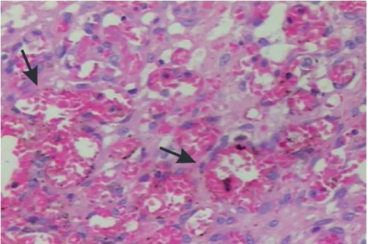
consistent with papillary endothelial hyperplasia (Figure 1). The patient recovered uneventfully and was discharged on the 2ndpostoperative day. The patient is now in the 16th postoperative month, with clinical improvement of the pain and complete weight regain.
DISCUSSION
DISCUSSION
DISCUSSION
DISCUSSION
DISCUSSION
Papillary endothelial hyperplasia is an uncommon lesion. In a recent review by Aktar et al.1, six cases of this tumor were described affecting the renal sinus and only one was adrenal Russel et al. 4.
Lesions are mostly asymptomatic; however, depending on their location, they may affect the urinary system (macroscopic hematuria, intermittent abdominal pain), parapharyngeal space (vocal cord edema), CNS (headache and dyslalia)2,3.
Radiological features are small calcifications in a well-circumscribed mass. The lesion resembles a true neoplasm with degenerative changes including necrosis and thrombosis as the proliferative process outgrows its blood supply. It is believed that endothelial proliferation takes place in response to inflammation and stasis inside the vascular bed. In its pure form, the lesion is small (average of 2 cm), purple-red, a multicystic mass containing blood clots, surrounded by a fibrous pseudocapsule containing residual smooth muscle or smooth tissue from the wall of the preexisting vessel. In dilated smaller-caliber vessels, little or no muscle tissue is evident in the pseudocapsule. Rarely,
Figure 1 – Figure 1 –Figure 1 –
278
Rev. Col. Bras. Cir. 2009; 36(3): 277-278
N a s s i f N a s s i f N a s s i f N a s s i f N a s s i f Papillary endothelial hyperplasia of adrenal: case report
the rupture of the vessel of origin allows the spilling over of the process into surrounding tissue, which should not be understood as a sign of malignancy1-3.
Papillary endothelial hyperplasia lesions are qui-te ofqui-ten confused with angiosarcomas. They can be differentiated from the latter because PEH lesions are confined to the vascular lumen, and do not exhibit frank necrosis, marked pleomorphism nor a high mitotic index. Passive extension of this process into the surrounding soft tissue occurs as a result of vessel rupture.2,4.
Despite the benign nature of the tumor, weight loss is reported for 60% of the patients. Some authors
relate that symptom to the intermittent pain, eating difficulties as a result of the obstruction to the passage of food (for pharyngeal tumors) or malabsorption (small intestine and colon tumors). The significant protein loss (renal sinus tumors), hormonal changes caused by the destruction of the glands that secrete those hormones, and the production of endothelial factors promoting satiety are also mentioned as likely explanations for the weight loss3.
Prognosis is excellent for this lesion, which is cured by simple excision. In cases with recurrence, there is usually a concomitant vascular tumor4.
A B S T R A C T A B S T R A C T A B S T R A C T A B S T R A C T A B S T R A C T
Intravascular papillary endothelial hyperplasia is a benign and rare intravascular process thought to arise from an organizing thrombus. Involvement of the adrenal gland is extremely rare, with only one case reported in the literature. We report a case of this vascular lesion in the adrenal gland, treated with laparoscopic adrenalectomy.
Key words: Key words: Key words: Key words:
Key words: Hyperplasia. Adrenal glands. Endothelial cells.
REFERENCES
REFERENCES
REFERENCES
REFERENCES
REFERENCES
1. Akhtar M, Aslam M, Al-Mana H, Bamefleh H, Al-Khateeb SS, Lindstedt E. Intravascular papillary endothelial hyperplasia of renal vein: report of 2 cases. Arch Pathol Lab Med. 2005; 129(4):516-9. 2. Garber BB, Prestipino AJ, Pollack HM, Levine SR, Whitmore KE. Masson’s tumor of the kidney: a new renal lesion. J Urol. 1990; 143(2):344-6.
3. Johraku A, Miyanaga N, Sekido N, Ikeda H, Michishita N, Saida Y et al. A case of intravascular papillary endothelial hyperplasia (Masson’s tumor) arising from renal sinus. Jpn J Clin Oncol. 1997; 27(6):433-6.
4. Kawashima A, Johsen T, Murayama S, Russel WJ. Intravascular papillary endothelial hyperplasia of the adrenal gland. Br J Radiol. 1986; 59(702):610-3.
Received 22/03/2006
Accepted for publication 22/05/2006 Conflict of interest: none
Financial source: none
How to cite: How to cite:How to cite: How to cite: How to cite:
Nassif AE, Pozzobon HJ, Azevedo EZ, Taguchi WS, Gomes RX. Paplllary endothelial hyperplasia of the adrenal gland: a case report . Rev Col Bras Cir. [periódico na Internet] 2009; 36(3). Disponível em URL: http:/ /www.scielo.br/rcbc
Correspondence address: Correspondence address:Correspondence address: Correspondence address: Correspondence address: Aissar Eduardo Nassif