○ ○ ○ ○ ○ ○ ○ ○ ○ ○ ○ ○ ABSTRACT○ ○ ○ ○ ○ ○ ○ ○ ○ ○ ○ ○ ○ ○ ○ ○ ○ ○ ○ ○ ○ ○ ○ ○ ○ ○ ○
INTRODUCTION
Epidermal differentiation is a process in which keratinocytes are morphologically and biochemically modified. Leaving the stratum basale, they move through the stratum spinosum and stratum granulosum and stop at the upper layer (stratum corneum), thus con-stituting multilamellar structures of anucleated corneocytes surrounded by extracellular lipids. In addition to the keratinocytes, the basal membrane contains melanocytes, which are cells responsible for pigmenting the skin, with the synthesis of melanin that is progressively transferred to the keratinocytes.1-3
The dermis is composed of a dense tissue of collagen and elastic fibers produced by der-mal fibroblasts, which provides the physical consistency of the skin. It contains blood and lymph vessels as well as nerves, which inform the organism about its interaction with the environment. It also contains hair follicles, sweat and sebaceous glands.4
Degeneration of dermal and epidermal elements may occur in extensive, deep skin and mucosal lesions, without spontaneous tis-sue regeneration. In such cases it is possible to use autologous or allogenic transplants of fro-zen or lyophilized human or animal skin, syn-thetic tissues or biodegradable materials.1
An option for the in vitro culturing of autologous cells has recently emerged, with the aim of regenerating the destroyed cutane-ous tegument. Through technological ad-vances in epithelial cell culturing, models of the epidermis reconstructed in vitro have been achieved, presenting characteristics of mor-phological and biochemical differentiation
• Jussara Rehder
• Luís Ricardo Martinhão Souto
• Cláudia Maria Bernardino Magro Issa
• Maria Beatriz Puzzi
Model of human epidermis
reconstructed
in vitro
with
keratinocytes and melanocytes
on dead de-epidermized
human dermis
Skin Cell Culture Laboratory at Faculdade de Ciências Médicas,
Universidade Estadual de Campinas, Campinas, São Paulo, Brazil
CONTEXT: Recent progress in the field of epithelial culture techniques has allowed the development of culture systems in which the reconstructed epi-dermis presents characteristics of morphological differentiation similar to those seen in vivo. Hu-man epidermis reconstructed in vitro may be used as the best alternative for the in vitro testing of the toxicology and efficiency of products for topical use, as well as in the treatment of skin burns and chronic skin ulcers.
OBJECTIVE: To demonstrate a method for obtaining human epidermis reconstructed in vitro, using keratinocytes and melanocytes cultivated on dead de-epidermized human dermis.
TYPE OF STUDY: Experimental/laboratory.
SETTING: Skin Cell Culture Laboratory of the Faculdade de Ciências Médicas, Universidade Estadual de Campinas, Campinas, São Paulo, Brazil.
PROCEDURE: Human keratinocytes and melanocytes cultured in vitro were grown on a biological ma-trix (dead de-epidermized human dermis) and the system was kept at an air-liquid interface, in a suitable culturing medium, until a stratified human epidermis was formed, maintaining the histologi-cal characteristics of the epidermis in vivo.
RESULTS: It was histologically demonstrated that it is possible to reproduce a differentiated epidermis through keratinocytes and melanocytes cultured on dead de-epidermized human dermis, thus obtain-ing a correctly positioned human epidermis recon-structed in vitro with functional keratinocytes and melanocytes that is similar to in vivo epidermis.
CONCLUSIONS: It is possible to obtain a completely differentiated human epidermis reconstructed in vitro from keratinocyte and melanocyte cultures on a dead de-epidermized human dermis.
KEY WORDS: Epidermis. Melanocyte. Culture. Keratinocyte.
similar to those seen in vivo.5-9
Over the last few years, several laborato-ries have made continuous efforts to obtain living skin models in vitro, so as to investigate the regulation of keratinocyte proliferation and differentiation and for efficacy tests on toxi-cology and skin products.10,11
The method for keratinocyte culturing at the air-liquid interface was first described by Pruniéras et al. in 1983.12 Currently, several methods are available.5-10,12-14
At the Skin Cell Culture Laboratory of Faculdade de Ciências Médicas, Universidade Estadual de Campinas, the method for keratinocyte and melanocyte culturing and achievement of reconstructed epidermis at the air-liquid interface, which was developed by Pruniéras et al. and improved by Bessou et al. in 1995,15 has been modified, implemented and improved, with the aim of obtaining a reconstructed epidermis equivalent to in vivo epidermis.
○ ○ ○ ○ ○ ○ ○ ○ ○ ○ ○ ○ ○ ○ ○ ○ ○ ○ ○ ○
MATERIAL AND METHODS
Collection of material
Skin fragments from patients submitted to breast and abdomen surgical procedures at the University of Campinas Teaching Hospi-tal were collected. This procedure was in ac-cordance with the ethical standards of the Eth-ics Committee of Faculdade de Ciências Médicas, Universidade Estadual de Campinas, Campinas, São Paulo, Brazil.
Preparation of culture samples
The material was placed in sterile tem-pered glass jars and conserved in 0.9%
physi-Original Ar
ticle
São Paulo Medical Journal — Revista Paulista de Medicina
23
ological serum refrigerated to 4° C, without exceeding a limit of 12 hours until its ma-nipulation.
The skin fragments were separated from the adipose tissue, placed on a Petri dish (Corning) and sectioned into pieces of 2 to 3 mm, using a surgical instrument under a laminar flow culture hood, so as to keep the whole procedure sterile.
These fragments were placed on a new Petri dish with 10 ml of 0.25% trypsin solution and 1 mM of ethylenediamine tetraacetic acid (GIBCO BRL, Grand Island, New York, USA, cat. no. 25200-056), with the epidermis always facing upwards. They were then incubated in an oven at 37° C, with 5% CO2 tension for four hours. This procedure resulted in separa-tion of the epidermis from the dermis.
After this period, the trypsin was neu-tralized using the same volume of fetal bo-vine serum (GIBCO, cat. no. 10270-106) and the suspension was filtered in a 50 ml tube (Falcon) with a 40-mm nylon filter (Fal-con code 2340).
This suspension was centrifuged at 1,200 rpm and 4° C for 10 minutes and the supernatant was discarded, thus obtaining a cell “pool” con-taining keratinocytes and melanocytes, which were resuspended in 5 ml of 0.9% saline solu-tion. After this, one aliquot was removed for manual cell counting in a Neubauer chamber using the trypan blue exclusion method.
Cell culturing
The cells were divided among Corning culture flasks, with 1 x 10⁵ cells per cm2 and incubated at 37° C, with 5% CO2 tension, in a specific culture medium for keratinocytes and melanocytes.
Culture medium for keratinocytes Keratinocyte culture medium was used (GIBCO cat. no. 10724-011), complemented with L-glutamine 2 mM/ml, penicillin 100 UI/ml, streptomycin 0.1 mg/ml (GIBCO cat. no. 10378-016) and fetal bovine serum 10%. Cell adhesion to the culture flasks oc-curred within 48 hours, thus obtaining the primary keratinocyte culture (Figure 1).
Culture medium for melanocytes Melanocyte culture medium MCDB 153 was used (Sigma Chemical Co., St. Louis, Missouri, USA, M 7403), complemented with L-glutamine 2 mM/ml, penicillin 100 UI/ml, streptomycin 0.1 mg/ml, fetal bovine serum 10%, epidermis growth factor 5 µg/ml (GIBCO cat. no. 10450-013), bovine pitui-tary extract 50 µg/ml (GIBCO cat. no.
13028-014), hydrocortisone 0.6 µg/ml (Sigma H 0888) and bovine insulin 3 µg/ml (GIBCO cat. no. 13007-018).
Cell adhesion to the culture flasks oc-curred within 48 hours, thus obtaining the primary melanocyte culture (Figure 2).
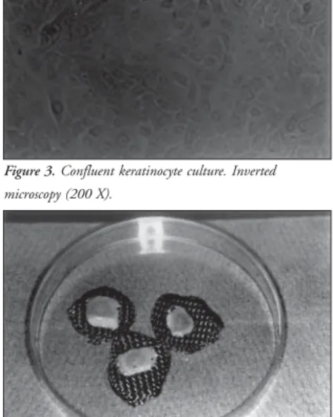
The culture medium (for keratinocytes and melanocytes) was changed every three days. When the flask wall was totally covered by cells (Figure 3), we cut them into small pieces.
Preparation of the dead de-epidermized human dermis
In order to obtain the reconstructed epi-dermis in vitro, melanocytes and keratinocytes need to be reproduced on a substrate. For this, we chose to use dermis, which we named dead de-epidermized human dermis, following the technique described by Pruniéras et al. (1979).16,17
The skin originated from patients submit-ted to corrective breast and abdomen surgery at the University of Campinas Teaching Hos-pital, it was cut into fragments of 2.0 x 2.0 cm. The skin squares were rinsed in 70º GL alcohol and then put in 0.9% saline solution with antibiotics (penicillin 100 UI/ml, strep-tomycin 0.1 mg/ml), and incubated for 10 days at 37° C. Then the epidermis was sepa-rated from the dermis.
Developing reconstructed epidermis
The keratinocyte and melanocyte cultures were prepared separately (centrifuged), to be seeded on the de-epidermized dermis. The melanocyte to keratinocyte ratio used was 1:40. The dead de-epidermized human dermis was placed on a grid and/or gauze and the mixed epidermal cells were seeded with 2 x 10⁶ cells per cm² on the dermis, in 150 ul of keratinocyte culture medium contained by a polypropylene ring. Then this seeded dermis was incubated at 37° C, with 5% CO2 ten-sion for 48 hours, which was the time needed for cell adhesion to the dermis.
After this period the polypropylene rings were removed and the system (dermis plus cells) was submersed in epidermis culture medium.
Culture medium for epidermis Three parts of Iscove’s Modified Dulbecco’s Medium (IMDM — GIBCO cat. no. 12200-036) and one part of keratinocyte culture medium (GIBCO cat. no. 10724-011) were used, complemented with L-glutamine 2 mM/ml, penicillin 100 UI/ml, streptomy-cin 0.1 mg/ml (GIBCO cat. no. 10378-016) and fetal bovine serum 10%.
Seventy-two hours later, the system was maintained at the air-liquid interface and the medium was complemented with Ca++ 1.5 mM and kept for 20 days, with three weekly changes.
Morphological studies of reconstructed human epidermis in vitro
The system was interrupted after being maintained at the air-liquid interface for 20 days (Figure 4), fixed in formaldehyde 10% and paraffin-embedded. Histological cuts colored with hematoxylin-eosin (HE) were made.
Figure 1. Primary keratinocyte culture. Inverted microscopy (200 X).
Figure 2. Primary melanocyte culture. Inverted microscopy (200 X).
Figure 3. Confluent keratinocyte culture. Inverted microscopy (200 X).
Figure 4. Dead de-epidermized human dermis with
epidermis reconstructed on steel grids.
São Paulo Medical Journal — Revista Paulista de Medicina
24
○ ○ ○ ○ ○ ○ ○ ○ ○ ○ ○ ○ ○ ○ ○ ○ ○ ○ ○ ○
RESULTS
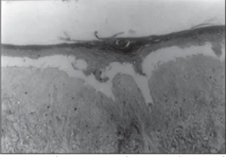
We were able to histologically demon-strate, through the hematoxylin-eosin (HE) staining, that it is possible to reproduce a com-pletely differentiated epidermis reconstructed
Figure 5. Human epidermis reconstructed in vitro on dead
de-epidermized human dermis. Optical microscopy. Hematoxylin-eosin (HE) staining (165 X).
constructed epidermis is physiologically com-patible with autografts.9,14,18
The use of autografts is limited by the extent of the donor site and the clinical con-dition of patients, in the case of large lesions. Allotransplants collected from cadavers or volunteers are rejected after one or two weeks and provide only temporary cover. Human or animal skin grafts that are devitalized, lyophi-lized or refrigerated in glycerol accommodate the connective tissue and stimulate blood ves-sel growth, but in general are prematurely de-graded. The treatment of large skin lesions with reconstructed autologous epidermis of-fers an attractive alternative to replace exist-ing therapies since, from a small skin fragment of the patient, we can obtain cell cultures that multiply rapidly and can be cryopreserved, thereby allowing their use for new treatments for an indeterminate time and making the re-moval of new skin fragments unnecessary.1
The real challenge in the twenty-first cen-tury will be to reproduce the whole skin. In fact, our interest in the present study was only the epidermis. It would be interesting to in-troduce the Langerhans cells into this model that is already quite advanced. Such a proce-dure would have the objective of restoring the immune function to the skin.7
The utilization of this model on dead de-epidermized human dermis facilitates the ad-hesion of keratinocyte and melanocyte through the preservation of the basal mem-brane constituents.19 However, it would also be interesting to reproduce this system on a more physiological dermis. The types of der-mis for such a proposal have not yet been well developed.
The model of human epidermis recon-structed in vitro presented herein has low pros-pects for clinical use in burns and chronic skin ulcers. This is not only because of the diffi-culty in removing the reconstructed epimis from dead de-epidermized human der-mis without causing lesions, but also because it does not present an associated dermis. Oth-erwise, as already mentioned, it possesses ex-cellent applicability for laboratory studies.
○ ○ ○ ○ ○ ○ ○ ○ ○ ○ ○ ○ ○ ○ ○ ○ ○ ○ ○ ○
CONCLUSION
It is possible to obtain a sufficient number of cells from human keratinocyte and melano-cyte cultures for emplacement in dead de-epidermized human dermis. This allows the formation of a completely differentiated hu-man epidermis reconstructed in vitro.
Our next step would be to improve this Figure 6. Human epidermis reconstructed in vitro on dead
de-epidermized human dermis. Optical microscopy. Hematoxylin-eosin (HE) staining (330 X).
Figure 7. Epidermis reconstructed in vitro, in the process of separation from the dead de-epidermized human dermis.
Optical microscopy. Hematoxylin-eosin staining (165 X).
Figure 8. Epidermis reconstructed in vitro, in the process of separation from the dead de-epidermized human dermis.
Optical microscopy. Hematoxylin-eosin (HE) staining (330 X).
in vitro from keratinocyte and melanocyte cultures on a dead de-epidermized human dermis (Figures 5 and 6), with functional keratinocytes and melanocytes that are cor-rectly positioned, equivalent to epidermis in vivo. The extent of the stratification and kerati-nization of human epidermis reconstructed in vitro had the same characteristics as found in vivo (Figures 7 and 8).
After developing the human epidermis reconstructed in vitro, we successfully tripli-cated the experiment to validate the technique.
○ ○ ○ ○ ○ ○ ○ ○ ○ ○ ○ ○ ○ ○ ○ ○ ○ ○ ○ ○
DISCUSSION
The present study, although it describes a method that is sophisticated and difficult to put into practice, showed that it is possible to obtain a model of reconstructed human epi-dermis using materials and methodology dif-ferent from those previously described, with the purpose of enabling laboratory investiga-tions and clinical treatments that have been difficult to obtain in our country up to the present day.
Over the course of a two-year period, we had attempted to reproduce in its totality the technique used by foreign authors. We did not obtain cell reproduction in the cultures until we standardized the addition of fetal bovine serum 10% directly to the melanocyte and keratinocyte culture media.
The model of human epidermis recon-structed in vitro provides a good system for studies, especially in relation to tests on the efficiency and toxicology of new chemicals and drugs in vitro.10,11
Ultraviolet rays affect epidermal differen-tiation. Therefore, it is possible to study the effects of solar radiation on an epidermis com-posed of melanocytes and keratinocytes. This model does not allow the study of the immu-nological effects of radiation measured by Langerhans cells or UV-induced macropha-ges.15 However, it does allow the study of the biological effects of irradiation, particularly lipid peroxidation.3 It also allows us to study the effect of sunscreens to validate the photoprotection model (non-immunological). This model will allow us to study the physiopathology and possible therapies for still-undetermined pigmentary affections such as vitiligo, melasma and the formation of melanocytic nevus.
The transplantation of cultured autolo-gous keratinocytes is the most advanced area of tissue engineering and it has an important application in the restoration of skin lesions such as burns and chronic ulcers.18 The
São Paulo Medical Journal — Revista Paulista de Medicina
25
system, with the purpose of reproducing hu-man dermis with viable fibroblasts inside it,
in order to facilitate the adhesion, multiplica-tion and differentiamultiplica-tion of the epidermal cells,
and to clinically use such dermis in associa-tion with this epidermis.
Modelo de epiderme humana reconstruída in
vitro com queratinócitos e melanócitos so-bre derme humana morta desepidermizada
CONTEXTO: Recentes progressos no campo das
técnicas de cultura epitelial têm levado ao desenvolvimento de sistemas de cultura nos quais a epiderme reconstruída obtida exibe características de diferenciação morfológica semelhantes àquelas vistas in vivo. Uma epiderme humana reconstruída in vitro pode ser utilizada como melhor alternativa para testes toxicológicos e de eficácia de produtos de uso tópico in vitro e ainda no tratamento de queimaduras e úlceras crônicas de pele.
OBJETIVO: Demonstrar um método de
obten-ção de epiderme humana reconstruída in vitro, utilizando queratinócitos e melanócitos cultivados sobre uma derme humana morta desepidermizada.
TIPO DE ESTUDO: Experimental laboratorial.
LOCAL: Laboratório de Cultura de Células da
Pele da Faculdade de Ciências Médicas da Universidade Estadual de Campinas, Cam-pinas, São Paulo, Brasil.
○ ○ ○ ○ ○ ○ ○ ○ ○ ○ ○ ○ ○ ○ ○ ○ ○ ○ ○ ○ ○ ○ ○ ○ ○ ○ ○ ○ ○ ○ ○ ○ ○ ○ ○ ○ ○ ○ ○ ○ ○ ○
RESUMO
Jussara Rehder, MD. Chief biologist of the Laboratory of Molecular Biology and Skin Cell Culture Laboratory, Universidade Estadual de Campinas, Campinas, São Paulo, Brazil.
Luís Ricardo Martinhão Souto, MD. Plastic surgeon and MSc student of Medical Sciences at Faculdade de Ciências Médicas, Universidade Estadual de Campinas, Campinas, São Paulo, Brazil.
Cláudia Maria Bernardino Magro Issa, MD. Derma-tologist and PhD student of Internal Medicine at Faculdade de Ciências Médicas, Universidade Estadual de Campinas, Campinas, São Paulo, Brazil.
Maria Beatriz Puzzi, MD, PhD. Professor of the Disci-pline of Dermatology, Department of Internal Medicine, and Head of the Skin Cell Culture Laboratory at Faculdade de Ciências Médicas, Universidade Estadual de Campinas, Campinas, São Paulo, Brazil.
Sources of funding: None
Conflict of interest: None
Date of first submission: May 9, 2003
Last of received: July 17, 2003
Accepted: August 27, 2003
Address for correspondence:
Luís Ricardo Martinhão Souto
Rua Coronel Quirino, 320 — Apto. 43 — Cambuí Campinas/SP — Brasil — CEP 13025-001 Tel. (+55 19) 3295-0902
Fax (+55 14) 432-3920 E-mail: [email protected]
COPYRIGHT © 2004, Associação Paulista de Medicina
○ ○ ○ ○ ○ ○ ○ ○ ○ ○ ○ ○ ○ ○ ○ ○ ○ ○ ○ ○
Publishing information
PROCEDIMENTOS: Queratinócitos e
mela-nócitos humanos cultivados in vitro foram semeados sobre uma matriz biológica (derme humana morta desepidermizada) e o sistema foi mantido em interface ar-líquido, em meio de cultura adequado, até haver a formação de uma epiderme humana estratificada, man-tendo as características histológicas da epi-derme in vivo.
RESULTADOS: Demonstramos,
histologica-mente, que é possível reproduzir uma epi-derme diferenciada, a partir da cultura de queratinócitos e melanócitos sobre uma derme humana morta desepidermizada, ob-tendo uma epiderme humana reconstruída in vitro, com queratinócitos e melanócitos funcionais, corretamente posicionados, equi-valente à epiderme in vivo.
CONCLUSÕES: É possível obter uma epiderme
humana reconstruída in vitro completamen-te diferenciada a partir da cultura de quera-tinócitos e melanócitos sobre uma derme humana morta desepidermizada.
PALAVRAS-CHAVES: Epiderme. Cultura.
Melanócitos. Queratinócitos. Cultura de celulas.
1. Boranic M, Jakic-Razumovic J, Stanovic S, Kljenak A, Fattorini I. Kultura koznih stanica: primjena u plasticnoj kirurgiji i laboratorijskom istrazivanju. [Skin cell culture: utilization in plastic surgery and laboratory studies]. Lijec Vjesn 1999;121(4-5):137-43.
2. Watt FM. The epidermal keratinocyte. Bioessays 1988; 8(5):163-7.
3. Taube MBP, Taieb A. Metabolismo lipídico na cultura de queratinócitos. [Lipid metabolism in cultured keratinocytes]. An Bras Dermatol 2000;75(1):75-84.
4. Huang Y, Ren L, Qin Y. Observation of cicatricial fibroblasts in culture and its biological properties. Zhongguo Xiu Fu Chong Jian Wai Ke Za Zhi 1998;12(6):332-5.
5. Valyi-Nagy IT, Murphy GF, Mancianti ML, Whitaker D, Herlyn M. Phenotypes and interactions of human melanocytes and keratinocytes in an epidermal reconstruction model. Lab In-vest 1990;62(3):314-24.
6. Bernerd F, Asselineau D. Successive alteration and recovery of epidermal differentiation and morphogenesis after specific UVB-damages in skin reconstructed in vitro. Dev Biol 1997;183(2):123-38.
7. Régnier M, Patwardhan A, Scheynius A, et al. Reconstructed
○ ○ ○ ○ ○ ○ ○ ○ ○ ○ ○ ○ ○ ○ ○ ○ ○ ○ ○ ○ ○ ○ ○ ○ ○ ○ ○ ○ ○ ○ ○ ○ ○ ○ ○ ○ ○ ○ ○ ○ ○ ○ ○ ○ ○ ○ ○ ○ ○ ○ ○ ○ ○ ○ ○ ○ REFERENCES○ ○ ○ ○ ○ ○ ○ ○
human epidermis composed of keratinocytes, melanocytes and Langerhans cells. Med Biol Eng Comput 1998;36(6):821-4. 8. Chistolini P, De Angelis G, De Luca M, Pellegrini G, Ruspantini
I. Analysis of the mechanical properties of in vitro reconstructed epidermis: preliminary results. Med Biol Eng Comput 1999;37(5):670-2.
9. Carsin H, Ainaud P, Le Bever H, et al. Cultured epithelial autografts in extensive burn coverage of severely traumatized patients: a five year single-center experience with 30 patients. Burns 2000;26(4):379-87.
10. Régnier M, Caron D, Reichert U, Schaefer H. Reconstructed human epidermis: a model to study in vitro the barrier func-tion of the skin. Skin Pharmacol 1992;5(1):49-56. 11. Régnier M, Asselineau D, Lenoir MC. Human epidermis
re-constructed on dermal substrates in vitro: an alternative to ani-mals in skin pharmacology. Skin Pharmacol 1990;3(2):70-85. 12. Pruniéras M, Régnier M, Woodley D. Methods for cultivation of keratinocytes with an air-liquid interface. J Invest Dermatol 1983;81(1 Suppl):28s-33s.
13. Ponec M, Gibbs S, Pilgram G, et al. Barrier function in recon-structed epidermis and its resemblance to native human skin. Skin Pharmacol Appl Skin Physiol 2001;14(Suppl 1):63-71.
14. van Dorp AG, Verhoeven MC, Nat-Van Der Meij TH, Koerten HK, Ponec M. A modified culture system for epidermal cells for grafting purposes: an in vitro and in vivo study. Wound Repair Regen 1999;7(4):214-25.
15. Bessou S, Surlève-Bazeille JE, Sorbier E, Taieb A. Ex vivo re-construction of the epidermis with melanocytes and the influ-ence of UVB. Pigment Cell Res 1995;8(5):241-9. 16. Pruniéras M, Régnier M, Schlotterer M. Nouveau procédé de
culture des cellules épidermiques humaines sur derme homo-logue ou hétérohomo-logue: préparation de greffons recombinés.[New procedure for culturing human epidermal cells on allogenic or xenogenic skin: preparation of recombined grafts]. Ann Chir Plast1979;24(4):357-62.
17. Régnier M, Pruniéras M, Woodley D. Growth and differentia-tion of adult human epidermal cells on dermal substrates. Front Matrix Biol 1981;9:4-35.
18. Terskikh VV, Vasiliev AV. Cultivation and transplantation of epidermal keratinocytes. Int Rev Cytol 1999;188:41-72. 19. Ponec M, Kempenaar J, Weerheim A, de Lannoy L, Kalkman I,
Jansen H. Triglyceride metabolism in human keratinocytes cul-tured at the air-liquid interface. Arch Dermatol Res 1995;287(8):723-30.