Sialic acid change s in D alto n’s
lym pho m a-be aring m ice afte r
cyclo pho spham ide and cisplatin
tre atm e nt
Cell and Tumour Biology Laboratory, Department of Zoology, School of Life Sciences, North-Eastern Hill University, Shillong, India B.M. Nicol and
S.B. Prasad
Abstract
Sialic acid changes in Daltons lymphoma cells and other tissues of 10-12-week-old Swiss albino mice were investigated in relation to tumour growth in vivo and following cyclophosphamide (ip, 200 mg/ kg body weight) or cisplatin (ip, 8 mg/kg body weight) treatment. Three to four animals of both sexes were used in each experimental group. The sialic acid level of tumour cells (0.88 µmol/g) increased with tumour progression (1.44-1.59 µmol/g; P£0.05) in mice. Sialic acid concentration in other tissues (liver, kidney, testes and brain) also increased (~40, 10, 30 and 58%, respectively) in the tumour-bearing hosts as compared with that in the respective tissues of normal mice. In vivo cyclophosphamide or cisplatin treatment resulted in an overall decrease of sialic acid contents in the tissues. Cyclophosphamide was more efficient in lowering tissue sialic acid than cisplatin (P£0.01, ANOVA). It is suggested that sialic acid residues could be an impor-tant factor contributing to the manifestation of malignant properties in cancer cells in general and Daltons lymphoma cells in particular. A significant decrease in the sialic acid content of Daltons lymphoma cells after cisplatin or cyclophosphamide treatment may bring about specific changes in tumour cells which could be associated with tumour regression.
Co rre spo nde nce
S.B. Prasad
Cell and Tumour Biology Laboratory Department of Zoology
School of Life Sciences North-Eastern Hill University Shillong-793 022 India
Fax: + 91-364-55-0076/55-0108 E-mail: sbpnehu@ hotmail.com or sbprasad@ nehu.ac.in
Research supported by North-Eastern Hill University, Shillong and University Grants Commission (under DRS, CO SIST programme), New Delhi.
Received June 20, 2001 Accepted March 6, 2002
Ke y wo rds
·Sialic acid ·Cisplatin
·Cyclophosphamide ·Dalton’s lymphoma
Cyclophosphamide (2-[bis-/2-chloro- ethyl-amino]-tetrahydro-2H-1,2,3-oxaza-phosphorine-2-oxide) is an alkylating che-motherapeutic drug used against a wide spec-trum of malignancies which include leuke-mia, breast cancer, lymphoma, lung cancer, prostate cancer and ovarian cancer (1). The cytotoxicity induced by cyclophosphamide is directly connected with its metabolism and toxic reactions of its metabolites. The parent compound is inactive in vitro and in vivo. Activation of cyclophosphamide by the
leading chemotherapeutic drugs being used effectively against various malignancies (4,5). It has been reported that cisplatin has an effect on the surface of the cells, and brings about definite changes in cell lectin agglutinability and in the topographical pat-tern of lectin-binding sites on the cell surface (6).
Sialic acids are derived from neuraminic acid whose main derivative is N-acetyl-neuraminic acid which is generally used as the synonym for sialic acid (7). Sialic acids are widely distributed in nature as non-re-ducing termini of glycoproteins and glycolip-ids. The presence of sialic acids has been reported in some viruses, bacteria, plants, different invertebrates and all vertebrate tis-sues (8). About 70% of the total sialic acids of eukaryotic cells are found on the cell surface and the remainder is distributed among the endoplasmic reticulum, mitochon-dria, lysosomes, etc. (8). Because of their acidic nature, they impart a negative charge to the cell surface and are important in cell-to-cell or cell-to-matrix interactions. Sialic acid residues on the cell surface may also be involved in the masking of cell surface anti-gens and may act as receptors for lectins, virus particles, some hormones and antibod-ies (9). There is a large body of evidence suggesting that the surface properties of tu-mour cells differ from those of their normal counterparts and that these changes are due in part to altered sialoglycoconjugates ex-pressed on the plasma membrane (10). It has been reported that with the progression of tumour development there is an increase in sialic acid content of Yoshida ascites sar-coma cells (11). Although elevated levels of sialic acid have been often associated with malignancy (12), a clear correlation of changes in sialic acid concentrations and malignancy has not emerged, because some reports (13) have shown a decrease and not an increase in sialic acid in association with malignancy. Thus, evaluation of sialic acid changes could be very helpful by
contribut-ing both to the diagnosis of patients and to monitoring their progression and response to treatment.
In view of the importance of sialic acid in the manifestation of the biological proper-ties of malignant cells, the present studies were undertaken to elucidate quantitative changes in sialic acid (N-acetylneuraminic acid) during ascites Daltons lymphoma tu-mour progression and after treatment with cyclophosphamide and cisplatin to illustrate the comparative effects of these drugs on the sialic acid content of various host tissues.
Ascites Daltons lymphoma was main-tained in vivo in 10-12-week-old inbred Swiss albino mice by serial intraperitoneal (ip) transplantations of 1 x 10⁷ tumour cells per
animal (0.25 ml volume in PBS). PBS was prepared by adding 0.15 M NaCl to 0.01 M sodium phosphate buffer, and pH was ad-justed to 7.4. Tumour-transplanted hosts usu-ally survived for 20-22 days.
The cyclophosphamide (14) and cisplatin (4) doses used were 200 and 8 mg/kg body weight, respectively, for the treatment of tumour-bearing mice. A single dose of cyclo-phosphamide or cisplatin was administered ip to tumour-bearing mice on the 10th day post-tumour transplantation which is approxi-mately the mid period of tumour growth. After 24, 48, 72 and 96 h of treatment (i.e., on the 11th, 12th, 13th and 14th day post-tumour transplantation) liver, kidney, testes, brain and ascites tumour were collected. The same tissues were also collected from the control tumour-bearing mice which had been injected with the same volume of 0.89% NaCl. Ascites tumour was centrifuged (800 g, 10 min, at 4ºC) to separate the tumour cell pellet. Liver, kidney, testes and brain were also collected from normal animals bearing no tumour and injected with 0.89% NaCl only. The drug treatments and sialic acid determinations were repeated independently 3-4 times.
centrifuged (800 g, 10 min, 4ºC) to obtain tumour cell pellets. The tumour cell pellets and other tissues were homogenized in 0.1 N H2SO4 (1.0 ml/10 mg tissue) and incubated in a water bath at 80ºC for 1 h with intermit-tent shaking. The resulting suspensions were centrifuged (800 g, 10 min) and the sialic acid contents were estimated in the superna-tants by the method of Warren (15).
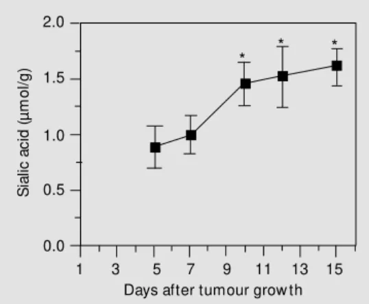
Sialic acids terminate oligosaccharide chains in the mammalian glycoconjugates and play a key role in the normal functions of these glycoconjugates. It has been reported that sialic acid could be used as a sensitive biomarker for lung cancer although their specificity is low (16). However, a definite correlation of the changes in sialic acid con-centration with malignancy has not been reported because of various reports indicat-ing an increase (12) or a decrease (13) of sialic acid in different malignancies. The present findings showed an increase in sialic acid concentrations in Daltons lymphoma cells with tumour growth in mice (Figure 1), which may be an important feature of this tumour. The increase of sialic acid in Daltons lymphoma cells may be due to enhanced activity of enzymes involved in sialic acid synthesis and/or transfer. Some reports have indicated a 3-5 times increased sialyl trans-ferase activity in various virally transformed cells as compared to the corresponding nor-mal cells, an event that may be associated with the increase in the amount of sialic acid in the transformed cells (13). The elevated sialic acid levels in malignant cells have also been observed for murine Yoshida ascites sarcoma (11). The influence of sialic acid on the oncogenicity of tumour cells may be based on i) a negative charge determining constituent on the cell surface, resulting in the loss of contact inhibition, ii) an antigen-masking agent, and iii) a component of the cell surface involved in the adherence of tumour cells to the mesothelial membrane prior to their dissemination to form metasta-ses (9).
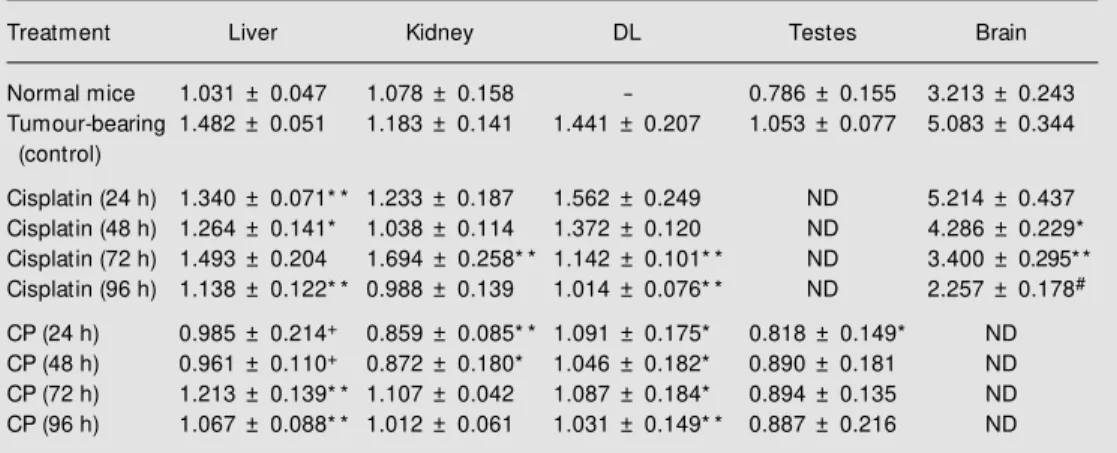
In our experiments, the determination of sialic acid in other tissues revealed that sialic acid content was much higher in brain (~3 µmol/g) than in liver, kidney and testes (~1 µmol/g) (Table 1). It was noted that sialic acid content increased in the tissues of tu-mour-bearing mice when compared with the respective tissue of normal mice (Table 1). This increase in sialic acid content was higher in the brain (~58%), liver (~40%) and testes (~30%) than in the kidney (~10%) (Table 1). In many diseases such as Salla disease, free sialic acid storage disease, and sialuria, an increased concentration of free sialic acid in various tissues and fluids has been observed which may be due at least in part to defective de novo synthesis, transport, storage, ca-tabolism, excretion and/or metabolic regula-tion of sialic acid in the cells (17), and a similar activity may have been involved here in Daltons lymphoma-bearing mice also. Furthermore, the observation of increased sialic acid content in the tissues of tumour-bearing mice could be helpful for Daltons lymphoma cells in the host since sialic acid has also been known to be important in the transport of proteins, amino acids and ions to the cancer cells (10). Therefore, it was quite natural to evaluate the activities of some anticancer agents in terms of their effects on the changes in sialic acid levels of tissues as well as Daltons lymphoma cells of tumour-bearing mice. For this purpose, in the present study cisplatin and cyclophosphamide were
S
ia
lic
a
c
id
(
µ
m
o
l/
g
)
2.0
1.5
1.0
0.5
0.0
1 3 5 7 9 11 13 15
Days after tumour grow th
* * *
Figure 1. Changes in the sialic acid content of Dalton’s lym-phoma cells w ith grow th of the tumour in mice. Data are re-ported as means ± SD (N = 4). * P<0.05 compared to the 5th day of tumour grow th (Student
used as the anticancer agents. Cisplatin or cyclophosphamide treatment of Daltons lymphoma-bearing mice causes tumour re-gression, suggesting an effective anticancer activity of these drugs in this murine tumour model (Prasad SB and Nicol BM, unpub-lished results). Although the ability of these drugs to interact with cellular DNA has been suggested to be the primary target in the mechanism of their anticancer activity (3,4), the association of additional components such as biochemical/enzymatic changes, cell sur-face, immune response of the cells/host is also known (6) and has led to the proposition of the involvement of multistep/multilevel effects in the hosts during cisplatin-medi-ated cancer chemotherapy (5). As far as their effects on quantitative changes in sialic acids of Daltons lymphoma cells and tissues is concerned, it was noted that cyclophospha-mide or cisplatin treatment of tumour-bear-ing mice for 24-96 h caused a decrease of sialic acid in the tissues (Table 1). The de-crease of sialic acid contents after cisplatin treatment was noted to be predominant in the brain. Furthermore, a comparative analysis of the effect of these two drugs revealed that
the sialic acid decrease in cyclophospha-mide-treated tissues was more marked than the decrease in cisplatin (Table 1, P£0.01, ANOVA) which suggests that sialic acid decrease may be playing a more substantial role in the cyclophosphamide-mediated antitumour effect than in the cisplatin-medi-ated effect. About 70% of total sialic acid is generally found on the cell surface (8) and the decrease of sialic acid in Daltons lym-phoma cells after cyclophosphamide or cis-platin treatment may be associated with an enhancement of the immune response of the host. It has been suggested that the loss of sialic acid should lower the negative surface charge and may lead to increased cell defor-mity and to enhanced cell susceptibility to phagocytosis (18). It has been suggested that although sialic acid itself is not antigenic, it may control the expression of surface anti-gens, and sialic acid release from tumour cells after cisplatin treatment may cause the possible exposure of certain antigenic sites on the tumour cell surface (19). In fact, other studies have also shown that specific release of sialic acid after neuraminidase treatment of many tumour cells such as Landschutz Table 1. Quantitative changes in the sialic acid content of different tissues of Dalton’s lymphoma-bearing mice after cisplatin or cyclophosphamide treatment.
Treatment Liver Kidney DL Testes Brain
Normal mice 1.031 ± 0.047 1.078 ± 0.158 - 0.786 ± 0.155 3.213 ± 0.243
Tumour-bearing 1.482 ± 0.051 1.183 ± 0.141 1.441 ± 0.207 1.053 ± 0.077 5.083 ± 0.344
(control)
Cisplatin (24 h) 1.340 ± 0.071* * 1.233 ± 0.187 1.562 ± 0.249 ND 5.214 ± 0.437
Cisplatin (48 h) 1.264 ± 0.141* 1.038 ± 0.114 1.372 ± 0.120 ND 4.286 ± 0.229*
Cisplatin (72 h) 1.493 ± 0.204 1.694 ± 0.258* * 1.142 ± 0.101* * ND 3.400 ± 0.295* *
Cisplatin (96 h) 1.138 ± 0.122* * 0.988 ± 0.139 1.014 ± 0.076* * ND 2.257 ± 0.178#
CP (24 h) 0.985 ± 0.214+ 0.859 ± 0.085* * 1.091 ± 0.175* 0.818 ± 0.149* ND
CP (48 h) 0.961 ± 0.110+ 0.872 ± 0.180* 1.046 ± 0.182* 0.890 ± 0.181 ND
CP (72 h) 1.213 ± 0.139* * 1.107 ± 0.042 1.087 ± 0.184* 0.894 ± 0.135 ND
CP (96 h) 1.067 ± 0.088* * 1.012 ± 0.061 1.031 ± 0.149* * 0.887 ± 0.216 ND
Sialic acid content is reported as µmol/g w et w eight. Data are reported as means ± SD (N = 3-4).
* P<0.10, * * P<0.05, +P<0.02, and #P<0.01 compared w ith the respective tumour-bearing control. The
ascites tumour, L1210 tumour, methylcholan-threne-induced fibrosarcoma, and 6C3 HED lymphosarcoma resulted in increased immu-nogenicity (20) and a reduced capacity of tumour cells to proliferate in vivo. Along with the sialic acid decrease in Daltons lymphoma cells this drug-mediated decrease of tissue sialic acid in tumour-bearing mice should also bring their functional activity to normal in the hosts, thereby facilitating tu-mour regression.
Thus, the present findings suggest that
sialic acid changes in tumour cells and tis-sues could be an important step during tu-mour growth/malignancy as well as tutu-mour regression after cisplatin or cyclophospha-mide treatment and they support the view of multilevel/multistep effects of the drugs in cancer chemotherapy.
Ackno wle dgm e nts
We thank Prof. S.K. Mishra and Dr. N.P. Goel for help with some calculations.
Re fe re nce s
1. Black DJ & Livingston RB (1990). Anti-neoplastic drugs in 1990 - a review (Part I). Drugs, 39: 489-501.
2. Friedman OM , Wodinsky I & M yles A (1976). Cyclophosphamide-related phos-phoramide mustards: recent advances and historical perspective. Cancer Treat-ment Reports, 60: 337-346.
3. Wang JY, Prorok G & Vaughan WP (1993). Cytotoxicity, DNA-cross linking, and DNA single-strand breaks induced by cyclo-phosphamide in a rat leukemia in vivo.
Cancer Chemotherapy and Pharmacolo-gy, 31: 381-386.
4. Rosenberg B (1985). Fundamental stud-ies w ith cisplatin. Cancer, 55: 2303-2316. 5. Prasad SB & Giri A (1994). Antitumour effect of cisplatin against murine ascites Dalton’s lymphoma. Indian Journal of Ex-perimental Biology, 32: 155-162. 6. Prasad SB & Sodhi A (1982). Effect of cis
-dichlorodiammine platinum (II) on surface of tumor and normal cells: biochemical, fluorescence and electron microscopical studies. Indian Journal of Experimental Biology, 20: 559-571.
7. Ledeen RW & Yu RK (1976). Chemistry and analysis of sialic acid. In: Rosenberg A & Schengrund C (Editors), Biological Roles of Sialic Acid. Plenum Press, New York, NY, USA, 1-57.
8. Warren L (1976). The distribution of sialic acids w ithin the eukaryotic cell. In: Rosen-berg A & Schengrund C (Editors), Biologi-cal Roles of Sialic Acid. Plenum Press,
New York, NY, USA, 103-121.
9. Jeanloz RW & Codington JF (1976). The biological role of sialic acid at the surface of the cell. In: Rosenberg A & Schengrund C (Editors), Biological Roles of Sialic Acid. Plenum Press, New York, NY, USA, 201-238.
10. M cDonagh JC & Nathan RD (1990). Sialic acid and the surface charge of delayed rectifier potassium channels. Journal of M olecular and Cellular Cardiology, 22: 1305-1316.
11. Rao VS & Sirsi M (1973). Studies on sialic acid in Yoshida ascites sarcoma cells. In-dian Journal of Biochemistry and Biophys-ics, 10: 37-41.
12. Ingraham HA & Alhadeff JA (1978). Char-acterization of sialyl-transferase in non-cancerous and neoplastic human liver tis-sue. Journal of the National Cancer Insti-tute, 61: 1371-1374.
13. Onodera K, Yamaguchi N, Kuchino T & Aoi Y (1976). Alterations in surface glyco-proteins and level of sialyltransferase of cells transformed by a temperature-sensi-tive mutant of SV40. Proceedings of the National Academy of Sciences, USA, 73: 4090-4094.
14. Czyzew ska A & M azur L (1995). Suppress-ing effect of WR-2721 on micronuclei in-duced by cyclophosphamide in mice. Ter-atogenesis, Carcinogenesis, and M u-tagenesis, 15: 109-114.
15. Warren L (1959). The thiobarbituric acid assay of sialic acid. Journal of Biological
Chemistry, 234: 1971-1975.
16. Kakari S, St ringou E, Toum bis M , Ferderigos AS, Poulaki E, Chondros K, Dema A, Kotsovoulou V & Pavlidis N (1991). Five tumour markers in lung can-cer: significance of total and lipid-bound sialic acid. Anticancer Research, 11: 2107-2110.
17. Thom as GH, Scocca J, M iller CS & Reynolds LW (1985). Accumulation of N-acetylneuraminic acid (sialic acid) in hu-man fibroblasts cultured in the presence of N-acetylmannosamine. Biochimica et Biophysica Acta, 846: 37-43.
18. Simmons RL & Rios A (1972). M odifica-tion of immunogenicity in experimental immunotherapy and prophylaxis. In: Day SB & Good RA (Editors), M embranes and Viruses in Immunopathology. Academic Press, New York, NY, USA, 563-576. 19. Sarna S, Bhola RK & Sodhi A (1988).
Re-lease of protein bound sialic acid from fibrosarcoma cells after cis dichlorodi-ammineplatinum(II) treatment: the pos-sible role in tumour regression. Polish Journal of Pharmacology and Pharmacy, 40: 73-80.
20. Bekesi JG, Roboz JP & Holland JF (1976). Characteristics of immunity induced by neuraminidase-treated lymphosarcoma cells in C3H (M TV+) and C3H (M TV-) mice.