Sensibility of the PCR technique in the detection of
Stenocarpella
sp.
associated with
maize seeds
1Ellen Noly Barrocas
2*, José da Cruz Machado
2, Mirella Figueiró de Almeida
2,
Luana Silva Botelho
2, Édila Vilela de Resende Von Pinho
3ABStRACt - Maize seeds, infected by Stenocarpella species, are important sources of inoculum for the introduction and dissemination of stalk and ear rot and macrospore leaf spot diseases. the use of healthy seeds is an important strategy for the preventive control of these diseases. However, one of the difficulties in the health quality control programs for maize seeds is the availability of a reliable and quick method for detecting these fungi during routine seed analyses. Therefore, the objective of the present study was to investigate the possibility of using the PCR technique as an alternative method for accurately detecting these pathogens in maize seed samples. Maize seeds were kept in contact with S. maydis colonie developed in PDA media containing mannitol at -1.4 MPa for 72 h. the seed samples used in this study were prepared with infected seeds at incidences of 100, 20, 10, 2, 1 and zero %.the primers used were able to detect S. maydis fungi in association with seeds with a maximum of 2% , however those primers were not able to differentiate between the two species.
Index terns: fungus, molecular detection, stalk and ear rot, macrospore leaf spot.
Sensibilidade da técnica PCR na detecção de
Stenocarpella
sp. associados às sementes de milho
RESuMo - Sementes de milho infectadas por Stenocarpella são importantes fontes de inóculo para a introdução e disseminação da podridão do colmo e da espiga e mancha foliar de macrospora em campos de produção. o uso de sementes sadias constitui uma importante estratégia para o controle preventivo dessas doenças. A disponibilidade de métodos rápidos para a detecção desses fungos em análises de rotina é uma das dificuldades em programas de controle de qualidade de sementes de milho, já que o método utilizado demanda longo tempo e sua identificação é bastante subjetiva. o objetivo desse trabalho foi investigar o uso da técnica PCR para a detecção desses patógenos em sementes de milho. Para isto sementes de milho inoculadas com S. maydis foram misturadas a sementes sadias de modo que formassem lotes com incidências de 100, 20, 10, 2, 1 e zero%. o par de primers utilizado para detectar S. maydis foi capaz de detectar o máximo de 2% de incidência, todavia os primers não foram suficientes para diferenciar as duas espécies.
termos para indexação:fungo, detecção molecular, podridão do colmo e da espiga, mancha foliar de macrospora.
1Submitted on 04/13/2011. Accepted for publication on 02/08/2012. 2Departamento de Fitopatologia, universidade Federal de Lavras,
37200-000 - Lavras, MG, Brasil
3Departamento de Agricultura, universidade Federal de Lavras,
37200-000 - Lavras, MG, Brasil.
*Corresponding author < [email protected]>
Introduction
the fungi Stenocarpella maydis (Berk.) Sacc. and Stenocarpella macrospora, Earle, are important pathogens
grain quality, besides producing mycotoxins, which are
an important concern worldwide (Eddins,1930; Dorrance et al., 1998).
the Stenocarpella species are necrotrophic, with a parasitic phase in the live tissues of the host and a saprophytic phase in the crop residue (Casa et al., 2003; Casa et al., 2006). As such, they can form pycnidia in the crop residue and may survive inside the seeds, which are important sources of primary inoculum (Casa et al., 1998), as a dormant mycelium. the infected seeds are the major source of introduction of the Stenocarpella complex in newly-planted areas.
In general, the use of healthy seeds is an important strategy in disease management and a reliable and fast diagnosis for diseases is necessary. the available methods to detect these species in routine seed analyses is based on morphological aspects of the fungal colonies, such as the color of colonies of infected seeds developed on a
filter paper substrate (Mario and Reis, 2001), and other
characteristics, as described in the methods recommended by the International Seed testing Association-IStA.
Nevertheless, the diagnosis of Stenocarpella sp. in maize seeds can be complicated by the presence of fast growing microorganisms, which are often associated with maize seeds (Phan et al., 2002). In addition, the period
required for these fungi to produce pycnidia on the seeds,
or in the substrate, may take too long thus making the method unsuitable for routine analysis purposes.
Due to its high importance for Brazilian agriculture, Stenocarpella maydis is now considered as a “non
quarantine regulated pest”, according to the Brazilian seed certification program and the pathogen is consequently subject to seed health standardization
(Machado and Pozza, 2005).
the detection of fungi based on DNA analysis, by means of the polymerase chain reaction (PCR), has been proved to be a feasible alternative for a fast and sensitive diagnosis of several diseases and can be adapted for routine analysis (Lee et al., 2002). this method has been used
for detection, identification and quantification of several plant pathogens. In the last decade, the PCR technique has
been successfully employed, in combination with other
techniques, for the detection of pathogens associated with
seeds, including bacteria and other pathogenic agents. the objective of this study was to evaluate the sensibility
of the of PCR technique for detecting Stenocarpella sp.
in maize seed samples, as required in seed health testing certification programs.
Material and Methods
Isolates origin and inoculum multiplication: pure colonies of the two species of Stenocarpella and two different species of Fusarium were obtained from different sources (table 1). they were initially transferred to potato dextrose agar medium (PDA) and incubated at a temperature of 25 ± 2 °C with a photoperiod of 12 h for seven days. the mycelium of each fungal isolate, formed on the medium surface, was scraped, washed with sterile
water, dried and stored in liquid nitrogen.
table 1. Pathogenic species of fungi associated with maize seeds used in this study.
Fungi Culture Code origin Institution
Stenocarpella maydis MY1 Sete Lagoas, Brazil CNPMSb
S.maydis MY2 Sete Lagoas, Brazil CNPMSb
S.maydis CML594 Brazil CML a
S.maydis CML 603 Brazil CML a
S.maydis CML 697 Brazil CML a
S.maydis CML 698 Brazil CML a
Stenocarpella macrospora MC1 Sete Lagoas, Brazil CNPMSb
S.macrospora MC2 Sete Lagoas, Brazil CNPMSb
S.macrospora MC-M Monsanto, Brazil
Fusarium graminearum CML 780 Brazil CML a
F. verticillioides CML 323 Brazil CML a
For the subsequent extraction of DNA, the mycelium
of each species was macerated with a pestle and mortar in
liquid nitrogen.
Preparation and inoculation of seeds: seeds of hybrid maize 3041 (Monsanto) were disinfested with 2% sodium hypochlorite for 1 min, shade dried and inoculated with isolate CML 697 of S.maydis, using the osmo-conditioning method (Machado et al., 2004). A conidial suspension of the pathogens was added to BDA medium in 15 cm Petri dishes, with the osmotic potential adjusted to -1.4 MPa, according to the SPPM software calculation (Michel and Radcliffe, 1995). the dishes were incubated at 25 ± 2 °C with a photoperiod of 12 h, for seven days, and then seeds were placed on the fungal colonies, where they were incubated for 72 h, under the same conditions as previously described. the seeds were then shade dried and kept for future mixing with healthy seeds.
DNA extraction: the seed samples and the fungal sample
were ground up in a refrigerated mill until a fine powder was
produced. Approximately 1 g of each sample was macerated
with liquid nitrogen and replicates of 0.04 g were weighed for
the immediate extraction of DNA using a Wizard® Genomic
DNA Purification kit (Promega). The primer pairs ITS1 / ITS4
(tCCGtAGGtGAACCtGCGG/tCCtCCGCttAtt-GAtAtGC) and P1/2 (GttGGGGGtttAACGGCACG/ GttGCCtCGGCACAGGCCGG) were used for the
ampli-fications of the target genomic regions for all fungal isolates. For the amplification of the fragments extracted from the in -oculated seeds, only the primers P1 and P2 were used.
The quality of the DNA obtained was checked in
a 0.8% agarose gel in tBE buffer stained with ethidium bromide (0.5 mgL-1). The DNA was quantified in a Nano Drop ND 1000 Spectrophotometer.
PCR Amplification: the universal primers ItS1 and ItS4 were used to amplify ItS regions of the rDNA of both Stenocarpella species used in this study. the primers P1/P2 described by Xia and Achar (2001) were used to amplify the regions present only in the S. maydis isolate.PCR was carried out in 25 µL of the reaction containing PCR buffer (supplied by the manufacturer), dNtPs (2.5 mM of each dNtP), primers (10 µM of each forward or reverse primer)
and 5 u/µL units of Taq DNA polimerase (Phoneutria,
Brazil), and using either 20 ng of DNA from fungal isolates or 2 µL of concentrated DNA extracted from inoculated seeds. Initial denaturation of DNA was conducted at 95 oC for 3 min, followed by 30 cycles at 94 oC for 30 sec, 60 oC for 1 min, 72 oC for 1 min and final extension of 72 oC for 10 min. PCR fragments were visualized with ethidium bromide in 0.8% agarose gel in tBE buffer.
Sensitivity and specificity of the PCR technique: to
determine the sensitivity and capacity of the specific primer
pair (P1/P2) in amplifying the reduction in the amount of
DNA, amplifications were performed with serial dilutions of
isolated CML 697 of S.maydis at concentrations of 20 ng.μL-1
to 2 pg.μL-1 in two replications.
To determine the specificity of the primers P1/P2, genomic DNA were tested (20 ng.μL-1) of 11 fungal isolates (table 1) and compared with the universal pair of primers.
Detection of Sternocarpella sp. in seed samples with
different fungal incidence: to determine the efficacy
or sensibility of the PCR technique in the detection
of Stenocarpella in maize seed samples, batches of 100 seeds were prepared by mixing artificially infected seeds and healthy seeds, in proportions to produce infection levels of: 100, 20, 10, 2, 1 and nil. the test was carried out with three replicates and the experiment was repeated three times for each level of seed infection.
Results and Discussion
Specificity of the P1/P2 primers for Stenocarpella sp. in pure cultures: electrophoretic analysis in agarose gel at
0.8% of DNA fragments obtained by PCR amplification
of fungi isolated from maize with the primers P1/P2, carried out by Xia and Achar (2001), generated bands of
approximately 375 base pairs, showing specificity only
to the genus Stenocarpella sp species. For Fusarium verticilllioides and F. graminearum amplification of fragments of genomic DNA was observed only in relation to ItS1 and ItS4 primers, as occurred for all the DNA isolates of Stenocarpella analyzed, with bands of about 575 base pairs (Figure 1). the universal primers ItS1 and ItS4
were used as a positive control to ensure DNA quality and specificity of the primers target.
The P1/P2 primer pair was specific for differentiating
species of Stenocarpella from other species, but was not
specific enough to differentiate S. maydis from S. macrospora.
Only the genomic DNA isolated MC 1 was not amplified with
primers P1/P2 (Figure 1).
Sensitivity of PCR to detect Stenocarpella sp. in pure culture: the amplification reactions were conducted with decreasing amounts of genomic DNA of S. maydis, isolated
to determine the minimum amount required to produce a
product which can be detected by the pair P1/P2. this pair
of primers was able to amplify the target DNA sequence
Figure 1. Amplification of genomic DNA of isolates of
Stenocarpellamaydis,S. macrospora, Fusarium verticillioides and F. graminearum. Columns 1,3,5,7,9,11,13,15,17,19,21 representing
amplification of genomic DNA of pure cultures
of the Stenocarpella isolates: MY1, MY2, CML697, CML698, CML594,. CML603, MC-M, MC 1, MC 2, CML 780, CML 323, using the universal primers ItS1/ItS4. Lanes 2,4,6,8,10,12,14,16,18,20,22 representing the
amplification of the genomic DNA of pure
cultures of the same isolates using the primer P1/ P2. Lane M – 100 bp ladder DNA.
500 bp
400 bp
Figure 2. Minimum level of the detection of genomic DNA of Stenocarpella maydis used with the pair of primers P1/P2. these pairs were used in the PCR with a serial dilution of the total genomic DNA of S maydis (CML 697). Lanes 1 to 7: 200ng, 20ng, 10ng, 200pg 20pg, 2pg, 200fg of the total genomic DNA. Lane M contains a ladder with 100 bp.
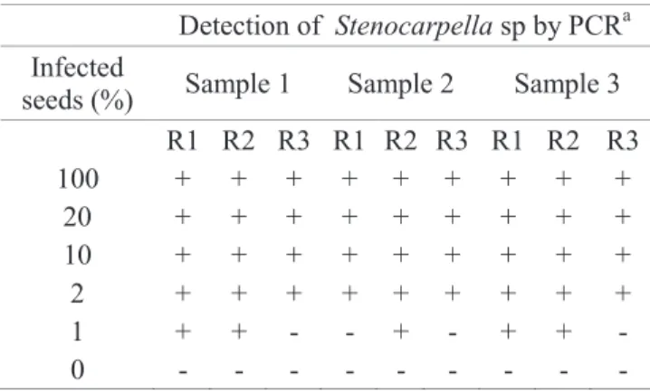
Detection of Stenocarpella sp. in infected maize seeds: clear bands of 325 base pairs were observed from maize
seed extracts, with the PCR technique, for seed samples
with levels of incidence higher than 2% for both species of Stenocarpella.
Maize seeds without infection by Stenocarpella sp. were tested in the present experiment as the control. For infection levels lower than 2%, the primers used in this
investigation also proved to be efficient, although visible
bands in DNA gel were not observed in some replicates at infection rates of 1% for both fungal species. the results of three replicates of each sample are summarized in table 2.
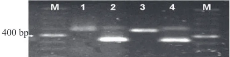
400 bp
Figure 3. Amplification of genomic DNA of Stenocarpella maydis at different levels of infection in maize seeds. Lane M= 100 bp DNA ladder 1-2=100%, 3-4=20%, 5-6= 10%, 7-8=2%, 9-10=1%, 11-12=0%.
table 2. Results of the detection of Stenocarpella sp. in
maize seeds infected artificially at different inoculum potentials using the PCR technique.
Detection of Stenocarpella sp by PCRa Infected
seeds (%) Sample 1 Sample 2 Sample 3 R1 R2 R3 R1 R2 R3 R1 R2 R3
100 + + + + + + + + +
20 + + + + + + + + +
10 + + + + + + + + +
2 + + + + + + + + +
1 + + - - + - + + -
0 - - - -
aPresence(+) or absence(-) of amplified bands with the primer P1/P2 for
the detection of Stenocarpella sp in maize seeds. R= replicates..
the internal transcribed spacer regions (ItS) of the rDNA have been widely used to differentiate and to detect closely related fungal species (Phan et al., 2002), as they are highly conserved parts of the genome within the same species and are variable among species (Lovic et al., 2002; Lee et al., 2002). Based on those regions, different primers have been designed and successful applications of them are
reported for detection and identification of several target
organisms (Lee et al., 2002; Iacomi-Vasilescu et al., 2009; Konstantinova et al., 2002; Rios et al., 2007; Haet al., 2009; Consolo et al., 2009 and Ioos et al.,2007).
For some species with very similar morphological
characteristics, designing specific primers is still a challenge. This mission may face difficulties for different
reasons as pointed out by Iacomi-Vasilescu et al. (2002), in studies focused on designing primers to differentiate Alternariabrassicae, A. brassicicola and A. japonica. In
this study the primers were only specific for distinguishing
A.brassicae from A. japonica.
According to Xia and Achar (2001), the use of primers allows an accurate detection of Stenocarpella maydis associated with maize seeds. In the present study, the
Stenocarpella maydis from S.macrospora. From the seed pathology point of view, both species are important pathogens, causing different symptoms and losses in the
same plants and are able to significantly reduce maize yields.
It should be noted that both species are often found
in the same seed sample of maize and their identification
by conventional methods is unreliable and laborious. In practice, both species should be detected separately considering that they are different pathogens. the results
of this study show that the development of specific primers
to distinguish both species of Stenocarpella is necessary
and should be investigated further. The use of sequences
or genes related to a pathogenic trait may be an alternative to be explored, as mentioned by Guillemette et al. (2004) in the differentiation of Alternaria brassicae from other species associated with Brassica seeds.
Regarding sensitivity, the PCR technique used was able
to reveal infection levels of both species of Stenocarpella at an incidence below 2% in the samples tested. Below that level of infection, bands were not seen in some replicates. As already mentioned, the molecular detection of fungi on
seeds can be influenced by different factors, such as low
concentration of target pathogens, the presence of other organisms and compounds like poly-phenols, tannins, polysaccharides and other natural components of the seeds. In other pathosystems, some compounds in the seed extraction have been demonstrated to interfere in the performance of
Taq polymerase enzyme. In the literature, there are reports of
the interference of polysaccharides from maize seeds in the DNA extraction.the increased viscosity of the extract makes
pipetting difficult and interferes in the DNA concentration
(Fang et al., 1992; Porebski et al., 1997).
An interesting point is the convenience of using commercial kits to extract DNA from plant tissues since it may prevent several factors interfering in the DNA extraction
and its amplification. (Phan et al., 2002; Konstantinova
et al., 2002; Guillemette et al., 2004; Ioos et al., 2007). In this study, the use of a commercial kit to extract DNA from
maize seeds was shown to be efficient as no influence on the
DNA extraction was observed. Because of this is important emphasize that this work the detection in pure culture and culture of fungus showed the same band (Figure 4).
According to a recent publications, the sensitivity of the detection of some pathogens by PCR can be considerably improved by using the real time PCR
technique (qPCR), as has been demonstrated for some pathosystems. It is possible to quantify the presence
of one single spore of pathogens associated with seeds
using such a technique (Chilvers et al., 2007). In work
conducted on the detection of Tilletia caries inwheat seeds, McNeil et al., (2004) report that less than one spore of this
pathogen was detected by that PCR technique at a 95% confidence level through calibration curves. The sensitivity of that technique in pure cultures was seen to be of 100 pg
for Alternaria brassicae (Guillemette et al., 2004) and 10 fg for Botrytis spp. (Chilvers et al., 2007).
400 bp
Figure 4. Eletrophoresis gel of the PCR obtained with the universal primer of the ItS region (ItS1 e ItS4)
and the specific primer (P1/P2) for the DNA
of Stenocarpella sp. DNA obtained from pure cultures.(1-2) and from infected seeds (3-4). Lane M – 100 bp ladder DNA (Amersham, u.S.A).
Another strategy to be considered for improving the
sensitivity of the PCR technique is to increase the biomass of
the pathogens associated with seeds before the extraction of the DNA. this can be done by incubating the seeds for some period of time under favorable conditions for the development of the pathogens in association with seeds. After incubation, seeds are used for DNA extraction. Such a procedure has
increased the sensitivity of the conventional PCR technique
in detecting some pathosystems, as reported by Guillemette et al. (2004) working with Alternaria radicina in seed samples of cabbage and radish incubated for 48 hours. through the
conventional PCR technique, Vechiattoet al. (2006) detected
Diaphorte meridionalis associated with a single soybean seed from a mixture of 399 healthy seeds, after an incubation period of 7 days by the conventional blotter method.
Early seed incubation may also be used to improve
the sensitivity of the real time PCR technique. Chadha and
Gopalakrishna (2006) were able to detect Magnaporthe grisea in rice seeds at levels of up to 0.2% incidence when seeds were incubated for 48 hours in PDA medium, whereas
by the conventional PCR technique this level was 5%.
Conclusions
The PCR technique as used in this study seems to be
compared to the biological methods based on filter paper incubation (blotter test) or on acidified PDA. However, an investigation of new and more specific and sensitive
primers, which are able to differentiate both species of Stenocarpella associated with maize seeds, should be
a priority for the Brazilian seed certification program.
In addition, the use of primers P1/P2 tested in this study can also be considered useful and may help in the initial screening for the presence of Stenocarpella in the seed lots commercialized.
Acknowledgements
thanks are due to FAPEMIG, to CNPq, to MAPA, to Dr. Anania Fessehaie of the Iowa State university- uSA, to EMBRAPA- Milho e Sorgo and to Dr. Ludwig H. Pfenning
of the “Universidade Federal de Lavras” for their support
of this research.
References
CASA, R.t.; REIS, E.M.; ZAMBoLIM, L. Decomposição dos restos
culturais do milho e sobrevivência saprofítica de Stenocarpella
macrospora e S. maydis. Fitopatologia Brasileira, v. 28, p. 355-361, 2003. http://www.scielo.br/scielo.php?script=sci_pdf&pid=S0100-41582003000400002&lng=en&nrm=iso&tlng=pt
CASA, R.t.; REIS, E.M.; ZAMBoLIM, L. Doenças do milho causadas
por fungos do gênero Stenocarpella. Revisão. Fitopatologia Brasileira,
v.31, p.427-439, 2006. http://www.scielo.br/pdf/fb/v31n5/01.pdf
CASA, R.t,; REIS, E.M.; ZAMBoLIM, L. Fungos associados a sementes de milho produzidas nas regiões Sul e Sudeste do Brasil. Fitopatologia Brasileira, v.23, p.370-373, 1998.
CHADHA, S.; GoPALAKRISHNA, t. Detection of Magnaporthe
grisea in infested rice seed using polymerase chain reaction. Journal of Applied Microbiology, v.100, p.1147-1153, 2006. http://www.ncbi.nlm. nih.gov/pubmed/16630016
CHILVERS, M.I.; Du toIt, L.J.; AKAMAtSu H, PEEVER, t.L.
A real-time quantitative PCR assay for Botrytis spp. that cause neck
rot onion. Plant Disease, v.91, p.599-608, 2007. http://ipm.wsu.edu/ seedcrops/Articles%20and%20Reports%20as%20PDFs/Chilvers%20 et%20al%20PCR%20seed%20assay%20Botrytis%20onion.pdf
CoNSoLo, V.F.; ALBANI, C.M.; BERÓN, C.M.; SALERMo, G.L.;
CORDO, C.A.A convencional PCR technique to detect Septoria tritici in
wheat seeds. Australasian Plant Pathology, v.38, p.222-227, 2009. http:// www.publish.csiro.au/paper/AP08099.htm
DoRRANCE, A.E.; HINKELMAN, K.H.; WARREN, H.L. Diallel analysis of Diplodia ear rot resistence in maize. Plant Disease, v.82,
p.699-703, 1998. http://apsjournals.apsnet.org/doi/abs/10.1094/ PDIS.1998.82.6.699
EDDINS, A.H. Dry rot of corn caused by Diplodia macrospora Earle.
Phytopathology, v.20, p.439-448, 1930.
FANG, G.; HAMMAR, S.; GRUMET, R. A quick and inexpensive method
for removing polysaccharides from plant genomic DNA. Biotechniques, v.13, p.52-56, 1992. http://www.ncbi.nlm.nih.gov/pubmed/1503775
GuILLEMEttE, t.; IACoMI-VASILESCu, B.; SIMoNEAu, P. Conventional and real-time PCR-based assay for detecting pathogenic
Alternaria brassicae in cruciferous seed. Plant Disease, v.88, p.490-496, 2004. http://apsjournals.apsnet.org/doi/abs/10.1094/PDIS.2004.88.5.490
HA, Y.; FESSEHAIE, A.; LING, K.S.; WECHtER, W.P.; KEINAtH, A.P.; WALCott, R.R. Simultaneous detection of Acidovorax avenae
subsp. citrulli and Didymella bryoniae in cucurbit seedlots using magnetic capture hybridization and real-time polymerase chain reaction.
Australasian Plant Pathology, v.99, p.666-678, 2009. http://www.ncbi. nlm.nih.gov/pubmed/19453225
IACoMI-VASILESCu, B.; BLANCARD, D.; GuÉNARD, M.; MoLINERo-DEMILLY, V.; LAuRENt, E.; SIMoNEAu, P. Development of PCR-based diagnostic assay for detecting pathogenic
Alternaria species in cruciferous seeds. Seed Science and Technology,
v.30, p.87-95, 2009. http://cat.inist.fr/?aModele=afficheN&cpsi
dt=13659044
IooS, R.; LAuGuStIN, L.; RoSE, S.; touRVIEILLE, J.; LABRouHE, t.D. Development of PCR test to detect the downy mildew causal agent Plasmopara hastedii in sunflower seeds. Plant Pathology, v.56, p.209-218, 2007. http://onlinelibrary.wiley.com/doi/10.1111/ j.1365-3059.2006.01500.x/pdf
KoNStANtINoVA, P.; BoNANtIS, P.J.M.; GENt-PELZER, M.P.E.; VAN DER ZouWEN, P.; BuLK RVAN DER. Development
of specific primers for detection and identification of Alternaria spp. in
carrot material by PCR and comparison with blotter and plating assays. Mycological Research, v.106, p.23-33, 2002. http://journals.cambridge. org/action/displayAbstract?fromPage=online&aid=98559
LEE, H.K.; TEWARI, J.P.; TURKINGTON, T.K. Quantification
of seedborne infection by Rhinchosporium secalis in barley using competitive PCR. Plant Pathology, v.51, p.217-224, 2002. http:// onlinelibrary.wiley.com/doi/10.1046/j.1365-3059.2002.00685.x/pdf
LOVIC, B.R.; MARTYN, R.D.; MILLER, M.E. Sequence analysis of
ItS regions of rDNA Monosporascus spp. to evaluate its potencial for PCR-mediated detection.Phytopathology, v.85, p.655-661, 2002. http://
cat.inist.fr/?aModele=afficheN&cpsidt=3621551
MACHADo, J.C.; GuIMARÃES, R.M.; VIEIRA, M.G.G.C.; SouZA,
R.M.; POZZA, E.A. Use of water restriction technique in seed pathology.
In:ISTA (ed.) Seed Testing International, v.128, p. 14-18, 2004.
estabelecimento de padrões de tolerância a patógenos em sementes. In: Zambolim L (Ed.). Sementes qualidade fitossanitária. Viçosa: uFV. 2005.
MARIo, L.J.; REIS, E.M. Método simples para diferenciar Diplodia macrospora de D. maydis em testes de patologia de sementes de milho.
Fitopatologia Brasileira, v.26, p.670-672, 2001. http://www.scielo.br/ pdf/fb/v26n3/a18v26n3.pdf
McNEIL, M.; RoBERtS, A.M.I.; CoCKERELL, V., MuLHoLLAND,
V. Real-time PCR assay for quantification of Tilletia caries contamination
of uK wheat seed. Plant Pathology,v.53, p.741-750, 2004. http:// onlinelibrary.wiley.com/doi/10.1111/j.1365-3059.2004.01094.x/pdf
MICHEL, B.E.; RADCLIFFE, D.A. Computer program relating solute
potential to solution composition for five solutes. Agronomy Journal,
v.87,n.131-136, 1995.
PHAN, H.t.t.; FoRD, R.; BREtAG, t.; tAYLoR, P.W.J. A rapid and sensitive polymerase chain reaction (PCR) assay for detection of
Ascochyta rabiei, the cause of ascochyta blight of chickpea. Australasian Plant Pathology, v.31, p.31-39, 2002. http://www.springerlink.com/ content/d2k5846602767713/
POREBSKI, S.; BAILEY, G.; BAUM, B.R. Modification of a CTAB
DNA extraction protocol for plants containing high polysaccharide and polyphenol components. Molecular Biology Reporter, v.15, p.8-15, 1997. http://www.springerlink.com/content/w450752253324r4j/
RÍoS, M.o; FERNÁNDEZ, P.; CARMoNA, M. Detection of
Rhynchosporium secalis in barley seeds from Argentina through
polymerase chain reaction technique. Fitopatologia Brasileira, v.32,
p.415-418, 2007. http://www.scielo.br/pdf/fb/v32n5/v32n5a07.pdf
VECHIAto, M.H.; MARINGoNI, A.C.; MARtINS, E.M.
Desenvolvimento de iniciadores para a detecção e identificação de Diaporthe phaseolorum var. meridionalis em sementes de soja.Summa Phytopathologica, v.32, p.161-169, 2006. http://www.scielo.br/pdf/sp/ v32n2/v32n2a11.pdf
XIA, Z.; ACHAR, N. Random amplified polymorphic DNA and