Gabapentin, a Synthetic Analogue of Gamma Aminobutyric
Acid, Reverses Systemic Acute Inflammation and Oxidative
Stress in Mice
Jordana Maia Dias,
1Tarcisio Vieira de Brito,
1Diva de Aguiar Magalhães,
1Pammela Weryka da Silva Santos,
1Jalles Arruda Batista,
1Eulina Gabriela do Nascimento Dias,
1Heliana de Barros Fernandes,
1Samara Rodrigues Bonfim Damasceno,
2Renan O. Silva,
2Karoline S. Aragão,
2Marcellus H. L. P. Souza,
2Jand-Venes R. Medeiros,
1and
André Luiz R. Barbosa
1,3Abstract—The aim of this study was to investigate the potential anti-inflammatory and anti-oxidant
e-ffects of gabapentin (GBP) in mice. The anti-inflammatory and anti-oxidant ee-ffects were evaluated using various mediators that induce paw edema, peritonitis model, myeloperoxidase (MPO) activity, proin-flammatory cytokine levels, glutathione (GSH) consumption, and malondialdehyde (MDA) production in mice. Pretreatment of mice with GBP (1 mg/kg) significantly reduced carrageenan or dextran-induced paw edema (P<0.05) when compared to vehicle group. Adding to this, GBP (1 mg/kg) significantly
inhibited paw edema induced by histamine, serotonin, bradikinin, 48/80 compound, and prostaglandin E2. In the carrageenan-induced peritonitis model, GBP significantly decreased total and differential
le-ukocyte counts and reduced the levels of MPO activity in the plantar tissue and IL-1βand TNF-α co-ncentrations in the peritoneal exudate. The same dose of GBP also decreased the MDA concentration and increased the levels of GSH into the peritoneal fluid. In summary, our results demonstrated that GBP exhibited anti-inflammatory activity in mice by reducing the action of inflammatory mediators, neutro-phil migration and proinflammatory cytokine levels, and anti-oxidant properties by decreasing the co-ncentration of MDA and increasing the GSH content. These observations raise the possibility that GBP could be used to improve tissue resistance to damage during inflammatory conditions.
KEY WORDS:gabapentin; anti-inflammatory effect; anti-oxidant action.
INTRODUCTION
Gabapentin (GBP) is an anti-convulsant drug struc-turally related toγ-aminobutyric acid. Evidence obtained
in a number of experimental models of neuropathic pain and inflammatory hyperalgesia [1,2] shows that GBP has an effective anti-nociceptive or anti-hyperalgesic action, in addition to being an anti-convulsant. In humans, GBP has become increasingly popular as a treatment for chronic neuropathic pain. Clinical studies have shown that GBP is an effective analgesic in different types of neuropathic pain syndromes, such as diabetic neuropathy [3], posther-petic neuralgia [4], and trigeminal neuralgia [5].
The literature data show that the GBP can also reverse the gastric inflammatory damage induced by indomethacin and ethanol and diminish the acute in-flammatory process induced by carrageenan into the
mice paw [6]. Paw edema induced by carrageenan can
1LAFFEX
—Laboratory of Experimental Physiopharmacology, Biotechnology and Biodiversity Center Research (BIOTEC), Federal University of Piauí, Parnaiba-PI, 64049-550, Brazil
2LAFICA
—Laboratory of Pharmacology of Inflammation and Cancer, Department of Physiology and Pharmacology, Federal University of Ceará, Fortaleza, CE, Brazil
3To whom correspondence should be addressed at LAFFEX—Laboratory of
Experimental Physiopharmacology, Biotechnology and Biodiversity Center Research (BIOTEC), Federal University of Piauí, Parnaiba-PI, 64049-550, Brazil. E-mail: andreluiz@ufpi.edu.br
0360-3997/14/0500-1826/0#2014 Springer Science+Business Media New York
be mediated by several mediators and intense neutro-phil influx [7,8]. However, the action of GBP against the inflammatory condition established by vascular inflammatory mediators and by the release of cyto-kines, free radicals, and neutrophil migration is still largely unknown.
The inflammatory process involves a complex cascade of biochemical and cellular events that occur in response to cellular injury [9], which is character-ized by neutrophil migration occurring in locally
produced inflammatory mediators, including TNF-α
and IL-1β, which activate neutrophils and promote
their migration to the inflammatory site [10, 11], as
well as a variety of chemical mediators such as histamines, serotonin, bradykinins, and prostaglandins
[12–14]. Those mediators cause increased vascular
permeability and promote extravasation of low levels of protein and neutrophils [15].
Knowing that the GBP can reduce some conditions of the inflammatory response, the aim of this study is to test whether GBP is able to inhibit the paw edema induced by several inflammatory mediators, production of free-radical scavengers, neutrophil infiltration, and release of proin-flammatory cytokines.
METHODS
Animals
Male Swiss mice (25–35 g) were sourced by the
Central Animal Facility of the Federal University of Piauí. The animals were housed at 25±2 °C under a 12:12-h light/dark cycle, and food and water were supplied
ad libitum. Experiments were conducted in accordance
with current established principles for the care and use of research animals (National Institutes of Health [NIH] guidelines) and were approved by the ethics committees
in research of Faculdade Integral Diferencial—FACID,
number of protocol: 002/13.
Drugs and Reagents
The following drugs and reagents were used: carra-geenan, dextran sulfate, histamin, serotonin, prostaglandin E2(PGE2), 48/80, aminoguanidine,L-arginine, and
indo-methacin (Sigma Aldrich, St Louis, MO, USA). These drugs were dissolved in sterile saline (0.9 % NaCl).
Experimental Protocol
Carrageenan-Induced Paw Edema
The animals were randomly divided into six groups (n=5), and edema was induced by the injection of 50μL of
a suspension of carrageenan (CG; 500μg/paw)
adminis-tered with a subplantar injection into the right paw (group I). The mice were pretreated intraperitoneally (i.p.) with either 0.9 % NaCl (group II, untreated control); 10 mg/kg indomethacin (group III, reference control); or 0.1, 0.5, or 1 mg/kg of GBP, i.p, respectively, 1 h before the carrageen-an injection. Paw volume was measured immediately be-fore (V0) and at 1, 2, 3, and 4 h after carrageenan treatment
(Vt) with a plethysmometer (PANLAB, LE7500). The effect of pretreatment was calculated as the percentage of inhibition of edema relative to the paw volume of the saline-treated controls by using the following formula [16]:
% inhibition of edema¼ðVt−V0ÞControl−ðVt−V0ÞTreated
Vt−V0
ð ÞControl 100
whereV0is the basal volume andVtis the final volume
measured at the indicated times
Paw edema Induced by Different Inflammatory Agents
To induce paw edema with different inflamma-tory agents, the animals received injections of
dex-tran (DXT; 500 μg/paw), serotonin (5-HT; 1 %w/v),
histamine (HIST; 100 μg/paw), bradykinin (BK;
6.0 nmol paw), PGE2 (3 nmol/paw), and 48/80
(12 μg/paw) into the right hind paw. One group
received 50 μL of 0.9 % sterile saline and served
as an untreated control group. GBP (1 mg/kg) or indomethacin (INDO; 10 mg/kg, reference control) was given i.p. 30 min before intraplantar injections of phlogistic agents. For paw edema induced by dextran, serotonin, histamine, bradykinin, PGE2, and 48/80, the paw volume was measured using plethys-mometer (PANLAB, LE7500) before the injection of inflammatory agents (time zero). Hence, the paw volume was measured for 30, 60, 90, and 120 min after the injection of those inflammatory agents, ex-cept for dextran or histamin, which was measured for 30 min and 1, 2, 3, and 4 h using the same plethysmometer.
Measurement of Myeloperoxidase Activity in Mice Paw
Briefly, 50–100-mg hind paw tissue was homogenized in
1-mL potassium buffer with 0.5 % hexadecyltrimethylam-monium bromide for each 50-mg tissue. The homogenate was centrifuged at 40,000gfor 7 min at 4 °C. MPO activity in the resuspended pellet was assayed by measuring the change in absorbance at 450 nm using o-dianisidine dihy-drochloride and 1 % hydrogen peroxide. The results were reported as the MPO units/milligram of tissue. A unit of
MPO (UMPO) activity was defined as converting 1μmol
hydrogen peroxide to water in 1 min at 22 °C.
Peritonitis Assay
Mice were pretreated with oral administration of
250 μL sterile saline or indomethacin 10 mg/kg or GBP
1 mg/kg. One hour later, the animals were injected i.p. with 250μL of the carrageenan (500μg/cavity). The mice were killed by cervical dislocation under anesthesia 4 h later, and the peritoneal cavity was washed with 1.5 mL heparinized phosphate-buffered saline (PBS) to count peritoneal cells. Total cell counts were performed in a Neubauer chamber, and differential cell (neutrophils) counts (total of 100 cells) were carried out on cytocentrifuge slides stained with hematoxylin and eosin. The results were presented as the number of total leukocyte cells or neutrophils per milliliter of peritoneal exudate.
Cytokine Measurements
The levels of IL-1βand TNF-αwere evaluated using
sandwich ELISA. Briefly, microliter plates were coated
overnight at 4 °C with antibody against mice IL-1β or
TNF-α(2μg/mL). Blocking of nonspecific binding sites
was accomplished by incubating the plates with PBS con-taining 2 % bovine serum albumin (BSA) for 90 min at 37 °C. After blocking the plates, the test samples and each standard at various dilutions were added in duplicate and incubated at 4 °C for 24 h. The plates were washed three
times with buffer. After washing the plates, 50 μL of
biotinylated sheep polyclonal anti-IL-1βand anti-TNF-α
(diluted 1:1000 with assay buffer 1 % BSA) was added to the wells. After a further incubation at room temperature for 1 h, the plates were washed, and 50μL of
streptavidin-HRP diluted 1:5000 was added to all wells. The reagent
o-phenylenediamine dihydrochloride (50 μL) was added
15 min later, and the plates were incubated in the dark at
37 °C for 15–20 min. After the color development, the
reaction was stopped with the addition of sulfuric acid (1 M), and absorbance was measured at 490 nm. The results are expressed as picogram per milligram of protein and reported as mean ± SD.
Measurement of Malondialdehyde
The malondialdehyde (MDA) concentration was measured using the method described previously with modifications [17].
Measurement of Levels of Glutathione
The glutathione (GSH) levels in the fragments of intestinal tissue were determined according to the method described previously with modification [18].
Statistical Analysis
Results are expressed as mean ± SEM from at least five animals per group. Statistical analysis was performed using analysis of variance followed by the Newman-Keuls
post hoctest, when appropriate. Statistical significance was
set atp<0.05.
RESULTS
Effect of Gabapentin on Carrageenan-Induced Paw edema in Mice
Table1shows that the administration of carrageenan into the plantar surface (500μg per paw) induced severe
paw edema within 1 h of injection and was maintained until 4 h after injection. Indomethacin (10 mg/kg) admin-istration significantly decreased paw edema throughout the experimental period (*p<0.05), with maximal inhibition of
100 %. Similarly, GBP (1 mg/kg, i.p.) inhibited edema formation in all times. At 2, 3, or 4 h, compared with the carrageenan group, the animals pretreated with 1 mg/kg of GBP showed 87.2, 77.8, and 69.4 % reduction in paw edema, respectively. GBP prevented carrageenan-induced
paw edema (500μg per paw/50μl) with maximal
inhibi-tory effect at dose of 1 mg/kg (2 h, 0.01±0.007 mL; 3 h, 0.022±0.001 mL; 4 h, 0.025±0.007 mL). Therefore, this dose was selected for studying the possible mechanisms of action involved in GBP-mediated decrease in inflammato-ry response.
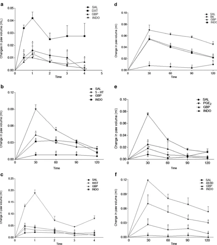
Effect of Gabapentin on Paw Edema Inflammation Induced by Different Inflammatory Agents
(0.0312±0.0030; Fig.1a). GBP also significantly inhibited the increase in paw volume of animals treated with serotonin (0.048±0.015; Fig.1b), histamine (0.103±0.025; Fig.1c),
bradykinin (0.058±0.005; Fig. 1d), PGE2 (0.033±0.014;
Fig.1e), and 48/80 (0.079±0.013; Fig.1f). On the other hand, the saline injected into the paw did not induce any effect. The values given are means ± SEM (n=5).
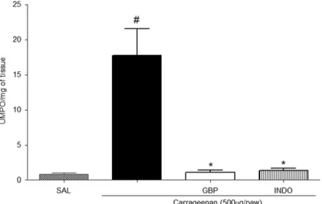
Effect of Gabapentin on Carrageenan-Induced Myeloperoxidase Activity in Paw Tissue
We can observe in Fig.2 that the carrageenan
sub-plantar injection elevated the concentration of MPO in the plantar tissue (17.73±3.85 UMPO/mg of tissue) when this group is compared to the saline group (0.84 ± 0.19 UMPO/mg of tissue). On the other hand, pretreatment with GBP (1 mg/kg) reduced the action of this tissue enzyme
(1.10±0.33 UMPO/mg of tissue; Fig.2).
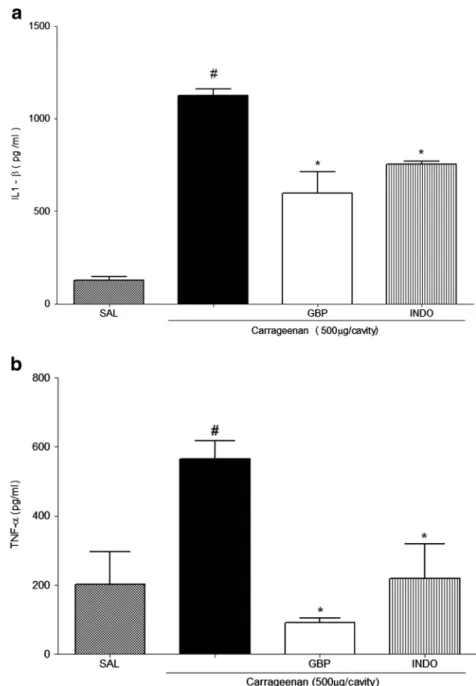
Effect of Gabapentin on Carrageenan-Induced Cytokine Production in Peritonitis
Figure 3 shows that intraperitoneal administration
of carrageenan was found to induce a marked increase
in IL-1β concentrations in the peritoneal exudates
(1.125±37.40 pg/mL). The level of IL-1β in the
peri-toneal cavity of control animals (saline group) was 127.0±19.04 pg/mL. Compared with the carrageenan group, the animals pretreated with GBP (1 mg/kg, i.p)
showed significantly decreased IL-1β peritoneal
con-centration (599.3±113.1 pg/mL; Fig.3a). Furthermore,
GBP treatment decreased the levels of TNF-α(90.12±
14.22 pg/mL) compared to the peritoneal carrageenan
group (564.9±52.32 pg/mL; Fig. 3b).
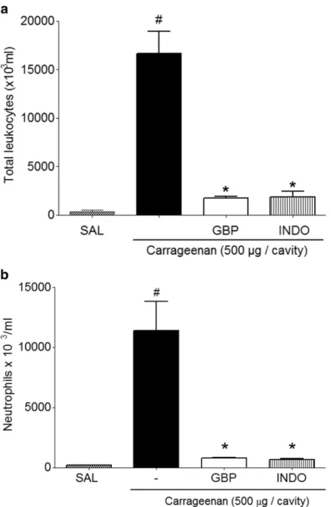
Anti-Inflammatory Effect of Gabapentin on Carrageenan-Induced Peritonitis in Mice
Figure4shows that the carrageenan group promoted
an increase in cell migration (leukocytes) into the
perito-neal cavity (16,675×103±2,252×103cells/mL). However,
GBP group showed significantly reduced peritoneal
leukocyte count (1.760 × 10³± 123.9 × 10³ cells/mL;
Fig. 4a). Furthermore, the same dose of GBP
significantly reduced neutrophil migration into the
peritoneal cavity (784.02×10³±55.02× 10³ cells/mL)
compared with that in the carrageenan group (11,410×
103± 2,392× 103 cells/mL; Fig. 4b). This result was
consistent with the fact that neutrophils are the most abundant cells in primary inflammatory exudates.
Effect of Gabapentin on MDA Levels in the Peritoneal Exudates of Mice
Figure 5 shows that the injection of carrageenan
(41.83±1.788) significantly increased the levels of MDA compared to the group that received only intraperitoneal saline (22.88±3.075). However, the group pretreated with GBP 1 mg/kg (24.55±1.192) had significantly reduced
MDA levels compared to the untreated group (Fig.5).
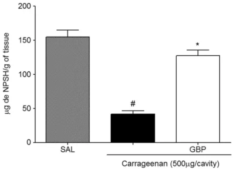
Effect of Gabapentin on Glutathione Levels in Peritoneal Exudate of Mice
Figure 6 shows that treatment with carrageenan
(41.98±4.515) increases the consumption of GSH com-pared to the saline group (155.0±10.02). It was also ob-served that the GBP (127.6±8.197) group significantly increased GSH levels compared to the untreated group.
Table 1. Effect of Gabapentin on Carrageenan-Induced Paw Edema in Mice
Treatment Dose (mg/kg) Paw edema in milliliters (time after inflammatory stimuli administration)
1 h 2 h 3 h 4 h
Control (Cg)(1)
– 0.067±0.002 0.078±0.006 0.101±0.010 0.081±0.001
Saline – 0.016±0.006* 0.013±0.004* 0.005±0.005* 0±0.0*
Indomethacin (INDO)
10 0.006±0.004* 0±0.0* 0.061±0.007 0.011±0.007*
GBP 0.1 0.065±0.005 (3.7 %) 0.067±0.007 (14.5 %) 0.061±0.007 (39.4 %)* 0.077±0.006 (5.1 %) 0.5 0.061±0.012 (8.6 %) 0.066±0.010 (14.9 %) 0.067±0.006 (50 %)* 0.075±0.009 (8.16 %)
1 0.03±0.004 (55.6 %)* 0.01±0.007 (87.2 %)* 0.022±0.001 (77.8 %)* 0.025±0.007 (69.4 %)*
Fig. 1. Effects of gabapentin on paw inflammation induced by different inflammatory agents. Edema was induced byadextran (DXT; 500μg/paw),b serotonin (5-HT; 1 %w/v),chistamine (HIST; 100μg/paw),dbradykinin (BK; 6.0 nmol/paw),ePGE2(3 nmol/paw), andf48/80 (12μg/paw). Animals were
pretreated with GBP (1 mg/kg i.p.), saline (SAL; control), or indomethacin (INDO; 10 mg/kg, i.p.). Eachpointrepresents the mean ± SEM of five animals.
DISCUSSION
The pharmacological approaches of GBP have drawn attention of researchers from basic and clinical areas. GBP, a drug used to improve neurological disorders such as epilepsy and seizures [19,20], reduces the inflammatory hyperalgesia induced by acid-acetic and formalin tests in mice [1,21–25]. Another study revealed that this substance
inhibited the acute inflammatory responses that occur in the indomethacin-induced gastropathy inflammation in rats
or carrageenan-induced paw edema [6]. However, the
mechanisms of this latter effect of GBP were not elucidated.
In this manuscript, we report new insights into the functions and possible mechanisms of GBP, including its anti-inflammatory ability demonstrated by decreasing the paw edema induced by carrageenan, dextran, and 48/80 and induced by several mediators, such as histamine, sero-tonin, PGE2, and bradikinin, the levels of proinflammatory
cytokines (TNF-αand IL-1β), the neutrophil infiltration,
and its anti-oxidant ability demonstrated by increased GSH levels and decreased MDA concentration.
Our results demonstrate that GBP was able to reduce paw edema induced by carrageenan or dextran. Paw edema in mice induced by carrageenan is used as a tool to inves-tigate potential anti-inflammatory agents [26]. In this acute inflammatory model, there are two phases. The first or
early phase is mediated by the release of histamine and serotonin, followed by the subsequent release of bradyki-nin and prostaglandins [25–27]. The late or second phase is
characterized by cytokine production and release of macro-phages and mast cells and intense neutrophil infiltration [28,29,8]. On the other hand, dextran-induced paw edema promotes inflammation by increasing vascular permeabil-ity dependent on mast cell degranulation and the subse-quent release of histamine and serotonin [30]. The extrav-asated fluid during dextran injection contains little protein and few neutrophils [31]. The results suggested that the anti-inflammatory effect of GBP seems to be mediated by the inhibition of neutrophil infiltration into the inflamma-tory site, as well as the inhibition of the release or activity of inflammatory mediators.
The vascular phenomenon that occurs during the acute phase of the inflammatory process is dependent on the release of several mediators that act on the vascular endothelium causing leakage of fluid and proteins into the interstitium [32]. This event can be mediated by the action of histamine, serotonin, bradikinin, PGE2, and/or induced
by 48/80 compound, which induces paw edema by mast cell degranulation with the release of histamine, serotonin, and bradikinin [33–35]. In this manuscript, GBP reduced
the paw edema induced by histamine, serotonin,
bradiki-nin, PGE2, and 48/80 compound. Thus, we can infer that
this drug affects the vascular component of edema, which
Fig. 2. Effect of gabapentin on carrageenan-induced myeloperoxidase activity in paw tissue. Saline or carrageenan (500μg per paw) was injected into the plantar surface of mice. One hour before this injection, animals had been treated with indomethacin (INDO; 10 mg/kg, i.p.) or gabapentin (GBP; 1 mg/kg, i.p.). Myeloperoxidase (MPO) activity was detected in the paw tissue after 4 h. The results are expressed as the mean ± SEM MPO units (UMPO)/milligram of tissue. *p<0.05 compared with carrageenan group;#p<0.05 compared with saline group. Statistical analysis was performed using analysis of variance
appears to be mediated by the decreased action of these several inflammatory mediators.
The carrageenan-induced inflammatory response in paw tissue is known to be accompanied by intense leuko-cyte migration, primarily neutrophils [12]. MPO activity has been found in neutrophil azurophilic granules, which is an indicator of neutrophil accumulation [36,37]. During
the neutrophil migration, MPO can be released on the inflamed tissue, inducing damage to adjacent cells and thus contributing to the pathogenesis of inflammatory process [38]. Our results obtained showed that GBP (1 mg/kg) or indomethacin (positive control) reduced the MPO concen-tration in paw tissue after carrageenan injection. Thus, we can suggest that GBP-reduced inflammatory process
Fig. 3. Effect of gabapentin on carrageenan-induced cytokine production in peritonitis.aThe level of interleukin (IL)-1βandbTNF-α. The level of IL-1βor TNF-αin the peritoneal cavity was measured 4 h after carrageenan injection. Mice were intraperitoneally administered with GBP (1 mg/kg) or indomethacin (INDO; 10 mg/kg), followed by injection of 250μL carrageenan (500μg per cavity, i.p.) after 1 h. Eachpointrepresents the mean ± SEM values obtained from five animals. *p<0.05 compared with carrageenan group;#p<0.05 compared with saline group. Statistical analysis was carried out using one-way
involves the inhibition of neutrophil migration into the inflammatory site.
The carrageenan-induced peritonitis has been linked to the neutrophil infiltration, the release of
neutrophil-derived mediators [27], and this
phenome-non occurs through an indirect mechanism that involves the activation of resident cells and the
re-lease of proinflammatory cytokines [39]. In the
Fig. 4. Anti-inflammatory effect of gabapentin carrageenan-induced peritonitis in mice.aTotal count of leukocytes.bCount of neutrophils per cavity. Mice received 250μL saline (i.p.), indomethacin (INDO; 10 mg/kg, p.o.), or gabapentin (GBP; 1 mg/kg, p.o.), followed by injection of 500μg carrageenan diluted in 250-μL saline solution (i.p.) after 1 h. Mice were killed 4 h later, and the peritoneal cavity was washed with 1.5 mL heparinized phosphate-buffered saline (PBS) to harvest the peritoneal cells. The values are represented as mean ± SEM. *p<0.05 compared to carrageenan group;#p<0.05 compared with saline
present study, we showed that GBP (1 mg/kg) dimin-ished the carrageenan-induced neutrophil migration into the peritoneal cavity. According to our findings,
we can infer that this compound decreased inflam-matory response by inhibiting the action and release
of cytokines such as IL-1β and TNF-α.
Fig. 5. Effect of gabapentin on MDA levels in the peritoneal exudate of mice. MDA levels in the peritoneal exudate were evaluated 4 h after carrageenan administration. Values are expressed as mean±EPM in nanomole per milliliter of MDA *p<0.05 compared to carrageenan group;#p<0.05 compared to
saline group. Statistical analysis was performed using analysis of variance followed by the Newman-Keuls test.
Fig. 6. Effect of gabapentin on GSH levels in peritoneal exudate of mice. The animals were killed 4 h after induction by carrageenan peritonitis. One hour before the experiment, they were pretreated with GBP (1 mg/kg). Values are expressed as mean ± EPM in microgram NPSH/gram of tissue. *p<0.05 co-mpared to carrageenan group;#p<0.05 compared to saline group. Statistical analysis was performed using analysis of variance followed by the
Recent studies have shown that administration of carrageenan into the peritoneal cavity induces the
release of TNF-α and IL-1β [40]. IL-1β and
TNF-α are potent proinflammatory cytokines that have
multiple effects, including the activation of inflam-matory cells, induction of several inflaminflam-matory
pro-teins, cytotoxicity, and neutrophil migration [41].
These cytokines have been recognized as a powerful chemotactic factor that activates the inflammatory cells, such as mature neutrophils, and induces the
diapedesis to the inflammatory site [42]. GBP also
inhibited the levels of IL-1β and TNF-α, and based
on our results, we could infer that the anti-inflamma-tory action of this anti-convulsant drug might occur through the inhibition of cytokines involved in car-rageenan-induced peritonitis.
Oxidative stress has been proposed to play an impor-tant role in the pathogenesis of inflammatory process and is related to promote the production of several cytokines,
including proinflammatory cytokines IL-1β, IL-6, and
TNF-α[43,44] and the recruitment of neutrophils during
inflammatory process. This pathological event is charac-terized by the overproduction of reactive oxygen resulting in tissue damage [44]. Thus, the present study also inves-tigated the effect of GBP on two oxidative stress markers: GSH and MDA.
Our results demonstrated that GBP increased the lev-els of GSH and decreased the MDA concentration in mice carrageeninduced peritonitis. GSH, an endogenous an-ti-oxidant, protects the cells against oxidative stress, keep-ing the sulfhydryl groups of proteins reduced and prevent-ing them from reactprevent-ing with free radicals [45] MDA is a product of lipoperoxidative processes that take place as a consequence of the tissue oxidative insult [46]. According to that result, we can infer that GBP decreased the tissue damage during the installation of the inflammatory process by stimulating the production and action of endogenous anti-oxidants and decreased the lipid peroxidation into the peritoneal cavity.
CONCLUSION
In summary, our results may show that GBP has an anti-inflammatory action to reduce the inflammatory re-sponse by inhibiting the action of various inflammatory mediators, neutrophil infiltration, proinflammatory cyto-kines, and oxidative stress. These observations raise the possibility that GBP could be used to improve tissue resis-tance to damage during inflammatory conditions.
ACKNOWLEDGMENTS
The authors gratefully acknowledge the National Council of Technological and Scientific Development, CNPq, (Brazil) and the Research Foundation for the State of Piauí-Brazil (FAPEPI) for financially supporting this work.
Conflict of Interest. The authors report no conflict of
interest.
REFERENCES
1. Shimoyama, M., N. Shimoyama, C.E. Inturrisi, and K.J. Elliott. 1997. Gabapentin enhances the antinociceptive effects of spinal morphine in the rat tail-flick test.Pain72: 375–382.
2. Hwang, J.H., and T.L. Yaksh. 1997. Effect of subarachnoid gabapen-tin on tactile-evoked allodynia in a surgically induced neuropathic pain model in the rat.Reg Anesth22: 249–256.
3. Backonja, M., A. Beydoun, K.R. Edwards, S.L. Schwartz, V. Fonseca, M. Hes, L. La Moreaux, and E. Garofalo. 1998. Gabapentin for the symptomatic treatment of painful neuropathy in patients with diabetes mellitus: a randomized controlled trial.JAMAb280: 1831–1836. 4. Singh, D., and D.H. Kennedy. 2003. The use of gabapentin for the
treatment of postherpetic neuralgia.Clin Ther25: 852–889. 5. Pöllmann, W., and W. Feneberg. 2008. Current management of pain
associated with multiple sclerosis.CNS Drugs22: 291–324. 6. Abdel-Salam, O.M.E., and A.A. Sleem. 2009. Study of the analgesic,
anti-inflammatory, and gastric effects of gabapentin.Drug Discov Ther3: 18–26.
7. Silva, V.G., R.O. Silva, S.R. Damasceno, N.S. Carvalho, R.S. Prudêncio, K.S. Aragão, M.A. Guimarães, S.A. Campos, L.M. Véras, M. Godejo-hann, J.R. Leite, A.L. Barbosa, and J.V. Medeiros. 2013. Anti-inflamma-tory and antinociceptive activity of epiisopiloturine, an imidazole alkaloid isolated from Pilocarpus microphyllus.J Nat Prod76: 1071–1077. 8. Vinegar, R., J.F. Truax, J.L. Selph, P.R. Johnston, A.L. Venable, and
K.K. McKenzie. 1987. Pathway to carrageenan-induced inflammation in the hind limb of the rat.Fed Proc46: 118–126.
9. Carvalho, W.A., and L. Lemônica. 1998. Mecanismos celulares e moleculares da dor inflamatória.Rev Bras Anestesiol48: 137–158. 10. Greer, C.W., I. Shomera, M.E. Goldsteinb, and W. Yaphe. 1984. Analysis
of carrageenan from Hypnea musciformis by usingκ- andι -carragee-nases and13C-N.M.R. spectroscopy.Carbohydr Res129: 189
–196. 11. Aziza, M., T. Givernaud, M. Chikhaoui-khay, and L. Bennasser. 2008.
Seasonal variation of the growth, chemical composition and carrageenan extracted from Hypnea musciformis (Wulfen) Lamouroux harvested along the Atlantic coast of Morocco.Sci Res Essays2: 509–514. 12. Carvalho, J.C., J.R. Teixeira, P.J. Souza, J.K. Bastos, D. dos Santos
Filho, and S.J. Sarti. 1996. Preliminary studies of analgesic and anti-inflammatory properties ofCaesalpinea ferreacrude extract.J Ethno-pharmacol53: 175–178.
13. Hajare, S.W., S. Chandra, J. Sharma, S.K. Tandan, J. Lal, and A.G. Telang. 2001. Anti-inflammatory activity of Dalbergia sissoo leaves.
Fitoterapia72: 131–139.
14. Srinivasan, K., S. Muruganandan, J. Lal, S. Chandra, S.K. Tandan, and V.R. Prakash. 2001. Evaluation of anti-inflammatory activity of
Pongamia pinnataleaves in rats.J Ethnopharmacol78: 151–157. 15. Calixto, J.B., M.M. Campos, M.F. Otuki, and A.R. Santos. 2004.
pro-inflammatory cytokines, chemokines and adhesion molecules.
Planta Med70: 93–103.
16. Lanhers, M.C., J. Fleurentin, P. Dorfman, F. Mortier, and J.M. Pelt. 1991. Analgesic, antipyretic and anti-inflammatory properties of Eu-phorbia hirta. Planta Med57: 225–231.
17. Sedlak, J., and R.H. Lindsay. 1968. Estimation of total, protein-bound, and nonprotein sulfhydryl groups in tissue with Ellman’s reagent.Anal Biochem24: 1992–2005.
19. Jensen, A.A., J. Mosbacher, S. Elg, K. Lingenhoehl, T. Lohmann, T.N. Johansen, B. Abrahamsen, J.P. Mattsson, A. Lehmann, B. Bettler, and H. Bräuner-Osborne. 2002. The anticonvulsant gabapentin (neurontin) does not act through gamma-aminobutyric acid-B receptors. Mol Pharmacol6: 1377–1384.
20. Bennett, M.I., and K.H. Simpson. 2004. Gabapentin in the treatment of neuropathic pain.Palliat Med18: 5–11.
21. Abdi, S., D.H. Lee, and J.M. Chung. 1998. The anti-allodynic effects of amitriptyline, gabapentin, and lidocaine in a rat model of neuro-pathic pain.Anesth Analg87: 1360–1366.
22. Yasuda, T., S. Miki, N. Yoshinaga, and E. Senba. 2005. Effects of amitriptyline and gabapentin on bilateral hyperalgesia observed in an animal model of unilateral axotomy.Pain115: 161–170.
23. Shimoyama, N., M. Shimoyama, A.M. Davis, C.E. Inturrisi, and K.J. Elliott. 1997. Spinal gabapentin is antinociceptive in the rat formalin test.Neurosci Lett222: 65–67.
24. Hunter, J.C., K.R. Gogas, L.R. Hedley, L.O. Jacobson, L. Kassotakis, J. Thompson, and D.J. Fontana. 1997. The effect of novel anti-epilep-tic drugs in rat experimental models of acute and chronic pain.Eur J Pharmacol324: 153–160.
25. Vinegar, R., W. Schreiber, and R. Hugo. 1969. Biphasic development of carrageenin edema in rats.J Pharmacol Exp Ther166: 96–103. 26. de Brito, T.V., R.S. Prudêncio, A.B.. Sales, F. Vieira Jr., S.J. Candeira,
Á.X. Franco, K.S. Aragão, R.A. Ribeiro, Ponte de Souza MH, L.S. Chaves, A.L. Freitas, J.V. Medeiros, and A.L.R. Barbosa. 2013. Anti-inflammatory effect of a sulphated polysaccharide fraction extracted from the red algae Hypnea musciformis via the suppression of neu-trophil migration by the nitric oxide signalling pathway. J Pharm Pharmacol65: 724–733.
27. Dawson, J., A.D. Sedgwick, J.C.W. Edwards, and P. Lees. 1991. A comparative study of the cellular, exudative and histological responses to carrageenan, dextran and zymosan in the mouse.Inter J Tissue Reac
13–14: 171–185.
28. Cragg, G.M., D.J. Newman, and K.M. Snader. 1997. Natural products in drug discovery and development.J Nat Prod60: 52–60. 29. De Smet, P.A. 2007. The role of plant-derived drugs and herbal
medicines in healthcare.Drugs54: 801–840.
30. Rowley, D.A., and E.P. Benditt. 1956. 5-hydroxytryptamine and his-tamine as mediators of the vascular injury produced by agents which damage mast cells in rats.J Exp Med103: 399–415.
31. Kulkarni, S.K., A.K. Mehta, and J. Kunchandy. 1986. Anti-inflamma-tory actions of clonidine, guanfacine and B-HT 920 against various
inflammagen-induced acute paw oedema in rats.Arch Int Pharmaco-dyn Ther279: 324–334.
32. Seibert, K., Y. Zhang, K. Leahy, S. Hauser, J. Masferrer, W. Perkins, L. Lee, and P. Isakson. 1994. Pharmacological and biochemical demon-stration of the role of cyclooxygenase 2 in inflammation and pain.
Proc Natl Acad Sci91: 12013–12017.
33. Lago, J., A. Alfonso, M.R. Vieytes, and L.M. Botana. 2001. Ouabain-induced enhancement of rat mast cells response. Modulation by pro-tein phosphorylation and intracellular pH.Cell Signal13: 515–524. 34. Okazaki, T., V.S. Ilea, A. Okazaki, K. Wicher, R.E. Reisman, and C.E.
Arbesman. 1976. Inhibition of antigen-induced histamine release by ouabain.J Allergy Clin Immunol57: 454–462.
35. Senol, M., I.H. Ozerol, A.V. Patel, and D.P. Skoner. 2007. The effect of Na+-K+ATPase inhibition by ouabain on histamine release from human cutaneous mast cells.Mol Cell Biochem294: 25–29. 36. Ajuebor, M.N., A. Singh, and J.L. Wallace. 2000.
Cyclooxygenase-2-derived prostaglandin D2 is an early anti-inflammatory signal in exper-imental colitis.J Physiol Gastrointest Liver Physiol279: 238–244. 37. Souza, G.E., F.Q. Cunha, R. Mello, and S.H. Ferreira. 1988.
Neutro-phil migration induced by inflammatory stimuli is reduced by macro-phage depletion.Agents Actions24: 377–380.
38. Klebanoff, S.J. 1999. Myeloperoxidase.Proc. Assoc. Am. Physicians
111: 383–389.
39. Gupta, M., U.K. Mazumdar, T. Sivakumar, M.L. Vamsi, S.S. Karki, R. Sambathkumar, and L. Manikandan. 2003. Evaluation of anti-inflam-matory activity of chloroform extract of Bryonia laciniosa in experi-mental animal models.Biol Pharm Bull26: 1342–1344.
40. Chaves, L.S., L.A. Nicolau, R.O. Silva, F.C. Barros, A.L. Freitas, K.S. Aragão, R.A. Ribeiro, M.H. Souza, A.L. Barbosa, and J.V. Medeiros. 2013. Antiinflammatory and antinociceptive effects in mice of a sulfated polysaccharide fraction extracted from the marine red algae Gracilaria caudate.Immunopharmacol Immunotoxicol35: 93–100. 41. Metcalfe, D.D. 2008. Mast cells and mastocytosis.Blood15: 946–
956.
42. Sutbeyaz, Y., B. Yakan, H. Ozdemir, M. Karasen, F. Doner, and I. Kufrevioglu. 1996. Effect of SC-41930, a potent selective leukotriene B4 receptor antagonist, in the guinea pig model of middle ear inflam-mation.Ann Otol Rhinol Laryngol105: 476–480.
43. Josse, C., J.R. Boelaert, M. Best-Belpomme, and J. Piette. 2001. Importance of post-transcriptional regulation of chemokine genes by oxidative stress.Biochem J360: 321–333.
44. Cuzzocrea, S., D.P. Riley, A.P. Caputi, and D. Salvemini. 2001. Antioxidant therapy: a new pharmacological approach in shock, in-flammation and ischemia/reperfusion injury.Pharmacol Rev53: 135– 159.
45. Amirshahrokhi, K., S. Bohlooli, and M.M. Chinifroush. 2011. The effect of methylsulfonylmethane on the experimental colitis in the rat.
T Appl Pharmacol253: 197–202.