BACILLUS CEREUS STRAIN MQ23 ISOLATED FROM SOPHORA ALOPECUROIDES ROOT NODULES
Longfei Zhao1, 2; Yajun Xu2; Ran Sun1; Zhenshan Deng1; Wenquan Yang1; Gehong Wei1*
1
College of Life Sciences, Shaanxi Key Laboratory of Molecular Biology for Agriculture, Northwest A & F University, Yangling,
Shaanxi, 712100, China; 2College of Life Sciences, Shangqiu Normal University, Shangqiu, Henan, 476000, China.
Submitted: July 01, 2010; Returned to authors for corrections: October 05, 2010; Approved: January 13, 2011.
ABSTRACT
Endophytes MQ23 and MQ23R isolated from Sophora alopecuroides root nodules were characterized by
observing their ability to promote plant growth and employing molecular analysis techniques. Results showed
that MQ23 and MQ23R are potential N2-fixing endophytes and belong to the same species as Bacillus cereus. MQ23 was shown to be able to produce siderophores, IAA, and demonstrate certain antifungal activity to
plant pathogenic fungi. Co-inoculation with MQ23+MQ23II showed a more significant effect than
inoculation alone in vitro for most of positive actions suggesting they have a cooperative interaction. Results
of plant inoculation with endophytes indicated that the growth indexes of co-inoculated MQ23+MQ23II were
higher than those of inoculated alone (p<0.05) (the exception being for root fresh weight) when compared to
negative control. There have been little of any studies of nonrhizobial putative endophytes with
growth-promotion attributes in S. alopecuroides root nodules. This could be exploited as potential bio-inoculants and
biocontrol agents in agriculture.
Key words: Plant growth promoting characterization; Sophora alopecuroides; Root nodules; Endophytic
bacteria; Bacillus cereus
INTRODUCTION
In the last few decades, endophytic bacteria have
attracted more and more attention as novel resources as a
method of biocontrol for plant diseases and promoters of plant
growth (18, 22). Generally, the endophytic bacteria live inside
the plant tissues and do not cause visible damage or
morphological change on their hosts. They can benefit the host
plants by the production of IAA (indoleacetic acid,
phytohormones), siderophores, and antibiotic compounds,
through nitrogen fixation, by phosphate solubilization, and
with the suppression of phytopathogens through competition
(16, 27). Additionally, they may help the symbiotic rhizobia
form nodules with non-specific hosts, further aiding plant
growth (20). The occurrence of Bacillus species as endophytes
has been reported from different plants such as pigeon pea (8),
wheat, kudzu (31), and soybean nodules (26). They have been
shown to benefit to their hosts by promoting nodulation and
growth. Moreover, plant studies have shown that the beneficial
effects of plant growth promoting microorganisms can be
enhanced by co-inoculation with other microorganisms.
Co-inoculation frequently increased growth and yield compared to
single inoculation (4, 8, 10). However, up to now, nodule
endophytes of S. alopecuroides have not yet been specifically
studied.
In a recent study, we collected and characterized nodule
endophytic bacteria from the legume S. alopecuroides (41).
The aims of this experiment were (i) to identify the
phylogenetic position of Bacillus cereus MQ23 and MQ23R,
and (ii) to determine their promoting plant growth
characterization via plant inoculation tests.
MATERIALS AND METHODS
Isolation of nodule endophytes
Healthy S. alopecuroides plants were collected from
Northwestern China. Three nodules were randomly selected
from each plant, washed with sterile distilled water to remove
soil particles, and surface sterilized with 95% alcohol for 30s
and with 0.1% (w/v) HgCl2 for 2 min, rinsed 6-8 times to eliminate thoroughly HgCl2 with sterile distilled water. The surface sterilized nodules were crushed and streaked on
yeast-extract-mannitol agar (YMA) plates for the isolation of
endophytic bacteria with the standard methods described
previously (36). The plates were incubated at 28°C for 5 days
and single colonies were further purified by repeatedly
streaking on the same medium. In order to verify surface
sterilization, aliquots of water from final rinse were spread onto
the Nutrient Agar (NA) medium and incubated. Plates without
any contaminants were considered effectively surface sterilized
and their corresponding YMA plates were used for the
isolation of endophytes. Nodulation capability was verified for
nodule isolates by inoculating on surface sterilized and
pre-germinated seeds. The inoculated S. alopecuroides seedlings
were grown for six weeks under greenhouse conditions and
nodules were checked and isolated its endophtes.
The nifH gene amplification
The nifH gene encoding the Fe protein subunit of
nitrogenase was used a molecular markers to estimate the
symbiotic gene diversity and phylogeny (7). The forward
primer nifH40F (5’-GGNATCGGCAAGTCSACSAC-3’),
reverse primer nifH817R (5’-TCRAMCAGCATGTCCTCSA
GCTC-3’) and procedure described by Vinuesa et al. (37) were
used for the nifH gene specific PCR assay. The PCR products
were separated by horizontal electrophoresis in 1% (w/v)
agarose gels and the patterns were visualized as described (38).
SDS-PAGE of whole cell protein
Solubilized proteins were subjected to SDS-PAGE in gel
slabs of 1 mm thickness (4% acrylamide stacking and 12.5%
acrylamide resolving gels) as described by Laemmli (17).
Electrophoresis was performed in a Mini Protean II apparatus
(Bio-Rad) with a discontinuous buffer system. The gel was run
at 30 mA until the bromophenol blue marker had reached the
bottom of the gel. Gels were then stained with Coomassie
Brillant Blue R-250 0.125% (w/v) in methanol/acetic
acid/water (5:1:4 v/v) and de-stained in the same solvent
mixture before being swollen to their original sizes in acetic
acid 7% (v/v). Molecular weight marker polypeptides
(prestained SDS-PAGE Standards-Low Range, Bio-Rad) were
run in the same gels to allow the estimation of molecular
weights. Visual comparisons of band patterns were made on
the wet gels with transillumination.
PCR-RFLP based on 16S rRNA gene
The total genomic DNA was extracted from each of the
nodule isolates outlined previously (24). In the present study,
the 16S rRNA gene was selectively amplified from the
genomic DNA with the universal forward primer P1
(5’-CGGGATCCAGAGTTTGATCCTGGCTCAGAACGAACGC
gTTACgACTTCACCCC-3’) (34) as described (33). An
aliquot of PCR product was digested separately with restriction
endonucleases HhaI, HaeIII, MspI, HinfI according to the
producer’s guide. The restricted bands were separated by
horizontal electrophoresis in 2% (w/v) agarose gels and the
patterns were visualized as described previously (38).
Sequencing and phylogenetic analysis
Aliquots of PCR products of isolate MQ23 and MQ23R
were directly sequenced using the same primers for PCR-RFLP
analyses as described by van Berkum et al. (34). The acquired
and related sequences were matched with ClustalX1.81
software, imported into Bioedit 4.8.4 and manually corrected.
Phylogenetic tree were constructed using the Jukes–Cantor
model and neighbor-joining (NJ) method (28) in TREECON
package, and computation of the similarity of each strain tested
was done with the DNAMAN application (version 6.0.3.40,
lynnon corporation). The 16S rRNA gene sequences obtained
were submitted to NCBI GenBank (http://www.nvbi.nlm.nih.
gov/) under the accession numbers HM241939, HM241940,
and HM241941.
Plant growth promoting characteristics of isolates
Siderophore examination
Bacterial cultures were multiplied in Lysogeny broth (LB,
10 g NaCl/L) for 48 h and aliquots of pure bacterial culture
were inoculated in plates containing agar Chrome Azurol S
(CAS) and incubated at 30 °C. Each plate was observed daily
for 7 days to detect the appearance of orange color around the
colony (30). Experiments were performed in triplicate.
Phosphate solubilization
Log phase LB pure bacterial cultures were spot inoculated
on Pikovasky’s inorganic and Mongina organic culture plates,
incubated at 30 °C, and observed daily for 7 days for
appearance of transparent halos (14). Experiments were
performed in triplicate.
Antifungal activity
Spores of fungal cultures (Fusarium oxysporum,
Magnaporthe grisea, Botrytis cinere Pers., Valsa mali Miyabe
et Yamada, Alternaria alternate) were grown on Potato
Dextrose Agar (PDA) plates and a small block of agar with
fungal growth was cut using sterile puncher ( Ø=4 mm ) and
placed in the centre of a fresh PDA plate. Tested strains were
spot inoculated on the PDA plate’s edge about 25 mm from the
centre, incubated at 30 °C for 7 days, and observed for zones of
clearance. Fungal mycelia cultivated for 7 days without spot
inoculation were used as control.
Organic acid production
Bacterial cultures were spot inoculated in MM9 (29) agar
medium and observed for a drop in pH using methyl red as an
indicator dye, which changes from yellow to pink below pH
5.0. Isolates having the ability to produce organic acid
displayed a pink zone around the colony.
IAA production
Indole acetic acid (IAA) production was estimated by
inoculating a bacterial suspension (3×10⁷ cfu ml-1) in 10 ml (LB) broth containing L-tryptophan (100 µg ml-1), and shaken in an incubator for 72 h. IAA concentration in the culture
supernatant was estimated using Sackowski´s reagent (9).
Plant inoculation experiment under greenhouse conditions
S. alopecuroides seeds were treated with 98% sulphuric
acid for 60 min, rinsed with sterile water to remove residual
sulphuric acid, surface sterilized with absolute alcohol for 1
min and 0.1% (w/v) HgCl2 for 2 min, finally rinsed 6 times with sterile distilled water. Surfaced sterile seeds were then
allowed to germinate axenically in Petri dishes filled with
moist filter paper at 28 °C, inoculated by immersing in the
liquid bacterial culture with a thick suspension (approx.109 -1010 cfu ml-1) for 3 h, and grown in pots filled with sterilized
solution as described by Vincent et al (36). Each isolate
cultured in 5 ml of YM broth to the exponential phase was used
as inoculant. The inoculated seedlings were cultured under
greenhouse conditions and programmed for a 14 h/d
photoperiod at a constant temperature of 28 °C during the day
and 20 °C during the night with about 50% relative humidity.
Mesorhizobium sp. MQ23II was acquired from our laboratory,
and 16S rRNA sequence confirmed it as Mesorhizobium sp.
All pots experiments were performed in ten repetitions with
five seedlings per pot; variables used were seedlings with
Mesorhizobium sp. MQ23II alone as positive control (PC),
seedlings without any bacteria alone as negative control (NC),
seedlings with isolate MQ23, and seedlings co-inoculated with
Mesorhizobium sp. MQ23II and isolate MQ23 (1:1 v/v). The
plants were harvested after six weeks when well developed
nodules could be detected. Parameters such as plant biomass
and dry weight, shoot and root length, and nodule number per
plant were estimated compared with control plants that were
not inoculated.
RESULTS
Isolation and determination of endophytes
Twenty-eight endophytes were isolated from S.
alopecuroides root nodules. Physiological and biochemical
tests were conducted (data not shown), including temperature,
pH values and salt tolerance, antibiotic sensitivity, metal
susceptibility, and dye resistance. On the basis of these
characteristics, a selected strain was termed MQ23, and strain
reisolated from S. alopecuroides nodule in plant inoculation
test was termed MQ23R.
The nifH gene amplification
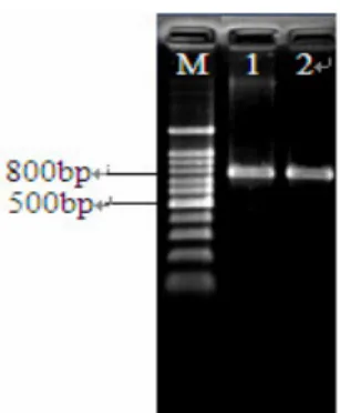
The results of nifH gene PCR amplification showed that
isolates MQ23 and MQ23R both could produce single product
bands on agarose gel by electrophoresis at the expected nifH
gene fragment size, about 785bp (Figure 1), with nifH PCR
primers nifH40F and nifH817R. This indicated these two
isolates share common nifH gene. Since nifH encodes the
highly conserved Fe protein of nitrogenase and has been used
as a marker gene for nitrogenase, the presence of same nifH
gene fragments in MQ23 and MQ23R strains under different
environment in this study provided genetic evidence of
nitrogen fixation capabilities, and a way to analyze the nitrogen
fixation potential. But, nif-DNA only shows the strains that
contain nitrogenase genes and does not indicate whether or not
these nitrogenase genes are expressed and these strains are
active and fix nitrogen. Therefore, the fact may play a
significant role in nitrogen fixation under field and greenhouse
environment. Further work will be necessary to address this
question.
Figure 1. The nifH gene amplification of inoculated and
reisolated strains (M: 100bp marker; 1:MQ23; 2:MQ23R)
Identification and phylogenetic analysis of endophytes
MQ23 and MQ23R
There were some differences between MQ23 and MQ23R
in its host growth environment. For example, strain MQ23 was
isolated from healthy S. alopecuroides plants growing in
Northwestern China. Where there have a dry climate, an
alkaline soil profile, host S. alopecuroides shows the excellent
performance of drought and alkaline tolerance and
anti-sandstorm for its developed root system under field
environment. However, strain MQ23R was isolated from
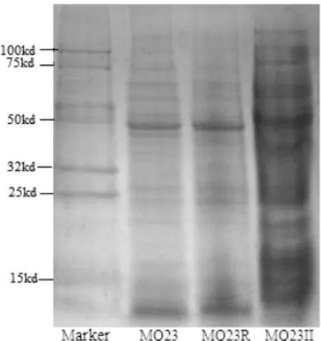
under greenhouse conditions. Though its host growth environment existed differences, our experiment results of SDS-PAGE (Figure
2) showed that whole-cell protein profiles generated by endophytes MQ23 and MQ23R were very similar or identical,
indicated exist close phylogenetic relationship between them,
perhaps they have same origins. However, compared with profiles of strain MQ23II, there were many differences among them,
whether the number or density of bandings. On the other side, the analysis of PCR-RFLP based on 16S rRNA gene (Figure 3) in
present study indicated that restriction patterns of MQ23 and
MQ23R are identical, while strain MQ23II show distinct banding patterns from them.
The phylogeny of 16S rRNA genes has been used as one of the main criteria for the differentiation of species, genera and
higher taxa in current bacterial taxonomy. In this work, upon constructed phylogenetic relationship of 16S rRNA genes
sequences (Figure 4) and rRNA sequence analysis we could see
that isolates MQ23 and MQ23R had 100% sequence homology, belonged to the same species. They were most related to Bacillus
cereus ATCC14579T and Bacillus thuringiensis ATCC10792T (with 100% and 99.9% similarity, respectively), they formed a
Bacillus sub-clade. Therefore, the nodule endophytes were designated as Bacillus cereus MQ23 and Bacillus cereus MQ23R.
Figure 2. Protein electrophoresis pattern of SDS-PAGE. Lane’s order
from left to right: marker, MQ23, MQ23R, MQ23II. MQ23 was
original strain, and MQ23R was reisolated from S. alopecuroides root
nodule of plant reinoculation. MQ23II was a Mesorhizobium sp.
Figure 3. Electrophoretic pattern of restriction endonuclease (HaeIII,
MspI, HinfI, HhaI) to strain MQ23, MQ23R and MQ23II. Lane’s
order from left to right: M: 100bp marker; 1: MQ23; 2: MQ23R; 3:
MQ23II.
Figure 4. Neighbor joining tree based on alignment of nucleotide
sequences of the 16S rRNAgene from tested strains (shown in bold)
and reference strains. GenBank accession numbers were placed in
parentheses. Bootstrap values greater than 50% were indicated. Scale
bar represents the number of substitutions per site.
Plant growth promoting characteristics of isolates
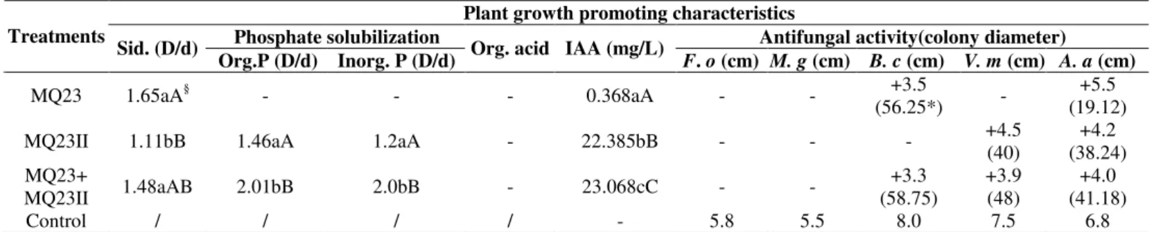
All of three treatments MQ23, MQ23II and MQ23+MQ23II
(Table1) gave a positive CAS assay showing that they all
phosphate), isolate MQ23 showed negative while coinoculation
MQ23+ MQ23II were positive. All treatments were negative
for organic acid product, positive for IAA product, and the
amount of IAA produced by three treatments was 0.368 mg/l,
22.385 mg/l and 23.068 mg/l, respectively. Another, three
treatments showed certain antifungal activity to plant
pathogenic fungi, and inhibition ratios was given in Table 1. As
a whole, for positive action coinoculation MQ23+MQ23II
showed better effect than inoculation alone in vitro. Therefore,
Bacillus cereus MQ23 and Mesorhizobium sp. MQ23II have
better cooperative interaction. But definite mechanism of their
interaction is further work.
Table 1. Plant growth promoting characteristics of endophytes isolated from root nodules
Plant growth promoting characteristics
Phosphate solubilization Antifungal activity(colony diameter)
Treatments
Sid. (D/d)
Org.P (D/d) Inorg. P (D/d) Org. acid IAA (mg/L) F. o (cm) M. g (cm) B. c (cm) V. m (cm) A. a (cm)
MQ23 1.65aA§ - - - 0.368aA - - +3.5
(56.25*) -
+5.5 (19.12)
MQ23II 1.11bB 1.46aA 1.2aA - 22.385bB - - - +4.5
(40)
+4.2 (38.24) MQ23+
MQ23II 1.48aAB 2.01bB 2.0bB - 23.068cC - -
+3.3 (58.75)
+3.9 (48)
+4.0 (41.18)
Control / / / / - 5.8 5.5 8.0 7.5 6.8
Sid. -Siderophore; Org.P-Organic Phosphate; Inorg. P-Inorganic Phosphate; Org.acid-Organic acid; F. o- Fusarium oxysporum; M. g-Magnaporthe grisea; B. c- Botrytis cinere Pers.; V. m- Valsa mali Miyabe et Yamada; A. a- Alternaria alternate.
D/d means the ability to produce siderophores. D-Diameter of Colony and halo; d- Colony diameter.
+ positive action; - negative action; / blank; Control for IAA assay was LB(10g NaCl/L) without inoculated bacterial suspension under same incubation condition; Control for antifungal activity assays were fungal mycelia cultivated for 7 days on PDA plates without tested strains under the same incubation condition.
§
The same letter means no significant difference between treatments, the capital letter indicates significant level at 0.01 while lowercase letter indicates significant level at 0.05. The data in columns is average values of five repetitions.
*
Inhibition ratio=(Control colony diameter-treatment colony diameter)100% / Control colony diameter .
Plant inoculation assay
To further confirm plant growth promoting characteristics,
we performed plant inoculation assay. Results in Table 2
showed that each growth indexes of inoculated MQ23 except
root fresh weight, slightly higher than those of negative control
(NC), but only root fresh weight and root dry weight showed
significant difference (p<0.05). Compared with MQ23II (PC),
plant inoculated MQ23 only presented significant difference in
nodule numbers. However, growth indexes of coinoculated
MQ23+MQ23II were higher than those of inoculated alone
(MQ23, PC, NC), except for root fresh weight, and showed
significant difference (p<0.05) compare with NC.
Table 2. Effect of Bacillus cereus MQ23 on growth and nodulation of S. alopecuroides
Treatments Shoot length (cm)
Root length (cm)
Shoot fresh weight (g/plant)
Root fresh weight (g/plant)
Shoot dry Weight (g/plant)
Root dry weight (g/plant)
Nodule number /plant MQ23 8.6666±0.6118ab* 9.9977±0.9636ab 0.0797±0.0378ab 0.0722±0.0119b 0.0078±0.0041ab 0.0137±0.0037a 0b MQ23II
(PC) 8.2317±1.4739ab 10.011±0.7859ab 0.069±0.0178ab 0.1159±0.0307ab 0.0092±0.0025ab 0.0131±0.001ab 2.333±0.5774a MQ23II+
MQ23 9.9833±0.2021a 11.9613±1.6113a 0.1276±0. 0314a 0.087±0.0116b 0.0123±0.0012a 0.0159±0.0015a 4.333±1.5275a Native
control (NC) 6.38±1.0868b 9.171±0.5688b 0.0519±0.0029b 0.1542±0.0057a 0.0036±0.0009b 0.0079±0.0015b 0b *The letters a and b indicate different Tukey grouping. The same letter means no difference among treatments, while different letters means significant difference (p<0.05). The value in the column is the averages of ten repetitions.
DISCUSSION
Endophytic Bacillus cereuses are potential N2-fixing strains The results of nifH gene PCR amplification (Figure 1)
indicated these two isolates MQ23 and MQ23R share common
nifH gene under different environment. The fact may play a
significant role in nitrogen fixation under field and greenhouse
environment. Ding et al. (6) reported that bacteria belonging to
Bacillus sp. have been identified as plant-growth-promoting
rhizobacteria, and nifH genes have been detected in some
species of Bacillus cereus. Zehr et al. (40) showed that
molecular tools for detection and characterization of the
nitrogenase gene (nifH) can provide new information on
activity of diverse nitrogen fixing organisms (12). Another, the
coexistence of endophytic Bacillus cereus and symbiotic
rhizobia in root nodules is an important ecological event
because the coexisting bacteria may have more opportunities to
exchange their core genomic and symbiont information, as
revealed in other report (39).
Identification of endophytes MQ23 and MQ23R
Based on whole-cell protein profiles (Figure 2) generated
by endophytes MQ23 and MQ23R, our experiment results
further supported this viewpoint that combination of protein
SDS-PAGE with computerized analysis of cellular protein
profiles provide an effective relatively simple and reproducible
approach to investigate of taxonomic relationships among
many bacterial species (5,15, 23), such as Bacillus (13).
Previous reports also showed protein profiles of SDS-PAGE as
having a high degree of correlation with DNA PCR-RFLP
analysis, rRNA sequence analysis, and DNA-DNA
hybridization for several bacterial species (3, 11, 25, 35).
In this work, analysis of 16S rRNA gene PCR-RFLP, gene
sequencing and phylogeny further confirmed that defining a
bacterial species based upon the consensus of combined results
of several analyses can overcome the potential prejudicial
grouping by each of the single analysis, supported previous
studies (19).
Co-inoculation enhanced plant growth
Previous reports informed that the cooperative interactions
between rhizobia and other plant root colonizing bacteria play
a role in the improvement in nodulation and N2 fixation in legume plants (2); other such examples include when rhizobia
are coinoculated with Rhizobium leguminosarum bv trifolii and
either B. insolitus or B. brevis (32), and with Bacillus spp. and
the soybean endosymbiont Bradyrhizobium japonicum (1, 21).
Geetha et al. (8) reported that co-inoculation enhanced growth
and nodulation of the pigeon pea with Bacillus strains and
Rhizobium spp. Similarly, Selvakumar et al. (31) showed that
the non-rhizobial plant growth promoting bacteria Bacillus
thuringiensis KR-1 from the nodules of Kudzu promoted
growth and positively influenced nutrient uptake in wheat
seedlings. Therefore, this report extends similar observations to
another legume-rhizobium system that of S. alopecuroides.
To the best of our knowledge, this is the first report about
the presence of putative non-rhizobial endophytes having
growth-promotion attributes in S. alopecuroides root nodules.
Further studies are required to prove the endophytic nature of
Bacillus cereus MQ23 and to harness their potential as
bio-inoculants and biocontrol agents in agriculture.
ACKNOWLEDGEMENTS
This work was supported by projects from the National
Science Foundation of China (30970003, 30900215), RFDP
(20050712013), PCSIRT of China, and Youth scientific
research foundation of Shangqiu Normal University.
REFERENCE
1. Bai, Y.; Zhou, X.; Smith, D.L. (2003). Crop ecology, management and quality. Enhanced soybean plant growth resulting from coinoculation of
Bacillus strains with Bradyrhizobium japonicum. Crop Sci., 43:1774-1781.
Microbial cooperation in the rhizosphere. J. Exp. Bot.,56:1761-1778. 3. Berber, I.; Cokmus, C.; Atalan, E. (2003). Characterization of
Staphylococcus species by SDS-PAGE of Whole-Cell and Extracellular Proteins. Microbiology. 72:42-47.
4. Chandra, S. N.; Puneet, S.C.; Sangeeta, M.D.; Karishma, S.; Ajit, V.; William, J. S. (2010). Tripartite interactions among Paenibacillus lentimorbus NRRL B-30488, Piriformospora indica DSM 11827, and
Cicer arietinum L. World J. Microbiol. Biotechnol., 26:1393-1399. 5. Costas, M. (1992). Classification, identification and typing of bacteria by
the analysis of their one-dimensional polyacrylamide gel electrophoretic protein patterns. In: Advances in Electrophoresis., V.5. Chrambach A, Dunn NJ and Radola BJ (Ed):351-408.
6. Ding, Y.; Wang, J.; Liu, Y.; Chen, S. (2005). Isolation and identification of nitrogen-fixing Bacillius from plant rhizospheres in Beijing region.
J.Appl.Microbiol., 99:1271-1281.
7. Franck, P.; Lucile, J. M.; René, B. (2001). Improvement in the RFLP procedure for studying the diversity of nifH genes in communities of nitrogen fixers in soil. Res.Microbiol.,152: 95-103.
8. Geetha, R.; Falguni, S.; Anjana, J. D.; Archana, G. (2008). Enhanced growth and nodulation of pigeon pea by co-inoculation of Bacillus strains with Rhizobium spp.. Biores. Technol., 99:4544-4550.
9. Gordon, A.S.; Weber, R.P. (1951). Colorimetric estimation of indoleacetic acid. Plant Physiol., 26: 192–195.
10. Hildebrandt, U.; Ouziad, F.; Marner, F.J.; Bothe, H. (2006). The bacterium Paenibacillus validus stimulates growth of the arbuscular mycorrhizal fungus Glomus intraradices up to the formation of fertile spores. FEMS Microbiol. Lett,. 254:258–267.
11. Hook, L.A.; Odelson, D.A.; Bogardt, A.H.; Hemmingsen, B.B.; Labeda, D.P.; MacDonell, M.T. (1991), Numerical analysis of restriction fragment length polymorphisms and whole-cell protein banding patterns: a means of bacterial identification at the species and subspecies level.
USFCC News lett., 21: 1-10.
12. Hou, J.; Huang, B. (2005). Overview of nitrogen fixation of marine cyanobacteria. Advance in earth science.,20: 312-319.
13. Ismet, B. (2004). Characterization of Bacillus species by numerical analysis of their SDS-PAGE protein profiles. J. Cell Mol. Biol., 3:33-37. 14. Katznelson, H.; Bose, B. (1959). Metabolic activity and phosphate
dissolving capability of bacterial isolates from wheat root, rhizosphere and non-rhizosphere soil. Can.J.Microbiol., 5: 79-85.
15. Kersters, K. (1985). Numerical methods in the classification of bacteria by protein electrophoresis. In: Computer Assisted Bacterial Systematic. Goodfellow M, Jones D and Priest FG (Ed). London: Academic Pres. pp. 337-368.
16. Khan, Z.; Kim, S.G.; Jeon, Y.H.; Khan, H.U.; Son, S.H.; Kim, Y.H. (2008). A plant growth promoting rhizobacterium, Paenibacillus polymyxa strain GBR-1, suppresses root-knot nematode. Biores.
Technol.,99: 3016-3023.
17. Laemmli, U.K. (1970). Cleavage of structural proteins during the assembly of the head of bacteriophage T4. Nature., 227:680-685. 18. Lin, L.; Qiao, Y.S.; Ju, Z.Y.; Ma, C.W.; Liu, Y.H.; Zhou, Y.J.; Dong,
H.S. (2009). Isolation and characterization of endophytic Bacillus subtilis Jaas ed1 antagonist of eggplant Verticillium wilt. Biosci. Biotechnol. Biochem., 73:1489-93.
19. Liu, J.; Wang E.T.; Chen, W.X. (2005). Diversit rhizobia associated with woody legumes Wisteria sinensis, Cercis racemosa and Amorpha fruticpsa grown in the temperate zone of China. Syst. Appl. Microbiol., 28:465-477.
20. Liu, J.; Wang, E.T.; Ren, D.W.; Chen, W.X. (2010). Mixture of endophytic
Agrobacterium and Sinorhizobium meliloti strains could induce nonspecific nodulation on some woody legumes. Arch Microbiol.,192: 229-234.
21. Liu, Z.L.; Sinclair, J.B. (1993).Colonization of soybean roots by Bacillus megaterium B153–2-2. Soil Biol. Biochem.,25:849–855.
22. Marois, J.J.; Johnston, S.A.; Dunn, M.T.; Papavizas, G.C.(1982). Biological control of Verticillium wilt of eggplant in the field. Plant Disease.,66:1166-1168.
23. Merquior, V. L. C.; Peralta, J. M.; Facklam, R. R.; Teixeira, L. M. (1994). Analysis of electrophoretic whole-cell protein profiles as a toll for characterization of Enterococcus species. Curr. Microbiol., 28:149-153.
24. Moulin, L., Béna, G.; Boivin-Masson, C.; pkowski, T. St. (2004). Phylogenetic analyses of symbiotic nodulation genes support vertical and lateral gene co-transfer within the Bradyrhizobium genus. Mol. Phylogenet. Evol., 30:720-732.
25. Niemi, R.M.; Niemela, S.I.; Bamford, D.H.; Hantula, J.; Hyvarinen, T.; Forsten, T.; Raateland, A. (1993). Presumptive fecal Streptococci in environmental samples characterized by one-dimensional sodium dodecyl sulfate-polyacrilamide gel electrophoresis. App. and Env.
Microbiol., 59:2190-2196.
26. Oehrle, N.W.; Karr, D.B.; Kremer, R.J.; Emerich, D.W. 2000. Enhanced attachment of Bradyrhizobium japonicum to soybean through reduced root colonization of internally seedborne microorganisms. Can. J. Microbiol., 46:600-606.
27. Ryan, R.P., Germaine, K.; Franks, A.; Ryan, D. J.; Dowling, D.N. (2008). Bacterial endophytes: recent developments and applications.
FEMS Microbiol. Lett., 278:1-9.
28. Saitou, N., Nei, M. (1987). The neighbor-joining method: a new method for reconstructing phylogenetic trees. Mol. Biol. Evol., 4: 406-425. 29. Sambrook, J.; Russell, D.W. (2001). Molecular Cloning: A Laboratory
Manual, 3rd ed, v.1.Cold Spring Harbor, New York.
31. Selvakumar, G.; Kundu, S.; Anand, D. G.; Yogesh, S. S.; Hari, S. G. (2008).Isolation and characterization of nonrhizobial plant growth promoting bacteria from nodules of kudzu (Pueraria thunbergiana) and their effect on wheat seeding growth. Curr. Microbiol., 56:134-139. 32. Sturz, A.V.; Christie, B.R.; Matheson, B.G.; Nowak, J. (1997).
Biodiversity of endophytic bacteria which colonize red clover nodules, roots, stems and foliage and their influence on host growth. Biol. Fertil.
Soil., 25:13-19.
33. Tan, Z.Y., Xu, X.D., Wang, E.T., Gao, J.L., Martínez-Romero, E., Chen, W.X. (1997). Phylogenetic and genetic relationships of Mesorhizobium tianshanense and related rhizobia. Int. J. Syst. Bacteriol., 47:874-879. 34. Van, B., Beyene, P. D.; Eardly, B. D. (1996). Phylogenetic relationships
among Rhizobium species nodulating the common bean (Phaseolus vulgaris L.). Int. J.Syst. Evol. Microbiol., 46:240-244.
35. Vauterin, L.; Vantomme, R.; Pot, B.; Hoste, B.; Swings, J.; Kersters, K. (1990). Taxonomic analysis of Xhantomonas campestris pv. begonidae
and X. campestris pv. pelargonii by means of phythopathological, phenotypic, protein electrophoretic and DNA hybridization methods.
Syst. Appl. Bacteriol.13,166-167.
36. Vincent, J.M. (1970). The cultivation, isolation and maintenance of
rhizobia. A Manual for the Practical Study of the Root-Nodule Bacteria. pp.1-13.
37. Vinuesa, P.; Leon-Barrios, M.; Silva, C.; Willems, A.; Jarabo-Lorenzo, A.; Perez-Galdona,R.; Werner, D; Martínez-Romero, E. (2005).
Bradyrhizobium canariense sp. nov., an acid-tolerant endosymbiont that nodulates endemic genistoid legumes (Papilionoideae: Genisteae) from the Canary Islands, along with Bradyrhizobium japonicum bv. genistearum, Bradyrhizobium genospecies alpha and Bradyrhizobium
genospecies beta. Int. J. Syst. Evol. Microbiol., 55:569-575.
38. Wang, E.T.; Berkum, P.; Sui, X.H.; Beyene, D.; Chen, W.X.; Martinez-Romero, E. (1999). Diversity of rhizobia associated with Amorpha fruticosa isolated from Chinese soils and description of Mesorhizobium amorphae sp nov. Int. J. Syst. Bacteriol., 49:51-66.
39. Wang, L.L.;Wang, E.T.; Liu, J.; Li, Y.; Chen, W.X. (2006). Endophytic occupation of root nodules and roots of Melilotus dentatus by
Agrobacterium tumefaciens.Microbial. Ecol.,52:436-443.
40. Zehr, J.P., Braun, S.; Chen, Y.; Mellon, M. (1996). Nitrogen fixation in the marine environment: relating genetic potential to nitrogenase activity.
J. Exp. Mar. Biol. Ecol., 203:61-73.
41. Zhao, L.F.; Deng, Z.S,; Yang, W.Q.; Cao, Y.; Wang, E.T.; Wei, G.H. (2010). Diverse rhizobia associated with Sophora alopecuroides grown in different regions of Loess Plateau in China. Syst. Appl. Microbio. doi:10.1016/j.syapm.2010.08.004.