AR
TICLE
Non-ionizing method of screening adolescent idiopathic scoliosis
in schoolchildren
Abstract Adolescent idiopathic scoliosis (AIS) af-fects 2% to 4% of young people in Brazil. Repeat-ed exposures to radiation usRepeat-ed in the monitoring of the deformity can be harmful to the health. This study aimed to present a photogrammetry proto-col as a non-ionizing method to quantify sproto-colio- scolio-sis and relate it to the Cobb radiological method. Sixteen individuals with idiopathic scoliosis (age: 21.4 ± 6.1 years, body mass index: 19.8 ± 0.2 kg/ m2) underwent standing posteroanterior X-ray examination of the trunk. Additionally, markers were placed on the spinal processes of the C7 to L5 vertebrae, and posterior trunk photographs were taken. All images were sent for independent analysis by two examiners who were trained in the quantification of scoliosis. The average of the thoracic curvature evaluated through the photo-grammetry and Cobb methods were 36.43° and 36.14°, respectively. With an average difference of 4.1°, the methods were not statistically different (p < 0.05). As a non-ionizing method that is low cost and portable, photogrammetry may represent a suitable alternative to the radiological method. Further studies are needed for the improvement of non-ionizing techniques in AIS screening. Key words Adolescent idiopathic scoliosis, Diag-nosis, Non-invasive method, Adolescent health
Rozilene Maria Cota Aroeira 1
Jefferson Soares Leal 2
Antônio Eustáquio de Melo Pertence 3
Estevam Barbosa de Las Casas 1
Marcelo Greco 1
1 Departamento de
Engenharia de Estruturas, Escola de Engenharia, Universidade Federal de Minas Gerais (UFMG). Av. Antônio Carlos 6627, Pampulha. 31270-901 Belo Horizonte MG Brasil. [email protected]
2 Departamento de
Ortopedia e Traumatologia, Escola de Medicina, UFMG. Belo Horizonte MG Brasil.
3 Professor,Departamento
A
ro
eir
a RMC
Introduction
Vertebral deformity is highly prevalent among young people and has been the subject of nu-merous investigations. Scheuermann’s kyphosis, spondylolisthesis, and adolescent idiopathic sco-liosis (AIS) are deformities related to vertebral growth that can affect healthy children at the
be-ginning of the growth spurt1. Overall, AIS is the
most common vertebral deformity found world-wide and comprises 80% of all types of scoliosis. The Scoliosis Research Society defines AIS as an unknown spinal deformity, characterized by changes in three planes: lateral curvature with
a Cobb angle ≥ 10°, thoracic lordosis, and axial
rotation. This disease primarily affects children of 10 years and older whose skeletons are still
developing2. Although scoliosis has a very low
life-threatening index, social, familial, and sur-gical factors can lead patients to develop mental disorders and can even increase the risk of
sui-cide3,4. A study by Payne et al.3 indicated that the
presence of spinal deformity in adolescents con-stitutes a risk factor for psychological depression independent of the treatment received by the
patient. According to Han et al.2, adolescence is
a sensitive period of personal and psychological maturation, and both the vertebral deformity and the physical discomfort generated by it can affect the patients’ quality of life.
Konieczny et al.5 study showed an
over-all prevalence of AIS between 0.47 - 5.2%. The prevalence and severity are higher in girls than in boys at a rate of 3:1 between 11 and 12 years and increase with age. In Brazil, according to Souza et
al.6, the prevalence of AIS ranges from 2% to 4%
between the ages of 10 and 16 years. According to
Konieczny et al.5 there are few studies that
pro-vide relevant data in relation to the prevalence of AIS. These studies present varied definitions for scoliosis in addition to several study protocols and age groups with inclusion of scoliotic curves < 10 °, although it is an international consensus that, by definition, scoliosis is a deformity with
an angle ≥10 °. In addition, according to the
au-thors, the efficacy of the widely used Adams7 test
was considered low and would be more effective if associated with trunk rotation or Moiré
topog-raphy evaluations. Fong et al.8 state that no
evalu-ation of scoliosis has yielded substantial benefits and sufficient levels of evidence to date.
The Cobb method, developed in 1948 by a researcher of the same name, is still considered the “gold standard” for scoliotic curve
measure-ment9. This method is used for diagnosis,
fol-low-up, and definition of the treatment to be instituted. However, several studies have present-ed the deleterious effects of numerous ionizing radiation sessions on young people with incom-plete bone maturation during the follow-up
pe-riod for idiopathic scoliosis10-15. During this
fol-low-up, the adolescent may be submitted to more than 25 trunk radiographs. One study revealed that approximately 15% of the patients under-went 50 or more radiographic examinations with an accumulation of estimated ionizing radiation
doses of 20 cGy or more12. Non-radiographic
ex-aminations aimed at postural evaluation, which allow for the topographic investigation of asym-metries related to vertebral deformities, are not common practice in the daily clinic. According
to Brink16, postural assessment should be a
rou-tine examination for individuals with neuromus-culoskeletal disorders. In the study by Kowalski
et al.17, postural monitoring tests performed in
schoolchildren revealed that 50-60% of the ad-olescents had postural abnormalities, 10% of them being at risk of progressive deformity of the
spine. For Cheung et al.18, early postural
screen-ing and observation of scoliosis may apparently mitigate the surgical risk. In addition, early di-agnosis of AIS can prevent excessive progression and pathological postural adaptations. In the
study by Aroeira et al.19, individuals with similar
Cobb angle values may present different asym-metries throughout the body; on the other hand, significant asymmetries may be present in those with low Cobb angle values. As a consequence, there is a gap in the proposed treatment for cases of scoliosis classified as “mild”; that is, between 10 and 25 ° of Cobb value. Because they are not eligible for treatment according to radiological criteria, they are deprived of an early therapeu-tic approach and adequate functional treatment.
Han et al.2, in a quality of life study of
post-op-erative scoliosis patients, stated that radiological examination should not be the only therapeutic indicator in AIS, and that new evaluation systems should be developed with a focus on the quality of life of patients.
aúd
e C
ole
tiv
a,
24(2):523-534,
2019
used by the proposed technologies20. Five
tech-nologies have stood out: 3D ultrasound system, 2D computerized photogrammetry, technologies based on laser projection or structured light, and
Moiré projection19. The great motivation of the
new studies lies in the search for a reduction in the number of radiographic examinations in the monitoring of these deformities that reach young people without complete bone maturation. In addition, the search for exams that assess asym-metries in their entirety, not only in the trunk, is equally relevant.
Thus, the objective of this study was to pres-ent a non-ionizing method, based on 2D photo-grammetry by computer vision, for the angular measurement of vertebral deviations, aiming at the diagnosis and follow-up evaluation of AIS.
Materials and methods
The method of measurement proposed in this study for the calculation of vertebral deviations in scoliosis using computerized
photogramme-try was patented at INPI under n. 1411000233521.
An observational and cross-sectional study was conducted to compare this new non-radiograph-ic method with the traditional Cobb method us-ing a direct numerical correlation between both methods. In addition, a nonparametric test was performed to describe the intensity of agreement between the Cobb methods and computerized photogrammetry at the location of the apical vertebra of the scoliotic curve.
Field study
After approval by the Research Ethics Com-mittee (COEP), patients who were in conserva-tive treatment for AIS during the study period and who had a medical request for radiological monitoring of scoliosis were invited to partici-pate in the study. All included participants gave their free and informed consent. After the adop-tion of the exclusion criteria for individuals sub-mitted to resection surgery of spinal processes of the vertebrae and those who had difficulty main-taining orthostatism, 16 patients were included in the study.
Radiological examination
A single anteroposterior x-ray of the trunk was obtained in the standing posture using the Toshiba KXO15R radiographic generator
(Toshi-ba, Tokyo, Japan) and the Agfa Health System digital radiography (AGEA, Mortsel, Belgium). The scanned images were printed on 35.6 × 43.2 cm film and sent to the Cobb angle measure-ment. This angle is obtained by the intersection of two lines perpendicular to the tangents of the upper plateau of the upper terminal vertebra and the lower plateau of the lower terminal vertebra of the curve presented by the vertebral column. With the objective of not marking on the printed radiography, which could interfere with the other measurements, the lines drawn with a ballpoint pen and a 30-cm ruler aid were made on trans-parent sheets positioned on the radiographs. A protractor was used to obtain the angle of cur-vature. Five measurements were performed for each patient at different times, and the mean of the five values were used in the statistical analysis. In addition to the Cobb angle measurement, the apical vertebra of the curve was identified; that is, the vertebra that presented the greatest distance from the vertical axis of the spine. All measure-ments were performed by a single examiner.
Computerized photogrammetry examination
Immediately after the radiological examina-tion, an examiner experienced in palpatory anat-omy performed the marking of the spinal pro-cesses of the vertebrae from C7 to L5, using der-mographic pencil. Next, to promote the external identification of each vertebra and its displace-ment in the three planes of the space, anatomical landmarks vector type 45 mm long and 5 mm in diameter, specially developed for this study, were positioned in the respective spinous processes of said vertebrae. After this procedure, each pa-tient was photographed standing in the front position, with the arms hanging over the body and standardizing the position of the feet at 36º (Piok position). A transparent acrylic symmetry graph was used as background, measuring 2.05 m in height and 0.72 m in width and squared in the dimensions 10 × 10 cm mark CARCI® for calibration. Photographs were taken using a tripod-mounted (GREIKA WT3750) Olympus 7.1-megapixel camera at 3072 × 2304 resolution, without zoom positioned at 1.3 m distance. The images were sent for the independent analysis by the examiner trained in the protocol for the quantification of scoliosis by photogrammetry as proposed in this study. A two-dimensional vector
drawing software, CorelDRAW13®, was used to
A
ro
eir
a RMC
angular measurement. The first step was to accu-rately highlight the center of the surface markers using the ellipse feature in the CorelDraw13® toolbar. Next, the first phase of the photointer-pretation was carried out, consisting of the iden-tification of the apical vertebra and the upper limit vertebra of the scoliotic curve. This proce-dure was performed by tracing two vertical lines, one tangential to the convex face of the curve and another to the vertical axis of the C7 vertebra, as shown in Figure 1. The apical vertebra of the curve gives rise to the phenomenon of scoliosis. It is the vertebra farthest from the vertical axis of C7 and, generally, is the one that presents great-er rotation of its body, which is visualized by the change of direction of the body of the vector. The upper limit vertebra is the first vertebra emerging from the vertical alignment of C7 and undergo-ing rotation, identified by the positionundergo-ing of the landmark.
Then, angular measurements of the vertebral deviations with the vertical Y axis of each seg-ment of two adjacent vertebrae from the upper limit vertebra to the apical vertebra, referred to
as angles R1, R2, R3, etc., were performed. The sum of the angles collected determines the angle of scoliosis, termed MR. This angle corresponds to the Cobb angle measured by radiography (Fig-ure 2).
Computed photogrammetry, through the identification vectors of each vertebra, also al-lowed the generation of three-dimensional virtu-al images of the spativirtu-al behavior of the vertebrvirtu-al
column22.
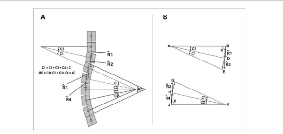
It was possible to mathematically correlate the angular measurements of the scoliotic curve obtained by the Cobb method (MC) and by the method of the present study (MR). If we define the scoliotic curve as consisting of segments of circle arcs, as shown in Figure 3, it is possible to consider that the MC measure is the sum of the angles C1, C2, C3, and C4 obtained between the consecutive spinous processes found in this mea-surement. Thus, if the angles C1, C2, C3, and C4 are equal to each other and equal to C, then the sum of these angles obtained in the Cobb meth-od interval will be equal to 4C (Figure 3a and b). Considering that the scoliotic curve is com-posed of segments of circle arcs, the triangles ABD, ADE, FGH, and FHJ will be isosceles tri-angles (Figure 3b). Therefore, the angle X will be indicated by EQ (1):
X = (180-C) / 2 (1)
In addition, the relationships between the an-gles of deviation of the Y-axis (R1, R2, R3, and R4) obtained by the method proposed in this study with the angles (C) of each vertebral seg-ment obtained by the Cobb method are shown, as indicated by EQs (2), (3), (4) and (5).
R1 = 90-X = 90 - (180-C) / 2 = C / 2 (2) R2 = R1 + C = C / 2 + C = 3C / 2 (3) R3 = R4 + C = C / 2 + C = 3C / 2 (4) R4 = 90 - X = 90 - (180 - C) / 2 = C / 2 (5) Thus, the MR measurement will be indicated by EQ (6):
R 2 = C 4 + C 2 + C 2 = 4C (6)
Therefore, in this case, the value of the mea-surement obtained by the present study will be equal to that obtained by the Cobb method.
It is important to point out that this demon-stration does not represent a general solution; however, it provides an understanding of the phenomenon observed in this experimental and statistically proven study, highlighting the equiv-alence between the measurement obtained in this study and that obtained by the Cobb method.
Figure 1. 1st phase of the photointerpretation: the
aúd
e C
ole
tiv
a,
24(2):523-534,
2019
Agreement test for the location of the apical vertebra of scoliosis
The Kappa measure was used to describe the intensity of agreement between the two methods, Cobb and computerized photogrammetry, to lo-cate the apical vertebra of the curve. This mea-sure consists of the degree of interobserver agree-ment beyond what would be expected by chance alone. Its maximum value is 1, which represents
total agreement, and values close to and below zero indicate no agreement, or that agreement was exactly as expected by chance. The Kappa co-efficient was indicated by the EQ (7).
κ = (7)
In which:
Co Observed agreement
Ce Expected Concordance
Figure 2. 2nd phase of the photointerpretation, angular measurement, performed in the CorelDraw13® software,
of a volunteer AIS carrier with left main convex thoracic curve. Measure of the angles of deviation of the spinal balance in the Y axis between the upper limit vertebrae (T4) and the apical vertebra (T10): R1 (10°), R2 (4°), R3 (8°), R4 (12°), R5 (10°), R6 (9°) and R7 (4°). On the left, measure of the Cobb angle to the X-ray of the same patient.
Figure 3. (a) Schematic demonstration of the scoliosis arc and its Cobb angle equivalence (MC) and the respective angles R1, R2, R3 and R4, which together form the angle MR of the present method; (b) geometric demonstration of the equivalence of angles C and R.
Co - Ce
A
ro
eir
a RMC
The level of significance adopted for all tests was 5%.
Results
In a population composed of 16 individuals with AIS, with 21.4 ± 6.1 years of age, 52.9 ± 5.8 kg body weight, 1.63 ± 0.05 m height, and 19.8 ± 0.2 body mass index (BMI), 12 individuals pre-sented a double curve (dorsal and lumbar), three individuals with a single lumbar curve, and one individual with a single dorsal curve, with a dor-sal Cobb angular mean of 36.14° ± 16.38° and mean angle of Cobb lumbar of 27.20° ± 10.05°. General characteristics and measurements using the Cobb method and the computerized pho-togrammetry method for lumbar and thoracic curves are presented in Table 1.
According to the descriptive statistics and the graphs presented in Figure 4, there was no sta-tistically significant difference between the mea-surements in the two methods. Thus, it can be stated that regardless of the adopted method, the measurements of both dorsal and lumbar curva-ture will be similar.
The Cohen’s Kappa statistical study revealed a general index of 0.92 for the location of the thoracic apical vertebra and 0.825 for the loca-tion of the lumbar vertebra. With a significance level of 5% the null hypothesis is rejected, which shows that the agreement between the two tests is different from zero; that is, they are concordant. The values obtained for the thoracic and lumbar Kappa index were very high and were considered excellent.
Discussion
A 2D computerized photogrammetry proto-col was proposed as a non-ionizing method for the diagnosis and follow-up of AIS in school-children. Other non-ionizing technologies have been proposed as an alternative to radiological
examinations for this purpose19. However, no
method has so far reached routine clinical use. The lack of objectivity and numerical correlation with Cobb’s “gold standard” method may be con-tributing to the low adherence to the proposed
methods23.
The values of the curves obtained by the com-puterized photogrammetry method were similar to the values obtained by the Cobb method. The difference between the two methods was lower
for thoracic curves than for lumbar curves, with values of 2.9° and 5.1°, respectively. The mean dif-ference of 4.1° for both methods, which includes all thoracic and lumbar curves, was compatible with the intrinsic error of the Cobb method in
an inter- and intra-observer analysis24, in which
the variability of Cobb angle measurements was
between 4° and 8°25,26. The location of anatomical
structures by palpatory anatomy is impaired in individuals with a very high BMI, due to the diffi-culty in identifying the bony prominences located below the skin. The location of the bony prom-inence of the spinal processes of the vertebrae is indispensable in the application of the method proposed in this study. During the recruitment of the study volunteers, two subjects with AIS had
a BMI above 24 kg/m2 (above ideal weight) but
were included in the study. Subjects who had pre-vious surgery of resection of these processes were excluded from the study due to the impossibility of applying these landmarks. It would be plau-sible, then, to suppose that the most discordant results in the measurements of the lumbar angles were related to the individuals with higher BMI, as in the case of subjects of n.1 and n.4 shown in the table of results. Despite the great difference presented in the lumbar angles of these two sub-jects (8°), the greatest disagreement between the lumbar results was found in the subject of n.5 (23.8°), whose body mass index is within nor-mal limits. Although the sample of this study is considered representative of the scoliotic popu-lation, which is sent to public medical services in the southeast region of the country, it may differ from other regions or countries where childhood obesity is more prevalent. Thus, a study with a larger number of subjects would be necessary for a conclusive evaluation of the possible causes of the difference between the means of the thoracic and lumbar angles presented in this study. A new study using the same methodology presented in
this article was conducted by Leal27 that included
re-aúd
e C
ole
tiv
a,
24(2):523-534,
2019
spectively. The differences between the means of the two measurements were 0.34 [95% = -0.153 to 1.807], close to zero. The Pearson coefficient for the 161 volunteers was r = 0.82 [95% CI = 0.77 – 0.86]. These results corroborate with the findings presented in the current study. Photo-grammetry showed an accuracy of 86.7%, sensi-tivity of 94.4%, specificity of 86.7% and predic-tive value of 75.5% in the aforementioned study. The differences between the angular means of the thoracic and lumbar curves were not reported.
The identification of the apical vertebra of the scoliotic curve was an important step in the proposed method, and the use of this specific landmark to identify vertebral alterations in the various planes of space had an important role in this analysis. The statistical study to evaluate the agreement of both methods in the identifica-tion of the apical vertebra of the scoliotic curve presented excellent concordance between them. However, the Kappa index was higher in cases of thoracic scoliosis when compared to lumbar scoliosis (0.92 and 0.825, respectively). Consid-ering the Kappa index for the general location of all the vertebrae involved in the study, the T5 and T8 thoracic vertebrae and the L3 lumbar vertebra had a value of 1, that is, maximum agreement. In addition, the possibility of finding three-di-mensional coordinates from the base to the tip of this landmark vector type made it possible to
visualize the angular variation of this vector in any of the three spatial planes, enabling the 3D reconstruction of the deformity. Thus, a major step was taken to reproduce the deformity across the surface, usually visualized only by invasive methods.
Although the cost comparison between the two methods is not an objective in this study, some considerations are relevant. The total cost of photogrammetric equipment (computer, soft-ware license, camera, tripod, and markers) was valued at USD 1,826. Strictly for comparative use, the cost of installing radiological equipment used in this study was estimated at USD 216,238. The basic skills for applying the proposed method are: basic knowledge of palpatory vertebral anatomy, photography, and image software. The time spent with the radiographic method in a typical patient was 13 min (positioning, exposure to radiologi-cal imaging, and Cobb measurement), while the time spent in the photogrammetric method was 28 min (landmark positioning, photographic ex-posure, and curve measurement).
Mrozkowiak et al.28 made the following
considerations about methods of measuring vertebral deformities: 1- the measurement ex-amination should work according to how the measurement result is used (e.g. as a basis for decision-making for surgery) and should be por-table for preventive examinations (detection of
Table 1. General characteristics of each individual included in the study and their angular measurements obtained by the Cobb method and by the computerized photogrammetry method in the thoracic and lumbar regions.
Subjects Age Sex BMI Scoliosis
level
Cobb Method Photogrammetry Method
Thoracic Lumbar ATV ALV Thoracic Lumbar ATV ALV
1 39 F 28.8 T e L 59.8° 26.0° T10 L4 57.4° 17.6° T10 L3
2 15 M 17.1 T e L 32.0° 15.0° T9-T10 L2-L3 34.4° 17.2° T9 L3
3 22 F 18.3 T 27.0° _ T9-T10 _ 25.8° _ T9 _
4 13 F 25.5 T e L 30.4° 32.0° T7-T8 L3 22.6° 24.0° T8 L2
5 12 F 21.7 T e L 39.6° 33.8° T8 L2 40.8° 10.0° T7 L2
6 19 F 22.9 T e L 28.2° 38.4° T8 L1 22.4° 35.4° T8 L2
7 16 F 22.2 T e L 42.2° 22.8° T9 _ 47.0° 22.8° T7 L2
8 14 F 17.4 T e L 22.4° 27.0° T9 L2-L3 23.0° 30.0° T9 L3
9 11 F 19.0 T e L 50.4° 48.6° T7 L2 51.0° 55.0° T7 L2
10 25 F 19.8 T e L 21.4° 18.8° T8 L2 26.4° 16.8° T9 L3
11 45 F 23.4 T e L 25.6° 26.4° T7 L2 29.0° 27.8° T7 L3
12 46 F 18.9 T e L 73.0° 41.8° T6-T7 L2 74.2° 39.4° T7 L2
13 14 F 17.8 L _ 19.8° _ L2-L3 _ 22.0° _ L3
14 13 F 15.9 L _ 14.0° _ L1 _ 26.0° T12 _
15 15 M 16.9 L _ 18.4° _ L1 _ 17.8° _ L1
16 25 F 23.1 T e L 17.8° 25.5° T5 L2 19.6° 23.8° T5 L1
A
ro
eir
a RMC
scoliosis in pre-school or school children); 2- the acquisition of images and the processing of data should be fast enough to provide results during
the visit of the health professional responsible for the case; 3- the measurement results should be presented in a readable form, not only for the
Figure 4. Comparison between the measurements of the scoliotic curves obtained by the Cobb method and the computerized photogrammetry method for (A) thoracic curves and (B) lumbar curves.
Volunteers
1 2 3 4 5 6 7 8 9 10 11 12 13 14 15 Thoracic Cobb
Photogrammetry Thoracic 80,00
60,00
40,00
20,00
A
ve
rag
e (d
eg
re
es)
(A)
Volunteers
1 2 3 4 5 6 7 8 9 10 11 12 13 14 15 Lombar Cobb
Lumbar Photogrammetry 60,00
50,00
40,00
30,00
20,00
10,00
A
ve
rag
e (d
eg
re
es)
aúd
e C
ole
tiv
a,
24(2):523-534,
2019
health professional but also for the patient, and should be compared with specific medical knowl-edge standards, preferably in terms of graphics and images; 4- the measurement should be sim-ple enough, particularly in terms of automation, so that it can be operated by a health profession-al; 5- the system must be reliable, error-resistant in its operation, and should not require constant intervention from a specialist.
In a review study, Aroeira et al.19 presented
the five main non-ionizing technologies for the evaluation of scoliosis in studies obtained in the literature from 2011 to 2015. The study showed that the same technology was used with different measurement protocols as, for example, the use of the 3D laser scanner without the use of a ref-erence marker, applied in the studies of Komeili
et al.20,29, and using 11 reference markers in the
studies of Parent et al.30. These results showed
analysis parameters with different maps of sur-face topography. Likewise, 2D computerized photogrammetry was used in four studies with different protocols. In this category, Fortin et
al.31 used topographic whole-body tracing, with
measurements of angular deviations in the trunk
and limbs. In a different way, Saad et al.32 used 2D
computerized photogrammetry only, in the pos-terior trunk, using different measurement proto-cols. These various protocols applied to different technologies make a comparative analysis diffi-cult. Studies using technologies based on ultra-sound, structured light, lasers, and fringe projec-tion have been shown to be of moderate to high quality. These techniques allow the evaluation of a large number of individuals in a reduced time, making them attractive for scoliosis screening in schoolchildren. However, the high cost and low objectivity of the results, based on color maps, can be a restriction factor for large scale use.
The 2D computerized photogrammetry protocol presented in this article provides the following advantages: the low cost of imple-menting the system; the transportability of the instruments that comprise it; the possibility of visualization of the whole body posture, allow-ing for correlation of dysfunctions associated to the body segments adjacent to the deformity of the spine, increasing the possibility of success in the diagnosis and the therapy used; and, finally, the reduction in the exposure of youths without complete bone maturation to the ionizing rays of the radiographs in the tracking and follow-up of the AIS. However, the method can be improved with its automation and, consequently, reduced exam time.
Recent technological advances point to deep cameras, which allow the acquisition of dense 3D scans of an area in real time, without the need for multiple cameras, at a low cost and
portabili-ty33-37. The use of 3D scanners offers new
oppor-tunities for the collection of anthropometric data in a wide range of applications. However, existing studies with the use of depth sensors in the anal-ysis of human activity have focused only on the qualitative estimation of body part positioning and on the knowledge of human action. A study using this technology for quantitative evaluation of the surface topography in AIS should be per-formed.
The search for non-ionizing methods of evaluation of vertebral deformities does not in-tend to replace radiological examination, which is important in the identification of congenital bone malformations and evaluation of skeletal maturity. However, new technologies introduce a new approach, not only for diagnosis, but mainly for the periodic monitoring of the developmen-tal deformities of the adolescent’s spine, with the possibility of also identifying compensatory asymmetries through reliable diagnoses. Low-cost and non-invasive diagnoses can be consid-ered of relevance in public policies considering, mainly, public health needs in Brazil. Considering the high prevalence of AIS and the fact that these are mainly preventive tests, which seek to avoid invasive and costly procedures such as surgeries, these tests are relevant in large-scale application for tracking vertebral alterations in schoolchil-dren as a measure of public health. The right to health of children and adolescents is State poli-cy, as the Brazilian constitution norm ratifies the principle of absolute priority of the protection of children and adolescents. The chapter on the right to life and health, in its Article 11, § discuss-es vertebral deviations in other areas of the body.
In this context, the 2nd paragraph of “Search for
technologies and methods” says:
“It is incumbent upon the public authority to provide, free of charge, to those in need, medi-cines, orthotics and other assistive technologies related to the treatment, habilitation or rehabili-tation for children and adolescents, according to the lines of care directed to their specific needs.”
Limitations
A
ro
eir
a RMC
by palpatory anatomy. In addition, the method cannot be applied in individuals who have un-dergone surgery for resection of the spinous
pro-cesses of the vertebrae involved in the analysis, due to the absence of the anatomical structure object of method analysis.
Collaborations
RMC Aroeira collaborated in the design, study design, data collection and final writing; JS Leal in the interpretation of the results of medical imaging tests; AEM Belongs in the delineation of the mathematical study, creation of figures and aid in the statistical analysis of the data; EB Las Casas in the critical review of the study; and M Greco in the critical review of the study and final approval of the version to be published.
Acknowledgments
aúd e C ole tiv a, 24(2):523-534, 2019 References
1. Castelein RM, van Dieen JH, Smit TH. The role of dor-sal shear forces in the pathogenesis of adolescent idio-pathic scoliosis - A hypothesis. Med Hypotheses 2005; 65(3):501-508.
2. Han J, Xu Q, Yang Y, Yao Z, Zhang C. Evaluation of quality of life and risk factors affecting quality of life in adolescent idiopathic scoliosis. Intractable & Rare Diseases Research 2015; 4(1):12-16.
3. Payne WK 3rd, Ogilvie JW, Resnick MD, Kane RL, Transfeldt EE, Blum RW. Does scoliosis have a psy-chological impact and does gender make a difference? Spine 1997; 22(12):1380-1384.
4. Tones M, Moss N, Polly Junior DW. A review of quality of life and psychosocial issues in scoliosis. Spine 2006; 31(26):3027-3038.
5. Konieczny MR, Senyut H, Krauspe R. Epidemiology of adolescent idiopathic scoliosis. J Child Orthop 2013; 7(1):3-9.
6. Souza FI, Ferreira RB, Labres D, Elias R, Souza APM, Pereira RE. Epidemiologia da escoliose idiopática do adolescente em alunos da rede pública de Goiânia-GO. Acta Ortop Bras 2011; 21(4):223-225.
7. Adams W. Lectures on the pathology and treatment of lateral and other forms of curvature of the spine. 2nd ed. London: J. & A. Churchill; 1882.
8. Fong DY, Lee CF, Cheung KM, Cheng JC, Ng BK, Lam TP, Mak KH, Yip PS, Luk KD. A meta-analysis of the clinical effectiveness of school scoliosis screening. Spine 2010; 35(10):1061-1071.
9. Cobb JR. Outline for the study of scoliosis. American Academy of Orthopaedic Surgeons Instr Course Lect 1948; 5:261-275.
10. Levy AR, Goldberg MS, Hanley JA, Mayo NE, Poitras B. Projecting the lifetime risk of cancer from exposure to diagnostic ionizin radiation for adolescent idiopathic scoliosis. Health Phys 1994; 66(6):621-633.
11. Goldberg MS, Maio NE, Levy AR, Scott SC, Poitras B. Adverse reproductive outcomes among women ex-posed to low levels of ionizing radiation from diag-nostic radiography for adolescent idiopathic scoliosis. Epidemiology 1998; 9(3):271-278.
12. Doody M, Lonstein JE, Stovall M, Hacker DG, Luck-yanov N, Land CE. Breast cancer mortality after di-agnostic radiography: finding from the U.S. Scoliosis Cohort study. Spine 2000; 25(16):2052-2063.
13. Ron E. Cancer risks from medical radiation. Health Phys 2003; 85(1):47-59.
14. Berrington de Gonzales A, Darby S. Risk of cancer from diagnostic X-rays: estimates for the UK and 14 other countries. Lancet 2004; 363(9406):345-351. 15. Enriquez G, Piqueras J, Catalá A, Oliva G, Ruiz A,
Ri-bas M, Duran C, Rodrigo C, Rodríguez E, Garriga V, Maristany T, García-Fontecha C, Baños J, Muchart J, Alava F. Optimization of radiological scoliosis assess-ment. Med Clin 2014; 143(Supl. 1):62-67.
16. Brink Y, Louw Q, Somers KG. The quality of evidence of psychometric properties of three-dimensional spi-nal posture-measuring instruments. BMC Musculoskel-et Disord 2011; 12:93.
17. Kowalski IM, Faldowska HP, Swornik M, Pierozynski B, Raistenskis J, Kiebzak W. Objective parallel-forms reliability assessment of 3-dimension real time body posture screening tests. BMC Pediatric 2014; 14:221.
18. Cheung CWJ. Ultrasound Volume Projection Imaging for Assessment of Scoliosis. IEEE Trans Med Imaging 2015; 34(8):1760-1768.
19. Aroeira RMC, Las Casas EB, Pertence AEM, Greco M, Tavares JM. Non-invasive methods of computer vi-sion in the posture evaluation of adolescent idiopathic scoliosis: a systematic review. J Bodyw Mov Ther 2016; 20(4):832-843.
20. Komeili A, Westover LM, Parent EC, Mareau M, El-Rich M, Adeed S. Monitoring for idiopathic scoliosis curve progression using surface topography asymme-try analysis of the torso in adolescents. Spine J 2015; 15(4):743-751.
21. Patente. Universidade Federal de Minas Gerais. Aroeira RMC, Pertence AEM, Leal JS. Dispositivo e método de medição utilizando fotogrametria computadorizada: n. 0000221106647968, 26 jul. 2011.
22. Aroeira RMC. Protocolo de fotogrametria computadori-zada na quantificação angular da escoliose [dissertação]. Belo Horizonte: Universidade Federal de Minas Gerais; 2009.
23. Krejci J, Gallo J, Stepanik P, Salinger J. Optimization of the examination posture in spinal curvature assess-ment. Scoliosis 2012; 7:10.
24. Mok JM, Berven SH, Diab M, Hackbarth M, Hu SS, Deviren V. Comparison of observer variation in con-ventional and three digital radiographic methods used in the evaluation of patients with adolescent idiopathic scoliosis. Spine 2008; 33(6):681-686.
25. Morrissy RT, Goldsmith GS, Hall EC, Kehl D, Cowie GH. Measurement of the Cobb angle on radiographic of patients who have scoliosis. Evaluation of intrinsic error. J Bone Joint Surg Am 1990; 72(3):320-327. 26. Carman DL, Browne RH, Birch JG. Measurement of
scoliosis and kyphosis radiographs. Intraobserver and interobserver variation. J Bone Joint Surg Am 1990; 72(3):328-333.
27. Leal SL. Validade da fotogrametria na detecção da pro-gressão da escoliose idiopática do adolescente [tese]. Belo Horizonte: Universidade Federal de Minas Gerais; 2004.
28. Mrozkowiak M, Miroslawa SE, Hanna Z, Waley Z. Re-view of Methods for Assessing Body Posture. J Health Sci 2014; 4(11):95-102.
29. Komeili A, Westover LM, Parent EC, Mareau M. El-Rivh M, Adeed S. Surface topography asymmetry maps categorizing external deformity in scoliosis. Spine J 2014; 14(6):973-983.
30. Parent EC, Chabot S, Westover L, Hill D, Moreau M, Hedden D, Lou E, Adeeb S. The ability of surface to-pography postural measurements to detect cobb angle progression in adolescents with idiopathic scoliosis (AIS) and a main thoracic curve: full torso scans com-pared to back only parameters. Scoliosis 2014; 9(Supl. 1):O10.
31. Fortin C, Feldman DE, Cheriet F, Gravel D, Gauthier F, Labelle H. Reliability of a quantitative clinical posture assessment tool among persons with idiopathic scolio-sis. Physiotherapy 2012; 98(1):64-75.
A
ro
eir
a RMC
33. Schwarz LA, Mkhitaryan A, Mateus D, Navab N. Hu-man skeleton tracking from depth data using geode-sic distances and optical flow. Image Vision Computer 2012; 30(3):217-226.
34. Clark RA, Pua YH, Fortin K, Ritchie C, Webster KE, Donehy L, Bryant AL. Validity of the Microsoft Kinect for assessment of postural control. Gait & Posture 2012; 36(3):372-377.
35. Chen L, Wei H, Ferryman J. A survey of human motion analysis using depth imagery. Patter Recognition Letters 2013; 34(15):1995-2006.
36. Bonnechère B, Jansen B, Salvia P, Bouzahouene H, Omelina L, Moiseev F, Sholukha V, Cornelis J, Rooze M, Van Sint Jan S. Validity and reability of the Kinect within functional assessment activities:comparison with standart stereophotogrammetry. Gait & Posture 2014; 39(1):593-598.
37. Straub J, Kading B, Mohammad A, Kerlin S. Charac-terization of a large, low-cost 3D scanner. Tecnologies 2015; 3:19-36.
Article submitted 05/03/2017 Approved 25/05/2017
Final version submitted 27/05/2017
This is an Open Access article distributed under the terms of the Creative Commons Attribution License BY