Computational improvement of small-molecule
inhibitors of Bacillus anthracis protective antigen
activation through isostere-based substitutions.
Sandra V. R. L. Silva1 and Pedro J. Silva 1,*
1 FP-ENAS/Faculdade de Ciências da Saúde, Universidade Fernando Pessoa, Rua Carlos da Maia, 296, 4200-150 Porto, Portugal
* Correspondence: pedros@ufp.edu.pt (P. J. S. )
Abstract: There has recently been interest in the development of small-molecule inhibitors of the oligomerization of Bacillus anthracis protective antigen for therapeutic use. Some of the proposed lead compounds have, however, unfavorable solubility in aqueous medium, which prevents their clinical use. In this computational work, we have designed several hundreds of derivatives with progressively higher hydro-solubility and tested their ability to dock the relevant binding cavity. The highest-ranking docking hits were then subjected to 125 ns-long simulations to ascertain the stability of the binding modes. Several of the potential candidates performed quite disappointingly, but two molecules showed very stable binding modes throughout the complete simulations. Besides the identification of these two promising leads, these molecular dynamics simulations allowed the discovery of several insights that shall prove useful in the further improvement of these candidate towards higher potency and stability.
Keywords: anthrax; lead compounds; molecular dynamics; docking 1. Introduction
The toxicity of Bacillus anthracis towards animal cells depends on the action of three proteins: a Zn2+ protease, an adenylcyclase and a transporting protein called
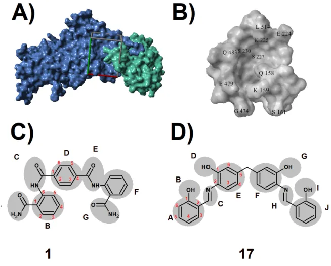
“protective antigen” due to its ability to elicit protective immunity against anthrax(Young et al., 2007). Upon infection of a host, the N-terminal domain of the protective antigen (Figure 1, panel A) is cleaved by a host-derived protease(Gordon et al., 1997; Gordon & Klimpel, 1995; Klimpel et al., 1992) and the remaining product assembles into heptamers (referred to as “prepores”) which bind the adenylcyclase or Zn2+ protease toxin components and are
endocyted(Abrami et al., 2003). In the endosome, acidification leads the heptamers into converting into membrane-spanning pores, through which the toxin components translocate into the cytosol, eventually leading to the symptoms of anthrax[6]. Treatment involves prolonged doses of antibiotics and the use of antitoxins which interact with the protective antigen and render it inoperative(Hendricks et al., 2014). Another possible treatment strategy consists in
1 2 3 4 5 6 7 8 9 10 11 12 13 14 15 16 17 18 19 20 21 22 23 24 25 26 27 28 29 30 31 32 33 34 35 36 37 38 39
the inhibition of the oligomerization of the protective antigen through the intervention of small molecules that bind the protein surfaces involved in the formation of the prepore(Omersa et al., 2019; Rubert Pérez et al., 2012; Wein et al., 2012). An in silico screen of a compound library identified(Wein et al., 2012) several molecules with the ability to bind a small cavity (Figure 1, panel B) in the protective antigen located at the oligomerization interface, close to the region where the N-terminal domain contacts the rest of the protein. Two of these molecules (Chembridge library members 5181401 and 5180717) were then experimentally shown to possess the ability to prevent the oligomerization and to protect cells from anthrax lethal toxin in vitro. Serum albumin, however, prevented these molecules from protecting the cells, suggesting that their high hydrophobicity will render them unsuitable for clinic applications. In this work, we have designed and analyzed computationally a large variety of less hydrophobic derivatives of these ligands to identify higher affinity analogues with improved clinical applicability.
Figure 1. A) Molecular surface of the protective antigen. The N-terminal portion cleaved prior to
activation is colored green. The docking site studied is highlighted with a rectangular box. B) Molecular surface of the binding site. C and D) Scaffolds used for the generation of novel inhibitor candidates: Chembridge library member 5181401 (C) and Chembridge library member 51810717 D). Regions subjected to bioisosteric modification(Meanwell, 2011; Patani & LaVoie, 1996) are
40 41 42 43 44 45 46 47 48 49 50 51 52 53 54 55 56 57 58 59 60 61
highlighted. The numbering scheme used throughout the manuscript is shown in each ring. In molecule 1, the numbering of moiety F is analogous to that of moiety B. In molecule 17 , the numbering of moieties F and J are analogous to E and A, respectively.
2. Materials and Methods
All computations were performed in YASARA(Krieger et al., 2004) using the 1. 45 Å resolution crystal structure of the protective antigen in which the membrane-insertion loop (residues 303–324) was replaced with the Pro-Gly dipeptide, at pH=8. 5 (PDB: 3TEW) (Figure 1)(Feld et al., 2012). New inhibitor candidates (Figure 1 and Supporting Information) were generated manually through bioisosteric replacement (Meanwell, 2011; Patani & LaVoie, 1996) at strategic positions of the two ligands(Wein et al., 2012) found previously (Chembridge library members 5181401, henceforth referred as 1, and 5180717, henceforth referred as 17), and then docked with AutoDock Vina(Trott & Olson, 2010) using default docking parameters. The docking region was confined to a large 51. 0×36. 7×34. 5 Å box centered on the crevice between segments 159-162 and 473-485 to confirm that binding is specific to the cavity and not due to unspecific adsorption to the protein surface. The cavity itself (Figure 1B) is about 8 Å wide and 20 Å long and can be divided in two regions: an “exposed” lobe open to bulk solvent lined by amino acids 224-230 and a “protected” lobe lined by amino acids 461-464 and 472-483, partially protected from bulk solvent by a “lid” formed by the side chains of N-terminal domain amino acids Q158 and K159. The docking box we used is considerably larger than the cavity to ensure that putative candidates that bound to only one of the lobes of the cavity and to the surrounding surface (instead of occupying the complete cavity) could be filtered out. The ligands with higher dissociation energies were selected for further study through molecular dynamics. All molecular dynamics simulations were run with the AMBER03 force field(Duan et al., 2003), using a multiple time step of 1. 25 fs for intramolecular and 2. 5 fs for intermolecular forces. Simulations were performed in cells 25 Å larger than the solute along each axis (final cell dimensions 159. 0 × 111. 9 × 124. 83 Å), and counter-ions (195 Cl- and 198 Na+) were added to a final concentration of 0. 9 %
NaCl. In total, the simulations contained approximately 221,770 atoms. A 8 Å cutoff was taken for Lennard-Jones forces and the direct space portion of the electrostatic forces, which were calculated using the Particle Mesh Ewald method(Essmann et al., 1995) with a grid spacing <1 Å, 4th order B-splines and a tolerance of 10-4 for the direct space sum. Simulated annealing minimizations
started at 298 K, velocities were scaled down with 0. 9 every ten steps for a total time of 5 ps. After annealing, simulations were run at 298 K. Temperature was adjusted using a Berendsen thermostat(Berendsen et al., 1984) based on the time-averaged temperature, i. e. , to minimize the impact of temperature control, velocities were rescaled only about every 100 simulation steps, whenever the average of the last 100 measured temperatures converged. Substrate parameterization was performed with GAFF(Wang et al., 2004) and charges were assigned through the AM1BCC protocol. All simulations were run for at least 125 ns. Unless otherwise noted, average interaction distances and standard deviations
62 63 64 65 66 67 68 69 70 71 72 73 74 75 76 77 78 79 80 81 82 83 84 85 86 87 88 89 90 91 92 93 94 95 96 97 98 99 100 101 102 103 104 105
were computed on the whole trajectory after discarding the initial 15 ns. Umbrella sampling was performed on the best ligands (as determined from their ability to remain bound to the protein throughout the molecular dynamics simulations) to obtain better estimates of their relative binding energies. 20 windows were used for each system to sample the distance between the center of the ligand and the bottom of the binding cavity. The centers of the umbrella potentials were spaced by 1. 0 Å. A harmonic potential of the form V=1/2 k
(
x − x0)
2with a force constant of 10. 0 kcal/ mol/Å2 was used to apply the distance restraints. Sampling was performed for 3. 6
ns in each bin. The first 0. 6 ns were eliminated from the analyses. The unbiased distributions were obtained through the weighted histogram analysis method (WHAM) (Grossfield, 2018; Kumar et al., 1992) using a bin size of 0. 2 Å. Solvation energies were computed with the Firelfy quantum chemistry package(Granovsky, 2013) using a polarizable continuum method(Cossi et al., 1998; Mennucci & Tomasi, 1997; Tomasi & Persico, 1994) with the PBE0-D3(BJ)/pcseg-2//PBE0-D3(BJ)/ 6-31G+(d,p) quantum chemical method(Adamo & Barone, 1999; Grimme et al., 2011; Jensen, 2014).
3. Results
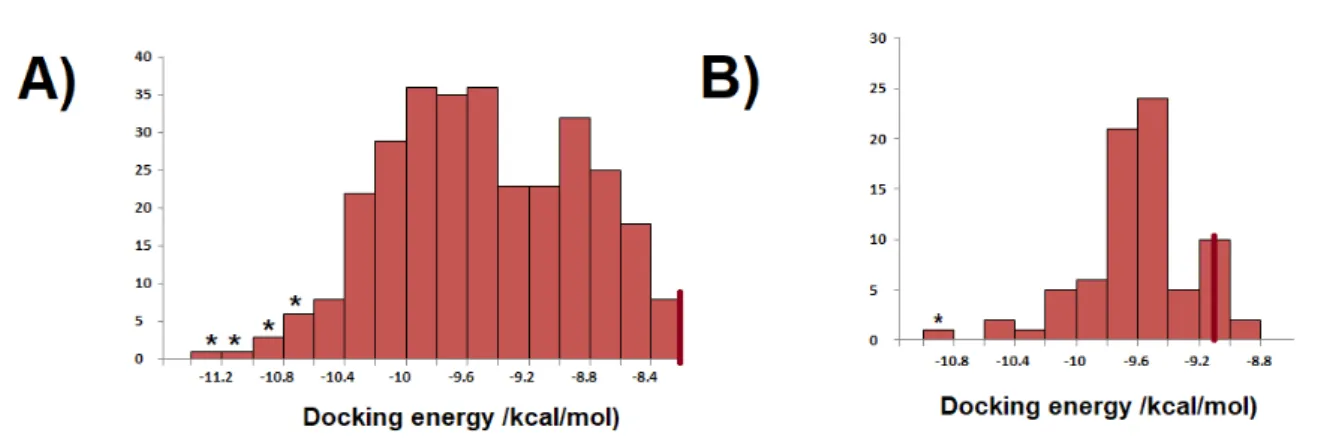
Figure 2. A) Distribution of the docking energies of the derivatives Chembridge library member
51810717. B) Distribution of the docking energies of the derivatives Chembridge library member 5181401. In both histograms, the energy of the unmodified library member is shown as a thick red line. In each histogram, an asterisk highlights the bins from which a candidate structure was analyzed through molecular dynamics simulations.
305 derivatives of Chembridge library member 17 and 76 derivatives of Chembridge library member 1 were designed and docked to the target cavity. Our modifications of 17 usually involved the increase of dipole moment of the rings through substitution of carbon by nitrogen or addition of halogen atoms, the reduction of one of the imine bonds or the replacement of phenolic hydroxyl groups by non-classical bioisosteres. The tested modifications invariably improved the docking energy (Figure 2A), showing that ample room exists for improvement of the binding capabilities of 17. Modifications of 1 were not always as effective in improving the docking energy: replacing one of the amides by an acetyl group, appending a single methylamine group on the D ring (Figure 1) or replacing one of the phenyl carbons by nitrogen yielded compounds with slightly worse binding
106 107 108 109 110 111 112 113 114 115 116 117 118 119 120 121 122 124 125 126 127 128 129 130 131 132 133 134 135 136 137 138 139
ability than the original compound. Only one derivative of 1 (1-67) had an improvement above 1. 5 kcal/mol over the original compound, whereas the improvement over 17 was much more extensive, with seven molecules (Table 1) showing an improvement above 2. 6 kcal/mol. Surprisingly, the effects of individual substitutions on the docking energies proved not to be additive, partly due to the flexibility allowed by the large size of the lobes of the binding cavity and partly due to the high symmetry of the parent molecules, which allows the generation of 2n-1 different molecules from individual changes at n
symmetry-unrelated positions.
The scoring functions used to rank the solutions from docking experiments are well-known to be imperfect and to be able, at most, to significantly improve the likelihood that a high-ranking molecule will be a strong ligand. We therefore proceeded to analyze the stability of the binding modes of our candidate molecules through molecular dynamics simulations.
Table 1. Substitution pattern in the derivatives of 17 with the best docking energies. Positions not
listed were kept unchanged relative to the parent compound.
Derivativ e Docking energy (kcal/mol) A B F G H I 17-061 -11. 34 N6 6-fluoro- 2,4-CHD 17-283 -11. 04 N6 6-hidroxy-2,4-CHD 17-275 -10. 90 N6 6-chloro-2,4 CHD 17-288 -10. 84 N6 -NH-C=O-O-17-239 -10. 83 N6 2,4 CHD 17-147 -10. 78 N6O5 -2,6 CHD F6 -NH-C=O-NH-17-222 -10. 77 HN6 =O 2,4-CHD =O -NH2+-CH2 -NH-C=O-O-Legend: 2,4-CHD: 2,4-cyclohexadiene; 2,6 CHD: cyclohexadiene with double bond between atoms 2 and 3 and between atoms 1 and 6; N6 : replacement of a -HC= at position 6 by -N=.
3. 1. Candidate 1-67 140 141 142 143 144 145 146 147 148 149 150 151 152 153 154 155 156 157 158 159 160 161
Figure 3. A) Evolution of the binding mode of 1-67 along two different simulations started from the
same binding pose. Ligand unbinding is evident at 125 ns in one simulation (upper panels), whereas stable binding is seen in the other simulation (lower panels) B) Chemical structure of candidate 1-67. C) Evolution of protein-ligand contacts along the two simulations (moving average of 200ps windows centered on the graphed points). Upper simulation: blue trace; Lower simulation: red trace
1-67 (Figure 3B) differs from the original ligand 1 in the replacement of both amide groups by carboxylates and by the presence of a nitrogen in the central ring, which increases its dipole moment. In the initial binding pose, strong hydrogen bonds are established between one of its carboxylate groups and the backbone amides of Lys159 (3. 63±0. 50 Å)and Gln158 (2. 73±0. 32 Å) while the other end of the molecule is nested in the “exposed” cavity lobe lined by residues 226-232 and the carboxylate in that end interacts with the backbone amides of Ser227 (2. 80±0. 19 Å)and Trp226(2. 39±0. 50 Å) (Figure 3A). Since two very similar binding poses were obtained in the docking procedure, we analyzed the stability of the complex with four 125 ns simulations (two from each binding pose). We found a similar behavior in each pair of simulations: in one instance, the complex remained stable throughout the 125 ns, whereas in the other instance the ligand started to unbind after around 80 ns (Figure 3A and 3C and Supporting Information). In both pairs of simulations, the separation of the ligand consistently proceeded through the motion of the exposed carboxylate group present in the 223-232 lobe of the cavity away from the tethering Ser226-Trp227 amides towards the bulk solvent. This separation is made possible by the open nature of this cavity, which enables large numbers of water molecules to compete with the backbone amides for the interaction with the carboxylate. In contrast, the interaction of the other carboxylate with the Lys159 (2. 77±0. 40 Å) and Gln 158 (2. 73±0. 32 Å) backbone amides is much more stable because the side chains of Lys159 and Gln158 largely shield it from bulk solvent. The observation of a stable interaction of the whole
162 163 164 165 166 167 168 169 170 171 172 173 174 175 176 177 178 179 180 181 182 183 184 185 186 187 188 189 190
molecule with the protein target in only half of the simulations implies that this candidate can not be relied upon to provide a dependable inhibition. Future attempts at improvement of 1-67 should therefore refrain from placing such strong hydrophilic groups in the end of the molecule interacting with this lobe of the cavity.
3. 2. Candidate 17-061
Figure 4. A) Evolution of the binding mode of 17-061 along two different simulations started from
the same binding pose. B) Chemical structure of candidate 17-061. C) Evolution of protein-ligand contacts along the two simulations (moving average of 200ps windows centered on the graphed points). Upper simulation: blue trace; Lower simulation: red trace
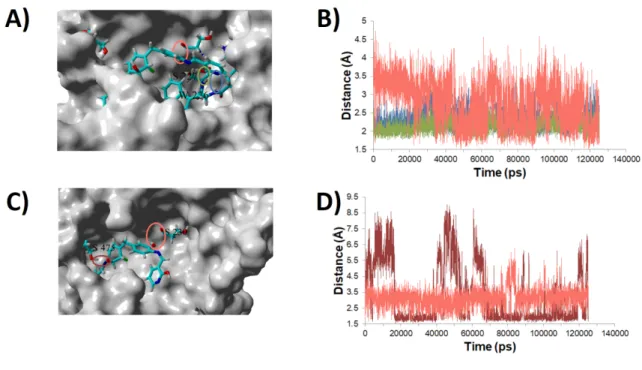
The first analogue of 17 we studied more thoroughly (candidate 17-061) differs from the original molecule by the elimination of aromaticity in moiety F, the introduction of a halogen in that ring to increase dipole moment and in the replacement of the benzene ring in moiety A by a pyridine. In the docking pose identified by VINA, the pyridine ring lies in the “open” lobe of the cavity and the hydroxyl in moiety D forms a hydrogen bond with the carbonyl of Ser230. The ring bearing the halogen is sandwiched between Gln158 and Lys159 and the terminal phenol ring lies in the “protected” lobe of the binding site (Figure 4). Two independent simulations started from this configuration showed that the binding is very stable, although the details may change considerably. In both instances the initial hydroxyl–Ser230 carbonyl interaction is maintained throughout the simulation (2. 70±0. 62 Å in the first simulation, 3. 17±0. 59 Å in the second simulation) but other interactions are different between simulations (Figure 5): in the first case, the pyridine nitrogen remains bound to the Trp226 backbone amide (2. 37±0. 27 Å) while the backbone amide of Ser227 locks the hydroxyl in moiety B in place (2. 14±0. 21 Å), while in the second simulation these interactions are absent but a transient hydrogen bond between Ser475 and the hydroxyl in moiety D is
191 192 193 194 195 196 197 198 199 200 201 202 203 204 205 206 207 208 209 210 211 212 213 214 215 216 217 218
present in more than 60% of the time (2. 90±1. 70 Å in the full simulation, 1. 98±0. 19 Å if the intervals with distance >3 Å are omitted).
Figure 5. A) Representative snapshot of the binding mode found in the first simulation of the
complex with 17-061. The binding cavity is shown from above, after Gln158 and Gln 483 have been removed for clarity. Binding interactions are highlighted with colored ellipses. B) Evolution of the stable hydrogen bonds in the first simulation. Color code is the same as the one used in panel A. C) Representative snapshot of the binding mode found in the second simulation of the complex with 17-061. The binding cavity is shown from above, after Gln158, Trp226, Ser227 and Gln 483 have been removed for clarity. Binding interactions are highlighted with colored ellipses. D) Evolution of the stable hydrogen bonds in the second simulation. Color code is the same as the one used in panel C
3. 3. Candidate 17-222 219 220 221 222 223 224 225 226 227 228 229 230 231 232
Figure 6. A) Evolution of the binding mode of 17-222 along two different simulations started from
the same binding pose. B) Chemical structure of candidate 17-222. C) Evolution of protein-ligand contacts along the two simulations (moving average of 200ps windows centered on the graphed points). Upper simulation: blue trace; Lower simulation: red trace
Candidate 17-222 differs from 17-061 in the tautomeric form of the iminol in one of the extremities and in the existence of a much more hydrophilic character in the opposite end, which bears a basic amine instead of an amine and an internal carbamate group instead of the phenol hydroxide. In the initial binding pose, this hydrophilic end binds to the protected lobe, while the other end binds to the solvent-exposed portion of the cavity (Figure 6). In the first simulation an extremely stable binding mode was observed, maintained (in the protected lobe) through hydrogen bonds between the polar hydrogen attached to the internal carbamate nitrogen and Thr461 (1. 97± 0. 15 Å) , the protonated amine and Ser475 (2. 02± 0. 41 Å), and between the protonated amine and the carbonyl group of Pro232 (2. 38± 0. 39 Å). As observed with other candidate molecules, considerable mobility is observed in the end of the candidate interacting with the open lobe. In the second simulation a similar behavior was observed, though only up until 49 ns: at this moment a rotation of the protonated amine C-N bond breaks the hydrogen bond between it and the Pro232 carbonyl, and the increased flexibility leads (1. 3 ns later) to the severing of the interaction of the internal carbamate moiety with Thr461. This hydrophilic site immediately interacts with the surrounding bulk water and promptly wriggles away from the cavity through the lateral opening between Gly474 and Ser161 (Figure 6).
3. 4. Candidate 17-239 233 234 235 236 237 238 239 240 241 242 243 244 245 246 247 248 249 250 251 252 253 254 255 256 257 258
Figure 7. A) Chemical structure of candidate 17-239. B) Evolution of the binding mode of 17-239.
The structure of candidate 17-239 is very similar to that of the promising 17-061 molecule discussed earlier: the only difference lies in the absence of a halogen in the ring we called “moiety F” (Table 1 and Figure 7A). Its behavior in the binding cavity is, however, dramatically different: two hydrogen bonds between the phenol hydrogen in moiety J and the hydroxyl in moiety G with the side chain of Ser475 are the only stable interactions between the ligand and the cavity surface. There interactions weaken dramatically after only 12 ns, and the molecule promptly leaves, though always interacting weakly with the protein surface instead of being released to the bulk.
3. 5. Candidate 17-275
Figure 8. A) Evolution of the binding mode of 17-275 along two different simulations started from
the same binding pose. B) Chemical structure of candidate 17-275. C) Evolution of protein-ligand contacts along the two simulations (moving average of 200ps windows centered on the graphed points). Upper simulation: blue trace; Lower simulation: red trace.
Molecule 17-275 differs from 17-061 and 17-239 in the presence of a chlorine atom in the position adjacent to the moiety G hydroxyl (Figure 8B). The initial
259 260 261 262 263 264 265 266 267 268 269 270 271 272 273 274 275 276 277 278
docking pose, like that of those candidates, has ring F lying between the side chains of Gln158 and Lys159 and the pyridine ring in the “open” lobe close to the Ser230 carbonyl. Both simulations yield stable, though different, binding modes. In the first simulation (Figure 9A) the hydroxyl in moiety G interacts with the side chains of Ser475 (O···O distance of 2. 96± 0. 73 Å) while the hydroxyl in moiety D establishes a hydrogen bond with the backbone amide of Asn476 (O···N distance of 3. 36± 0. 53 Å). Marginally less frequent interactions are present between the hydroxyl in moiety B and the Ser160 carbonyl (3. 18± 0. 76 Å) and between the chlorine and the Gln158 backbone amide (3. 11± 0. 35 Å). The second simulation visits two distinct binding modes: until 60 ns (Figure 9B), the ligand is hydrogen-bonded to the side chain of Thr461 (O···O distance of 3. 39± 0. 80 Å). After that moment, the molecule is kept in place mostly through steric interactions until a new hydrogen bond between Ser475 and the hydroxyl in moiety G (Figure 9C) forms at 86 ns (O···O distance of 2. 83± 0. 39 Å).
Figure 9. The three binding modes visited by candidate 17-275. Lys159 has been deleted for clarity.
A) Stable pose obtained in the first simulation. Stabilizing hydrogen bonds shown in orange. B) Binding mode obtained in the first 60 ns of the second simulation. C) Binding mode obtained after 86 ns of the second simulation.
4. Discussion
Our molecular dynamics simulations clearly show that several of the good candidates identified in the docking stage are not promising leads for further development. Problematic features include the presence of concentrated charges in the moiety which binds to the “open” lobe of the cavity (as seen with candidate 01-67) as well as excessive hydrogen-bonding potential in the moiety that binds the solvent-exposed opening between Ser161 and Gly 474 in the “protected” lobe of the cavity (as observed in candidate 17-222). These observations showed us that the high-ranking molecules 17-288 and 17-147 (Table 1) are most unlikely to be good candidates and therefore we did not expend any effort with expensive molecular dynamics simulations of their binding modes. In contrast, the transformation of the aromatic ring in moiety F into a cyclohexadiene bearing two electronegative substituents afforded molecules with very good binding behavior. The good results obtained with the fluoro- and the chloro-substituted molecules (17-061 and 275, respectively) strongly suggest that the hydroxy-substituted molecule 17-283, which we did not simulate due to its extreme stereo-electronic and geometric similarity to these molecules, should also be a very good candidate for further
279 280 281 282 283 284 285 286 287 288 289 290 291 292 293 294 295 296 297 298 299 300 301 302 303 304 305 306 307 308 309 310 311 312 313 314 315 316
development. The lack of the second electronegative substituent in the cyclohexadiene ring (as in 17-239) has dramatic effects on the binding ability. Such a strong effect shows that the dipole interactions of this moiety with Gln158 and Lys159 (which are present in 17-061 and 17-275 but greatly decreased in 17-239) function as a critical anchor imparting stability to the complexes.
To confirm the potential of the selected ligands for future clinical applications we compared their binding energies with that of the experimentally-confirmed parent compound 17 through umbrella-sampling simulations and subsequent computation of the potential of mean force. As depicted in Figure 10, both binding modes of 17-061 were more favorable than that of 17 by 2-4 kcal/mol. The three binding modes detected for 17-275 showed considerable more spread in their binding energies: the binding mode obtained in the first simulation (and depicted in Figure 9A) is more than 10 kcal/mol more favorable than that of the experimentally-confirmed ligand 17 , wheras the initial, transient binding mode, detected in the first part of the second simulation (Figure 9B) is shown to be less favorable than that of 17, and the binding mode (Figure 9C) obtained after the 85th
ns in this simulation, though less efficient than that seen in the first simulation, is still markedly more favorable than any of the ones seen for 17-061.
Figure 10. Potentials of mean-force for the ligand separation process. A) Comparison of the ease of
separation of ligand 17 (blue) with that of ligand 17-061 in the first (orange) and second simulations (yellow). B) Comparison of the ease of separation of ligand 17 (blue) with that of ligand 17-275 in three binding modes: that obtained in the first simulation (green line) and the two binding modes obtained from the second simulation (wine colored, prior to 60 ns, light blue, after 86 ns).
To quantify the extra hydro-solubility of these derivatives relative to the parent compound Chembridge 5180717, we computed the electrostatic stabilization provided by water and heptane on each of these molecules through the quantum chemical polarized continuum method (Table 2). Both 17-061 and 17-275 are confirmed by these computations to be more favorably stabilized in water relative to heptane than the parent compound. This method does not take into account the extra stabilization afforded by the presence, in the new compounds, of hydrogen-bonding ability, and therefore the values listed are an under-estimation of the real preference of the compounds for water vs. heptane.
Table 2. Electrostatic stabilization energies (kcal·mol-1) of the best derivative in water and n-heptane.
317 318 319 320 321 322 323 324 325 326 327 328 329 330 331 332 333 334 335 337 338 339 340 341 342 343 344 345 346 347 348 349 350 351 352 353
Molecule water n-heptane Preference for water over n-heptane
17 -17. 63 -5. 63 -12. 00
17-061 -22. 68 -7. 27 -15. 41
17-275 -19. 48 -5. 80 -13. 58
Additional consequences of the substitutions were analysed using the SwissADME server(Daina et al., 2017) . This model predicts that, in contrast to the parent compound, the newly proposed derivatives have highly improved gastrointestinal absorption and should not inhibit CYP1A2, CYP2C19 or CYP2D6, while 17-275 should inhibit CYP3A4, in contrast to the parent compound (Supporting Information).
5. Conclusions
Judicious bioisosteric substitutions led to the generation of several promising improved derivatives of two previously identified inhibitors, Chembridge library members 5181401 and 5180717. Molecular dynamics simulations of the obtained docking poses showed that several of the putative complexes were less stable than desired due to the excessive hydrophilicity introduced in the molecule by our substitutions. Two of the generated derivatives of Chembridge library member 5180717, which featured the replacement of a phenyl ring by a halogen-substituted cyclohexadiene, did result in very strong, stable, binding in repeated simulations, and show therefore good promise fro therapeutic use. Simulations of both the successful and the unsuccessful candidates additionally helped to identify several features of the active site important for the obtention of successful binding. This work further highlights the critical importance of molecular dynamics in the confirmation of the plausibility of a binding candidate identified through docking(Chen, 2015) .
Author Contributions: conceptualization, P. S. ; methodology, P. S. and V. S; formal analysis, P. S. and V. S;
writing—original draft preparation, P. S.
Funding: This work was performed using computational resources acquired under project
PTDC/QUI-QUI/111288/2009 and funded by FEDER through Programa Operacional Factores de Competitividade – COMPETE and by Portuguese Funds through FCT – Fundação para a Ciência e a Tecnologia.
Conflicts of Interest: The authors declare no conflict of interest. The funders had no role in the design of the
study; in the collection, analyses, or interpretation of data; in the writing of the manuscript, or in the decision to publish the results.
Abrami, L., Liu, S., Cosson, P., Leppla, S. H., & Van der Goot, F. G. (2003). Anthrax toxin triggers endocytosis of its receptor via a lipid raft-mediated clathrin-dependent process. Journal of Cell Biology. https://doi.org/10.1083/jcb.200211018
354 355 356 357 358 359 360 361 362 363 364 365 366 367 368 369 370 371 372 373 374 375 376 377 378 379 380 381 382 383 384 385 386 387 388 389 390 391 392
Adamo, C., & Barone, V. (1999). Toward reliable density functional methods without adjustable parameters: The PBE0 model. The Journal of Chemical Physics, 110(1999), 6158. https://doi.org/10.1063/1.478522
Berendsen, H. J. C., Postma, J. P. M., van Gunsteren, W. F., DiNola, A., & Haak, J. R. (1984). Molecular dynamics with coupling to an external bath. The Journal of Chemical Physics, 81(8), 3684. https://doi.org/10.1063/1.448118
Chen, Y. (2015). Beware of docking! Trends in Pharmacological Sciences, 36(2), 78–95. https://doi.org/10.1016/j.tips.2014.12.001
Cossi, M., Mennucci, B., Pitarch, J., & Tomasi, J. (1998). Correction of cavity-induced errors in polarization charges of continuum solvation models. Journal of Computational Chemistry, 19(8), 833–846. https://doi.org/10.1002/(sici)1096-987x(199806)19:8<833::aid-jcc3>3.0.co;2-q
Daina, A., Michielin, O., & Zoete, V. (2017). SwissADME: A free web tool to evaluate pharmacokinetics, drug-likeness and medicinal chemistry friendliness of small molecules. Scientific Reports, 7(October 2016), 1–13. https:// doi.org/10.1038/srep42717
Duan, Y., Wu, C., Chowdhury, S., Lee, M. C., Xiong, G., Zhang, W., Yang, R., Cieplak, P., Luo, R., Lee, T., Caldwell, J., Wang, J., & Kollman, P. (2003). A point-charge force field for molecular mechanics simulations of proteins based on condensed-phase quantum mechanical calculations. Journal of Computational Chemistry, 24(16), 1999–2012. https://doi.org/10.1002/jcc.10349 Essmann, U., Perera, L., Berkowitz, M. L., Darden, T., Lee, H., & Pedersen, L. G.
(1995). A smooth particle mesh Ewald method. The Journal of Chemical Physics, 103(19), 8577–8593. https://doi.org/10.1063/1.470117
Feld, G. K., Kintzer, A. F., Tang, I. I., Thoren, K. L., & Krantz, B. a. (2012). Domain flexibility modulates the heterogeneous assembly mechanism of anthrax toxin protective antigen. Journal of Molecular Biology, 415(1), 159–174. https://doi.org/ 10.1016/j.jmb.2011.10.035
Gordon, V., & Klimpel, K. (1995). Proteolytic activation of bacterial toxins by eukaryotic cells is performed by furin and by additional cellular proteases. Infection and Immunity, 63(1), 82–87. http://iai.asm.org/content/63/1/82.short Gordon, V., Rehemtulla, A., & Leppla, S. (1997). A role for PACE4 in the
proteolytic activation of anthrax toxin protective antigen. Infection and Immunity, 65(8). http://iai.asm.org/content/65/8/3370.short 393 394 395 396 397 398 399 400 401 402 403 404 405 406 407 408 409 410 411 412 413 414 415 416 417 418 419 420 421 422 423 424 425 426
Granovsky, A. A. (2013). Firefly 8.0.0. http://classic.chem.msu.su/gran/gamess/index.html
Grimme, S., Ehrlich, S., & Goerigk, L. (2011). Effect of the damping function in dispersion corrected density functional theory. Journal of Computational Chemistry, 32(7), 1456–1465. https://doi.org/10.1002/jcc.21759
Grossfield, A. (2018). “WHAM: the weighted histogram analysis method” (2.0.9).
Hendricks, K. A., Wright, M. E., Shadomy, S. V., Bradley, J. S., Morrow, M. G., Pavia, A. T., Rubinstein, E., Holty, J.-E. C., Messonnier, N. E., Smith, T. L., Pesik, N., Treadwell, T. A., & Bower, W. A. (2014). Centers for Disease Control and Prevention Expert Panel Meetings on Prevention and Treatment of Anthrax in Adults. Emerging Infectious Diseases, 20(2). https://doi.org/10.3201/eid2002.130687
Jensen, F. (2014). Unifying General and Segmented Contracted Basis Sets. Segmented Polarization Consistent Basis Sets. Journal of Chemical Theory and Computation, 10(3), 1074–1085. https://doi.org/10.1021/ct401026a
Klimpel, K. R., Molloy, S. S., Thomas, G., & Leppla, S. H. (1992). Anthrax toxin protective antigen is activated by a cell surface protease with the sequence specificity and catalytic properties of furin. Proceedings of the National Academy of Sciences, 89(21), 10277–10281. https://doi.org/10.1073/pnas.89.21.10277
Krieger, E., Darden, T., Nabuurs, S. B., Finkelstein, A., & Vriend, G. (2004). Making optimal use of empirical energy functions: Force-field parameterization in crystal space. Proteins: Structure, Function, and Bioinformatics, 57(4), 678–683. https://doi.org/10.1002/prot.20251
Kumar, S., Bouzida, D., Swendsen, R. H., Kollman, P. A., & Rosenberg, J. M. (1992). The Weighted Histogram Analysis Method for Free Energy Calculations on Biomolecules: I. The Method. J. Comp. Chem., 13(8), 1011–1021. https://doi.org/http://dx.doi.org/10.1002/jcc.540130812
Meanwell, N. A. (2011). Synopsis of Some Recent Tactical Application of Bioisosteres in Drug Design. Journal of Medicinal Chemistry, 54(8), 2529–2591. https://doi.org/10.1021/jm1013693
Mennucci, B., & Tomasi, J. (1997). Continuum solvation models: A new approach to the problem of solute’s charge distribution and cavity boundaries. Journal of Chemical Physics, 106(12), 5151–5158. https://doi.org/10.1063/1.473558
427 428 429 430 431 432 433 434 435 436 437 438 439 440 441 442 443 444 445 446 447 448 449 450 451 452 453 454 455 456 457 458 459
Omersa, N., Podobnik, M., & Anderluh, G. (2019). Inhibition of Pore-Forming Proteins. Toxins, 11(9), 545. https://doi.org/10.3390/toxins11090545
Patani, G. A., & LaVoie, E. J. (1996). Bioisosterism: A Rational Approach in Drug Design. Chemical Reviews, 96(8), 3147–3176. https://doi.org/10.1021/cr950066q Rubert Pérez, C., López-Pérez, D., Chmielewski, J., & Lipton, M. (2012). Small
Molecule Inhibitors of Anthrax Toxin-induced Cytotoxicity Targeted Against Protective Antigen. Chemical Biology & Drug Design, 79(3), 260–269. https://doi.org/10.1111/j.1747-0285.2011.01285.x
Tomasi, J., & Persico, M. (1994). Molecular Interactions in Solution: An Overview of Methods Based on Continuous Distributions of the Solvent. Chemical Reviews, 94(7), 2027–2094. https://doi.org/10.1021/cr00031a013
Trott, O., & Olson, A. J. (2010). AutoDock Vina: improving the speed and accuracy of docking with a new scoring function, efficient optimization, and multithreading. Journal of Computational Chemistry, 31(2), 455–461. https://doi.org/10.1002/jcc.21334
Wang, J., Wolf, R. M., Caldwell, J. W., Kollman, P. A., & Case, D. A. (2004). Development and testing of a general amber force field. Journal of Computational Chemistry, 25(9), 1157–1174. https://doi.org/10.1002/jcc.20035 Wein, A. N., Williams, B. N., Liu, S., Ermolinsky, B., Provenzano, D., Abagyan, R.,
Orry, A., Leppla, S. H., & Peredelchuk, M. (2012). Small molecule inhibitors of Bacillus anthracis protective antigen proteolytic activation and oligomerization. Journal of Medicinal Chemistry, 55(18), 7998–8006. https://doi.org/10.1021/jm300804e
Young, T., Abel, R., Kim, B., Berne, B. J., & Friesner, R. a. (2007). Motifs for molecular recognition exploiting hydrophobic enclosure in protein-ligand binding. Proceedings of the National Academy of Sciences of the United States of America, 104(3), 808–813. https://doi.org/10.1073/pnas.0610202104 460 461 462 463 464 465 466 467 468 469 470 471 472 473 474 475 476 477 478 479 480 481 482 483 484 485 486 487 488


