Development of a diagnostic procedure for VBNC cells of Listeria monocytogenes
62
0
0
Texto
(2) UNIVERSIDADE DE LISBOA FACULDADE DE CIÊNCIAS DEPARTAMENTO DE BIOLOGIA VEGETAL. DEVELOPMENT OF A DIAGNOSTIC PROCEDURE FOR VBNC CELLS OF LISTERIA MONOCYTOGENES Dissertação orientada por Prof. Doutora Lélia Chambel. Olívia Carina Campos da Costa. MESTRADO EM MICROBIOLOGIA APLICADA 2012.
(3) DEVELOPMENT OF A DIAGNOSTIC PROCEDURE FOR VBNC CELLS OF LISTERIA MONOCYTOGENES. Olívia Carina Campos da Costa 2012. This thesis was fully performed at Center for Biodiversity, Functional and Integrative Genomics (BioFIG- FCUL) under the direct supervision of Prof. Dra. Lélia Chambel in the scope of the Master in Applied Microbiology of the Faculty of Sciences of the University of Lisbon..
(4) Acknowledgements Someone once told me that we could be in a crowd and feel lonely and also be alone and not lonely. It was important to me, and to the final result of this work, that I did not felt lonely. Several persons gave me strength and encouraged me always to try the best that I could be. This thesis is not only the result of my efforts towards the objectives, but also of the positive influence that allowed the personal and professional growth. First I would like to thank my grandfather, Américo Costa, that is no longer between us, but to whom I own every day of my life and all that I was, am, and want to be. “May God keep you in heaven as I keep you in my heart”. I want to thank my supervisor, Dr.ª Lélia Chambel, for accepting me as her master student, for all comprehension and patience with me during the realization of this thesis. For all support and assistance during this last year, as well as for all the conversations that we had and that were essential to reaching our objective. To Daniela Pinto for her thoughtful explanations, help and answers to all of my questions. To Dr. º Mário Gadanho, from Biopremier, for the help and kind advises given. To my laboratory colleagues that helped whenever needed, especially Susana Marques and Tânia Ribeiro. To my son whose existence leads me to want to be always better and to live every day. To Victor Godinho for all the times that I was not completely present and for the patience that it demanded, especially during the writing phase. To Ana Catarina Dourado for all her friendship, attention and understanding for all the conversations and help, even when master thesis was my only subject. To my family, grandmother and uncles, that tried to understand me, even when it seemed impossible. Last but not least, I would like to thank Fundação para a Ciência e Tecnologia (FCT) for providing the financial support for the realization of the present work, performed under the project PTDC/AGR-ALI/098020/2008.. I.
(5) Abstract VBNC state represents a specific differentiation program into a survival state with several modifications in the cells walls and cytoplasmic membrane, a decrease in metabolic activity and maintaining gene expression. This state is induced upon exposure to stressful conditions, including nutrient starvation, different temperatures, pH, oxygen concentration and exposure to light or UV radiation. VBNC cells can represent an unknown public health risk, especially in the food industry, since they are unable to grow in routine analysis bacteriological media. This work aimed to develop a PCR based diagnostic procedure to detect VBNC cells of Listeria monocytogenes, a foodborne pathogen, able to resuscitate. It were defined as PCR targets lmo2522 and lmo0186 genes, due to their potential as Rpfs, and used for primers design. Two more genes were considered for detection, one for L. monocytogenes identification and one as internal amplification control. The Multiplex-PCR reaction established was characterized and had a 50 pg detection limit for the target genes. DNA extraction protocols were developed to yield enough DNA from low bacteria number for Multiplex-PCR detection. However the detection of just a few cells was never observed. Therefore it was adopted Lmo0186-PCR reaction with 125 fg DNA detection limit and the ability to detect 1 VBNC cell. Assays performed with PMA, which inhibits amplification from dead cells, revealed the same sensitivity, whereas inclusion of an amplification control decreased it. To overcome this issue a 5-fold increase of DNA sample in PCR reaction was necessary. Protocol application to spiked food revealed a 104 and 10 6 cells detection limits for active and VBNC cells respectively. In parallel, VBNC state induction was performed, with different cellular concentrations, in five different induction media. Smaller induction periods were verified in the less concentrated suspensions and a high persistence of injured cells was detected in the higher concentrated suspensions.. Keywords: VBNC; Listeria monocytogenes; Multiplex-PCR; DNA extraction; Food analysis.. II.
(6) Resumo Discrepâncias de viabilidade bacteriana entre os resultados de contagens realizadas a partir de ensaios de culturabilidade e ensaios indiretos com o objetivo de detetar membranas celulares intactas são relatadas desde os anos de 1800. No entanto, foi apenas na década de 1980 que Byrd and Colwell1 introduziram a hipótese da existência de um estado similar à dormência, no qual as células bacterianas poderiam subsistir a condições menos favoráveis à sua sobrevivência, no qual não ocorreria crescimento. Tal estado foi denominado de viável mas não cultivável (VBNC) e a primeira evidência experimental foi obtida por Xu et al. (1982)2. Segundo Oliver (1993)3 este estado pode ser definido como uma célula que, embora metabolicamente ativa, é incapaz de realizar a divisão celular necessária para o seu desenvolvimento num meio de cultura que normalmente suportaria o seu crescimento. No entanto, a hipótese da existência de um estado VBNC levantou alguma controvérsia entre a comunidade científica, pois este implicaria que os métodos microbiológicos convencionais, que assumem que a culturabilidade é um reflexo direto da viabilidade, não seriam adequados para estudos de quantificação de células viáveis. Existia de facto uma certa resistência de alguns autores em aceitar a existência de um estado VBNC que não seria idêntico a outros estados fisiológicos já descritos, nos quais as células bacterianas não seriam imediatamente cultiváveis em meio de cultura4–7. Tendo em conta que muitos dos métodos utilizados para quantificar a viabilidade celular eram indiretos, e como tal discutíveis pelos autores mais céticos, a evidência chave dependeria da capacidade de reanimação das células que haviam entrado no estado VBNC. Assumindo-se que este estado seria um meio de sobrevivência, as células deveriam ter a capacidade de reanimar quando as condições fossem apropriadas ao seu crescimento. Como tal diversas tentativas foram realizadas no intuito de obter a reanimação das células VBNC, mas muitas demonstraram-se infrutíferas dado que não excluíam a possibilidade de recrescimento de uma pequena fração de células cultiváveis ainda presentes na população de células VBNC4–6,8. A evidência conclusiva foi obtida por Whitesides and Oliver (1997)9 no estudo com Vibrio vulnificus, no qual a atribuição dos eventos de reanimação à presença de poucas células cultiváveis seria extremamente improvável. Apesar de alguma contestação este estudo, e outros que se seguiram, levaram ao desaparecimento da controvérsia existente, sendo já geralmente aceite que o estado VBNC existe. O estado VBNC representa um programa de diferenciação específico para um estado de sobrevivência após exposição a condições desfavoráveis. Uma das modificações mais associadas a este estado é a redução da dimensão celular, podendo ocorrer a transição de uma morfologia de bacillus para cocóide5,8,10. Algumas modificações nas paredes celulares foram também já descritas, nomeadamente o aumento no número total de interligações, incluindo interligações incomuns como DAP-DAP em Escherichia coli, e na quantidade de mucopéptidos covalentemente ligados a uma lipoproteína11,12. Tais alterações na parede celular são passíveis de contribuir para uma maior resistência a antibióticos, dado que estes têm como alvo principal as células em crescimento ativo, tendo tal já sido reportado5,8,13. Foi igualmente já descrito uma reorganização do subproteoma da membrana celular externa bem como um perfil proteico relativamente diferente aos das células ativas ou no estado de sobrevivência com baixo metabolismo (“starvation survival”)14. Modificações metabólicas também. III.
(7) ocorrem as quais incluem a diminuição da síntese de macromoléculas, do transporte de nutrientes e da taxa de respiração, mantendo-se a absorção de aminoácidos, os níveis de ATP e rRNA e a presença de plasmídeos5,7,8,15. A contínua expressão de genes foi também detetada, sendo que alguns autores reportaram a contínua expressão de genes de virulência neste estado, enquanto noutros estudos esta expressão não foi detetada, mas no entanto o potencial virulento era mantido e poderia ser retomado após a reanimação das células VBNC5,8,16,17. Assumindo o estado VBNC como uma estratégia de sobrevivência, a sua indução pode ser despoletada pela exposição a condições não favoráveis à subsistência da célula bacteriana, podendo ser estímulos químicos ou ambientais, tais como falta de nutrientes, diferentes temperaturas, pressões osmóticas, alterações do pH, concentração de oxigénio, presença de metais pesados, ou conservantes alimentares e ainda exposição à luz ou radiação UV. A reanimação das células VBNC demonstrou-se mais complexa, havendo alguns estudos cujos ensaios de reanimação se revelaram infrutíferos, indicando que para alguns microrganismos serão necessários outros fatores que não apenas a inversão do estímulo de indução, e que poderão mesmo incluir a presença de um organismo superior como mediador8,17–20. Os mecanismos responsáveis quer pela indução do estado VBNC quer pela reanimação ainda não são conhecidos, no entanto alguns dados relevantes foram já demonstrados. No caso da indução o peróxido de hidrogénio parece ter um papel importante, dado a incapacidade de metabolização deste composto, bem como o fator sigma alternativo RpoS que parece ser um regulador da indução do estado VBNC5,8,21. Na reanimação há evidências que um grupo de proteínas extracelulares, denominadas fatores de promoção de reanimação (“resuscitation promoting factors” - Rpfs), estarão envolvidas no processo de reanimação, tendo sido já identificadas em diferentes géneros. Estas proteínas apresentam semelhanças com transglicolases solúveis e estarão envolvidas em alterações da parede celular. Outro tipo de Rpfs possivelmente associadas serão as autoindutoras do crescimento estáveis ao calor (“heat-stable autoinducers of growth”) que aparentam ser moléculas de sinalização secretadas. Apesar de ainda existirem muitos parâmetros a desvendar no estado VBNC, a existência do mesmo já foi descoberta em vários microrganismos, incluindo patogéneos, podendo representar um reservatório desconhecido dos mesmos5,8. Tal pode representar um perigo para a saúde pública, dado que neste estado os microrganismos não serão detetados pelos métodos microbiológicos convencionais. No caso da segurança alimentar esta possível ameaça deveria ser considerada, tendo em conta que, quer os alimentos quer o processo da sua produção e acondicionamento, podem apresentar vários dos fatores de indução previamente mencionados. Outro fator importante é o facto do controlo biológico destes produtos ser ainda maioritariamente baseado na culturabilidade dos microrganismos. Um dos patogéneos importantes na segurança alimentar é Listeria monocytogenes. Este microrganismo é o único dentro do seu género, que engloba mais sete espécies, geralmente associado à ocorrência de listeriose humana. Esta doença apresenta sintomas deveras severos, com uma elevada taxa de mortalidade (cerca de 30%), principalmente nos grupos de risco determinados (adultos e jovens imunocomprometidos, grávidas, infantis e idosos). Apesar de uma baixa taxa de incidência, a severidade da doença derivou num nível zero de tolerância à sua presença nas amostras alimentares. Já foi igualmente reportada a entrada no estado VBNC e capacidade de reanimação por este patogéneo22–24. Desta forma a presença do. IV.
(8) mesmo em produtos alimentares deveria contabilizar a existência de células VBNC de forma a assegurar a segurança dos consumidores. O objetivo do presente trabalho foi o desenvolvimento de um método de diagnóstico baseado em PCR para a deteção de células VBNC de L. monocytogenes, com potencial de reanimação, em produtos alimentares. Este método deverá permitir a deteção da amplificação a partir de genes previamente escolhidos, fornecendo uma resposta específica, sensível e rápida, sendo que com a complementação. com. o. reagente. monoazida. de. propídio. (PMA),. os. resultados. obtidos. corresponderão a células viáveis, dado que este inibe a amplificação a partir de células não viáveis. Os genes alvo definidos foram o lmo2522 e lmo0186, cujas proteínas serão potenciais Rpfs uma vez que apresentam características semelhantes às descritas para estes fatores. Seguidamente, foi efetuado uma pesquisa (“blast”) da sequência das proteínas respetivas e, após a análise dos domínios das mesmas, foi efetuada uma comparação entre os alinhamentos preliminares das sequências em Listeria spp. e L. monocytogenes. A análise desta comparação permitiu verificar que ambas. as. sequências. proteicas. se. apresentavam. conservadas. para. o. género. Listeria,. principalmente no caso de Lmo0186. Foram então definidas zonas para o posterior desenho de primers, de forma a obter uma amplificação específica para L. monocytogenes, as quais se baseavam numa região variável para Lmo2522 e numa zona conservada que flanqueava uma zona variável em Lmo0186. Seguidamente, foram obtidas as sequências dos genes respeitantes a estas proteínas e realizado um alinhamento global por Clustal W, tendo sido obtidas as sequências consenso de ambos os genes. Estas foram utilizadas para o desenho de primers. Após o desenho dos pares de primers, o potencial destes formarem estruturas secundárias foi analisado, após o que foi escolhido um par para cada gene e um para o controlo interno de amplificação (IAC) - GFP. Adicionalmente, e tendo em conta que existia a possibilidade de não ocorrer especificidade apenas para L. monocytogenes, foram incluídos primers para o gene hlyA cuja amplificação é específica para esta espécie. Tendo em conta que se pretendia uma reação em Multiplex-PCR, conduzindo à simultânea amplificação destes alvos, foi efetuado um Gradiente-PCR, o qual permitiu analisar a eficiência destes primers isoladamente, a diversas temperaturas de ligação, e definir a temperatura de 51,6ºC para posteriores ensaios. Seguidamente, foram realizados ensaios para definir a especificidade e sensibilidade da reação. Para tal foram utilizadas amostras de DNA extraído através do método adaptado de Pitcher et al. (1989)25. No ensaio de especificidade foram testadas seis espécies do género Listeria, incluindo mais do que uma estirpe para contabilizar a variabilidade intraespécie, bem como seis estirpes de E. coli. Os resultados revelaram que os primers Lmo2522 seriam específicos para L. monocytogenes, enquanto que os primers Lmo0186 seriam específicos para Listeria spp., uma vez que apenas L. grayii e L. seeligeri não apresentaram amplificação. Tal não foi considerado problemático, dado que a análise de resultados deveria ser efetuada juntamente com os resultados obtidos para a amplificação de hlyA. Seguidamente. foi. realizado. o. ensaio. de. sensibilidade,. usando. concentrações. de. DNA. continuamente decrescentes, até 10 pg. Observou-se que até à concentração de 50 pg todos os produtos de amplificação eram distinguíveis o que não se verificava nas concentrações menores, pelo que este seria o limite de deteção da reação Multiplex-PCR. Foram igualmente realizados estes ensaios com cada par de primers isoladamente, tendo-se verificado limites de deteção de 5 pg para HlyA e 0,5 pg para Lmo2522 e Lmo0186, indicando uma diminuição de sensibilidade quando em Multiplex-PCR de 10x e 100x, respetivamente. V.
(9) O próximo passo foi a aplicação da reação Multiplex-PCR a DNA extraído a partir de culturas com concentrações de células progressivamente menores. O método de Pitcher et al. (1989)25 apesar de providenciar amostras de DNA muito puro, é laborioso e dispendioso. Tratando-se de um método de diagnóstico pretendia-se um método rápido, fácil de realizar e de baixo custo. Adicionalmente, o método a aplicar deveria apresentar elevada eficiência, tendo em conta que o mesmo deveria permitir uma extração de DNA suficiente para amplificação mesmo a partir de apenas algumas células. Este parâmetro seria crucial para o sucesso do protocolo uma vez que este seria aplicado para deteção direta de células VBNC em produtos alimentares, e como tal dependeria apenas da quantidade de células inicialmente presentes nesses produtos. Desta forma procedeu-se ao desenvolvimento de um protocolo de obtenção de DNA, tendo sido iniciados os ensaios com o método de lise das células por fervura. Estes ensaios demonstraram que este método conduzia a elevada variabilidade na quantidade de DNA obtido, independentemente da estirpe. Adicionalmente, verificou-se a existência de substâncias inibidoras na reação de PCR, tendo ocorrido a ausência de amplificação do IAC. Estes efeitos poderiam ser ligeiramente reduzidos pelo aumento da concentração de BSA na reação de PCR ou pela diluição das amostras, sendo que, no entanto, nenhum destes procedimentos, e outros testados para remoção dos detritos e proteínas, resultaram numa melhoria evidente da amplificação pretendida. Duas hipóteses foram colocadas para justificar a ausência de amplificação: inibição por excesso de DNA ou uma lise celular ineficiente com consequente baixa libertação de DNA. Para averiguar estas hipóteses foram sujeitas a lise suspensões com diferentes números de células, bem como o aumento da amostra aplicada no tubo de PCR. Os resultados mostraram que a lise celular era ineficiente resultando numa baixa concentração de DNA. Para aumentar a eficiência deste passo, foi efetuada a preparação das células numa solução de lise, TE+0,5% SDS, que pela ação do detergente aniónico poderia auxiliar na lise celular. O ensaio foi realizado nas mesmas condições anteriores, tendo sido obtidos os mesmos resultados negativos e demonstrando que o método se mantinha ineficiente. Para aumentar a referida eficiência de lise celular foi realizado um protocolo baseado na disrupção mecânica das células com microesferas de vidro na presença da solução de lise acima mencionada. Foram testadas duas suspensões correspondendo a aproximadamente 107 e 108 cfu. No entanto, foi apenas detetada amplificação do produto dos primers Lmo0186 ao testar a amostra obtida por diluição decimal, ocorrendo uma ausência completa de amplificação, incluindo do IAC, a partir das amostras originais. A inibição de amplificação foi atribuída, maioritariamente, à presença de substâncias inibidoras, dado que ambas as amostras apresentavam resultados semelhantes, o que não seria expectável considerando as diferenças no número de células utilizadas. Estes ensaios indicaram que a precipitação de DNA seria necessária, de modo a retirar quaisquer substâncias inibidoras das amostras. Tendo em conta que este passo iria aumentar a recuperação do DNA extraído, foi utilizado como método de lise o mais simples, a fervura das células, na presença de TE+2% SDS. Os resultados obtidos a partir de três amostras com cerca de 104, 106 e 108 cfu, revelaram que a amplificação apenas era detetável a partir da suspensão mais concentrada, indicando uma baixa concentração de DNA nas amostras com menor número de células. No entanto, tendo em conta que o SDS na reação poderia igualmente influenciar os resultados através da inibição da reação, também este efeito foi averiguado. Sendo assim foram utilizadas duas soluções de lise: TE+0,5% SDS e TE+2% SDS+10% Tripton X-100 como uma solução de lise mais forte. Os resultados mostraram que a solução de lise mais fraca não seria VI.
(10) suficiente, uma vez que apenas ocorreu amplificação com a solução de lise mais forte, e apenas na amostra de maior concentração celular. Tendo em conta que este novo protocolo se apresentava mais eficiente que os anteriormente utilizados, mas ainda ineficaz para a extração a partir de amostras de menor concentração celular, foi realizada uma otimização ao nível da precipitação de DNA. Para tal testou-se o efeito da incubação em gelo, durante 1 h, seguida de uma centrifugação de 15 ou 30 min a 4ºC. Os resultados demonstraram que a incubação em gelo permitia uma melhor recuperação do DNA libertado no decorrer da extração e que maior período de centrifugação não estava associado a maior rendimento de DNA. No entanto, apesar de ocorrer uma melhor recuperação de DNA com estas alterações, as mesmas não permitiam ainda a amplificação a partir de amostras de menores concentrações celulares, tendo sido então o protocolo alterado para uma lise mecânica com microesferas. Para auxiliar o processo foram utilizadas as duas soluções de lise com melhores resultados, no entanto, ambos os protocolos utilizados apenas apresentaram resultados positivos nas amostras de maiores concentrações celulares (cerca de 108 cfu). Para aumentar a eficiência do método o tempo de agitação foi aumentado para 20 min e foi ainda adicionado o reagente GES, cujo efeito desproteinizante poderia auxiliar no processo de lise. Para manter elevada a eficiência do método foi ainda efetuada uma comparação de ambas as soluções de lise, de modo a evitar qualquer possível efeito inibitório das mesmas, tendo sido escolhida como solução de lise o TE+2% SDS. Neste ponto a eficiência da lise e precipitação do DNA era já bastante elevada, pelo que não se justificava uma contínua ausência da amplificação das amostras de menores concentrações celulares, sendo tal atribuível à baixa sensibilidade da reação do Multiplex-PCR. De modo a aumentar a mesma foram realizadas tentativas de otimização ao nível da concentração de MgCl2, da concentração dos primers e ainda do tempo de ligação dos primers. As melhores condições detetadas foram a concentração de 0,4 pmol/ μL e o tempo de liga ção de 1 min, mantendo. -se a. concentração de MgCl2 inicialmente utilizada de 3 mM. Estas otimizações, apesar de apresentarem algumas melhorias face às anteriormente utilizadas, não conduziram ao aumento pretendido da sensibilidade do Multiplex-PCR. Sendo assim uma nova estratégia foi iniciada baseada na amplificação utilizando os pares de primers individualmente em cada reação. Foram efetuados ensaios para averiguar se, após estas alterações, a sensibilidade destes pares se mantinha e se estes seriam capazes de detetar baixas concentrações de DNA. Todos os pares permitiam amplificação a partir de amostras celulares com 102 cfu, sendo que a amplificação com Lmo0186 era mais facilmente visível em gel. Desta forma este par foi novamente sujeito a ensaios de sensibilidade, quer com DNA stock, quer com DNA extraído com o método desenvolvido. Os resultados revelaram um limite de deteção de 125 fg de DNA, e uma capacidade de amplificação a partir de amostras de concentrações celulares muito baixas, 10 e 1 cfu. Os mesmos resultados foram obtidos com amostras de células VBNC, revelando a eficiência do método. Seguidamente foi adicionado a esta reação o IAC, o qual será necessário para a validação dos possíveis resultados negativos provenientes de amostras alimentares. Para tal foi necessário determinar a concentração do plasmídeo pCambia 1302, contendo o gene gfp, na qual a amplificação do gene alvo não seria prejudicada. Foi escolhida inicialmente a concentração de 350 fg, no entanto esta levou a uma diminuição na eficiência da amplificação do gene alvo. Uma vez que a diminuição da concentração do plasmídeo para 3,5 fg obteve os mesmos resultados, um VII.
(11) equilíbrio seria necessário de forma a que a amplificação do gene alvo não fosse lesada. Diversas concentrações de plasmídeo foram testadas, tendo-se verificado que para valores inferiores a 90 fg ocorria uma maior inibição da reação. As concentrações com melhores resultados foram os 350 e 175 fg. Para contornar o efeito negativo na amplificação do gene alvo, foi efetuado o aumento do volume da amostra de DNA no tubo de PCR, tendo-se verificado que seria necessário um aumento de 5x, correspondendo a cerca de 250 fg. Adicionalmente foram realizados os ensaios com PMA. Estes demostraram que este reagente tem a capacidade de impedir a amplificação de DNA proveniente de células não viáveis, sendo apenas detetável a amplificação do DNA proveniente de células ainda viáveis. A aplicação deste reagente a suspensões de células VBNC, com diferentes concentrações, permitiu verificar que a amplificação a partir apenas de células viáveis incluindo VBNC era possível, verificando-se igualmente que a intensidade de amplificação era relativamente inferior à observada em ensaios anteriores ao usar as mesmas suspensões para deteção de células totais. Esta observação era mais evidente na amplificação correspondente às suspensões com maior concentração celular, relevando uma maior ocorrência de células não viáveis. A fase final deste trabalho consistiu na aplicação do método desenvolvido em amostras de queijo artificialmente contaminadas com L. monocytogenes. Nos ensaios realizados foi verificado que a matriz alimentar apresentava uma forte influência no resultado da amplificação, reduzindo o limite de deteção. Foi igualmente verificada a ocorrência de amplificação não específica, a qual poderia derivar de microrganismos presentes ou da própria matriz alimentar. De forma a atribuir a amplificação inespecífica foi efetuado uma pesquisa, através do NCBI, da sequência dos primers. Segundo as informações obtidas, uma das complementaridades possíveis corresponderia a Bos taurus, sendo que, no entanto, o tamanho previsto de amplificação não correspondia aos verificados. Não obstante, a amplificação inespecífica era distinguível da pretendida e como tal não foram efetuadas mais análises neste sentido. Os resultados dos ensaios efetuados revelaram que a amplificação era detetada até à concentração de 104 cfu em culturas overnight e 106 células nas suspensões de indução de células VBNC. A diminuição da deteção a partir das amostras de queijo poderia ser atribuível quer ao aumento da entropia durante a extração de DNA, derivado da presença de outros microrganismos bem como da própria matriz alimentar, quer ao esgotamento dos reagentes da reação de PCR, nomeadamente primers, pela amplificação inespecífica detetada. Para averiguar esta última hipótese, as amostras foram sujeitas a maiores temperaturas de ligação, para aumentar a restringência da reação, tendo sido igualmente testado o efeito do aumento da concentração dos primers para 0,8 e 1 pmol/ μL. Embora um aumento de sinal de amplificação fosse visível, as alterações não conduziram ao desaparecimento da amplificação inespecífica nem a uma alteração do limite de deteção nas amostras. Sendo assim, foi considerado que a melhor estratégia seria a remoção da matriz alimentar antes da realização do protocolo de extração de DNA através da realização de diferentes centrifugações, que permitissem a separação inicial da matriz alimentar das células bacterianas e seguidamente a recolha destas para posterior extração de DNA. Outra alternativa testada foi a utilização de uma matriz alimentar diferente: folhas de rúcula. Os ensaios realizados com estas amostras e procedimentos, revelaram uma amplificação apenas detetável nas amostras inoculadas com suspensões celulares com 106 cfu, indicando uma recuperação ineficiente das células. Não se observou amplificação inespecífica mas é necessário otimizar a recolha das células a partir deste tipo de alimento. VIII.
(12) Paralelamente a este trabalho foi ainda efetuada a análise do período de indução necessário para a perda de culturabilidade de suspensões com diferentes concentrações celulares nos mesmos meios de indução de VBNC. As concentrações celulares analisadas foram 106, 104, 102, 10 e 1 cfu/mL. Verificou-se que, nas suspensões de maior concentração celular, a indução poderia demorar mais de um ano, sendo que à data do término desta tese algumas suspensões mantinham-se ainda cultiváveis. Relativamente às restantes concentrações celulares, verificou-se que os tempos necessários para a indução eram relativamente menores. Na concentração de 104 cfu/mL a perda de culturabilidade, em TSA, variava entre o 14º e 99º dia e para as suspensões de 102 cfu/mL apenas seriam necessários cerca de 9 a 40 dias. Foi verificado que nas suspensões de menores concentrações celulares (10 e 1 cfu/mL) o tempo de indução era bastante semelhante ao verificado para as suspensões de 102 cfu/mL, e que nas concentrações de 104 e 102 cfu/mL a persistência de células injuriadas, detetadas pela culturabilidade em TSA+SP, era muito prolongada, com máximos detetados de 170 e 127 dias respetivamente, o que não ocorria nas concentrações de 10 e 1 cfu/mL. Tais observações levantam a hipótese de existir algum mecanismo de controlo de indução, mediante a concentração celular, subjacente. Atualmente estão a decorrer novos ensaios e após a análise de todos os dados espera-se poder compreender um pouco melhor o processo de indução de células de VBNC em L. monocytogenes. Palavras chave: VBNC; Listeria monocytogenes; Extração DNA; Deteção directa; Análise de alimentos.. IX.
(13) Index Figures and Tables index _______________________________________________________________ XI 1.. INTRODUCTION ___________________________________________________________ 1. 1.1. 1.2. 1.2.1. 1.2.2. 1.3. 1.4. 1.5. 1.6. 1.7. 1.8. 2.. MATERIALS AND METHODS _________________________________________________ 12. 2.1. 2.1.1. 2.2. 2.2.1. 2.2.2. 2.3. 2.4. 2.4.1. 2.4.2. 2.4.3. 2.4.4. 2.4.5. 2.5. 2.5.1. 2.5.2.. 2.5.3. 2.5.4. 2.6. 2.6.1. 2.6.2. 2.7. 2.8. 3.. VIABLE BUT NONCULTURABLE STATE : E XISTENCE AND CONTROVERSY ______________________ 1 VIABLE BUT NONCULTURABLE STATE : CHARACTERISTICS _______________________________ 3 ARE THEY V IRULENT ? ______________________________________________________________ 3 ANTIBIOTIC RESISTANCE ____________________________________________________________ 4 VIABLE BUT NONCULTURABLE STATE : D ETECTION METHODS _____________________________ 4 VIABLE BUT NONCULTURABLE STATE : I NDUCTION AND R ESUSCITATION _____________________ 5 VIABLE BUT NONCULTURABLE STATE : A PUBLIC HEALTH CONCERN ? ________________________ 6 L ISTERIA MONOCYTOGENES: A FOODBORNE PATHOGEN ________________________________ 7 POLYMERASE CHAIN REACTION : A MOLECULAR APPROACH ______________________________ 9 O BJECTIVES _____________________________________________________________ 11. S TRAINS AND MEDIA _______________________________________________________ 12 VBNC STATE INDUCTION AND CULTURABILITY ASSESSMENT_____________________________________ 12 S EQUENCES ANALYSIS AND ALIGNMENT __________________________________________ 13 PRIMER DESIGN _________________________________________________________________ 13 PRIMER QUALITY VERIFICATION _______________________________________________________ 14 I N VITRO PRIMER TEST _____________________________________________________ 14 DNA EXTRACTION ________________________________________________________ 14 CELLS LYSIS BY BOILING ____________________________________________________________ 15 CELLS LYSIS BY BOILING AND GLASS BEADS _______________________________________________ 15 CELLS LYSIS BY BOILING AND DNA PRECIPITATION ___________________________________________ 16 CELLS LYSIS BY GLASS BEADS AND DNA PRECIPITATION _______________________________________ 16 THE SELECTED DNA EXTRACTION PROTOCOL FROM CELLS SUSPENSIONS _____________________________ 16 PCR REACTIONS __________________________________________________________ 17 GRADIENT-PCR _________________________________________________________________ 17 MULTIPLEX-PCR _________________________________________________________________ 17 Multiplex-PCR specificity ___________________________________________________________ 18 Multiplex-PCR sensitivity ___________________________________________________________ 18 Multiplex-PCR optimizations _________________________________________________________ 18 LMO0186-PCR __________________________________________________________________ 19 Lmo0186-PCR sensitivity ___________________________________________________________ 19 Lmo0186-PCR specificity ___________________________________________________________ 19 LMO0186-IAC-PCR ______________________________________________________________ 19 PMA T REATMENT _________________________________________________________ 20 PMA ASSAYS WITH DNA ____________________________________________________________ 20 PMA ASSAYS WITH CELLS SUSPENSIONS _________________________________________________ 20 S PIKED FOOD SAMPLES _____________________________________________________ 21 METHODOLOGY OUTLINE ____________________________________________________ 22. RESULTS AND DISCUSSION _________________________________________________ 23. 3.1. 3.2. 3.2.1. 3.2.2. 3.3. 3.3.1. 3.4. 3.4.1. 3.5. 3.6. 3.7.. S EQUENCES ALIGNMENTS AND PRIMER DESIGN _____________________________________ 23 PCR REACTIONS _________________________________________________________ 24 GRADIENT-PCR _________________________________________________________________ 24 MULTIPLEX-PCR _________________________________________________________________ 24 DNA EXTRACTION AND PCR AMPLIFICATION ______________________________________ 26 OPTIMIZATION OF MULTIPLEX-PCR _____________________________________________________ 31 L MO 0186-PCR __________________________________________________________ 33 LMO0186-IAC-PCR ______________________________________________________________ 35 PMA-PCR______________________________________________________________ 36 S PIKED FOOD SAMPLES _____________________________________________________ 38 VBNC STATE INDUCTION ____________________________________________________ 42. REFERENCES _________________________________________________________________ 46. X.
(14) Figures and Tables index Figure 1 - A typical VBNC response ________________________________________________ 4 Figure 2 – A road map for methodology and results analysis ___________________________ 22 Figure 3 - Sensitivity assay with DNA concentrations between 10.5 ng and 10 pg ___________ 26 Figure 4 - Simple Boiling DNA extraction performed in EGDe overnight cells suspensions ____ 27 Figure 5 – Samples suspensions boiling with TE+2% SDS with DNA precipitation ___________ 30 Figure 6 - 108 cfu EGDe suspensions subjected to boiling in the presence of different lysis solutions, followed by DNA precipitation ____________________________________________ 30 Figure 7 - DNA precipitation optimization __________________________________________ 31 Figure 8 - Comparison between two lysis solutions ___________________________________ 31 Figure 9 - Primers concentration optimization _______________________________________ 32 Figure 10 - Lmo0186 primers assessment in pure cultures with different cells concentrations__ 33 Figure 11 – Sensitivity assay of Lmo0186 primers pair with decreasing DNA concentrations ___ 34 Figure 12 - Lmo0186-PCR performed in VBNC cells suspensions ________________________ 35 Figure 13 - PMA-PCR performed in 108 cells suspensions ______________________________ 37 Figure 14 - PMA-PCR applied to VBNC cells suspensions _______________________________ 38 Figure 15 - Spiked cheese PCR results: 107 cfu______________________________________ 39 Figure 16 - Spiked cheese PCR results: 105 and 103 cfu _______________________________ 40 Figure 17 - Spiked cheese PCR results: 104, 102 cfu and 106 VBNC cells __________________ 40 Figure 18 - Spiked PCR results in arugula and cheese ________________________________ 41 Figure 19 - TSA culturability analysis _____________________________________________ 43 Figure 20 - TSA+SP culturability analysis __________________________________________ 43 Table 1 - Pathogens known to enter the VBNC state ___________________________________ 7 Table 2 – General Gradient-PCR cycle conditions_____________________________________ 17 Table 3 – Initial conditions defined for Multiplex-PCR _________________________________ 18 Table 4 – Final conditions applied to Multiplex-PCR and Lmo0186-PCR ____________________ 19 Table 5 – Lmo0186-IAC-PCR defined conditions _____________________________________ 20 Table 6 - Specificity assay results ________________________________________________ 25. XI.
(15) 1.. INTRODUCTION. Bacteria are widespread throughout the world, living in diversified environments, like soil, water, air, animals or plants. They are resilient microorganisms, but in order to survive and further multiply they need to adapt to the ever-changing conditions that may surround them. Bacteria survival can be challenging in natural environments, where they can be exposed to a variety of sub-optimal conditions, as nutrient deprivation, light exposure and shifts on temperature or pH26. In order to resist under stressful conditions, bacteria have several stress avoidance strategies that allows them to detect, adapt and survive these conditions. Depending on the stressful conditions detected and on their persistence long-term survival strategies may occur that commonly involves a decrease on metabolic activity. If this decrease is accentuated it may lead to cell dormancy. For some gram positive bacteria there is even the formation of specialized forms, the endospore, resistant to most of stressful conditions and in which metabolic activity is undetectable. These specialized forms can therefore survive and germinate upon detection of favorable conditions for growth7. In nonsporulating bacteria, decrease on metabolic activity can also occur, reaching very low levels, and is associated with a decrease, or even loss, of culturability.. 1.1.. VIABLE BUT NONCULTURABLE STATE: EXISTENCE AND CONTROVERSY. The nonculturable state has been observed for many years, and dates back to the late 1800s 15. . Indeed, significant differences between plate counts results and indirect methods aiming to. detect intact membranes have been widely reported, being the later higher, especially in environmental studies. In these it was recognized that one of the greatest limitation was the inability to grow in culture the majority of bacteria that occur in nature7,15,23,27. So, in the last four decades, scientific observations lead to the rise of the hypothesis that bacterial cells could enter and subsist, when exposed to adverse conditions (most commonly nutrient depravation), in a state of dormancy-like in which, as mentioned above, growth and division do not occur but viability is retained. Such state was named viable but nonculturable (VBNC)27 and, accordingly to Oliver (1993)3, it can be defined as a cell that, while metabolically active, is incapable of undergoing the cellular division required for growth in or on a medium normally supporting growth of that cell 28. The possibility of a VBNC state existence was introduced by Byrd and Colwell 1 in the 1980s and experimental evidence was provided by Xu et al. (1982)2, where it was shown that Escherichia coli and Vibrio cholerae cells, suspended in artificial seawater, lost rapidly their ability to grow on routine media used for their detection4,5,15,28. Subsequent works followed the assumption that cells that did not grow on culture media were in VBNC state, maintaining their viability, whereas other authors assumed that the nonculturable cells were no longer viable 4,6,29. As one can imagine the VBNC hypothesis raised some controversy, since it implied that conventional microbiological methods, established over five decades ago, were not really accurate for viability measurements. Indeed these methods assume that growth and division are viability indicators, and hence culturability is considered to be a direct reflection of viability, where the first can be defined as the ability of a single cell to yield a population discernible by the observer, either by a visible colony on the surface of a nutrient agar plate 1.
(16) or turbidity in a liquid media4–7. Thus, the existence of a VBNC state was challenging to long established methods that had been extensively used and had provided abundant coherent information. This led to a profuse debate on whether or not bacterial cells could exist in such a state, some of which was revolved around the terminology used to describe the nonculturable state. As Kell et al. (1998)6 stated, critical terms such as viability, vitality, active, alive, nonviable and dead should be reviewed, since viability in VBNC hypothesis was not synonymous of culturability. Furthermore there was the need to distinguish VBNC cells from other type of already established physiological states where culturability could not be readily assessed. For instance, injured cells, a transient state, results from increasing cellular damage that can be reversed under appropriate conditions to resume growth but are nevertheless nonculturable on selective media. VBNC state is somewhat different from an injury state, being a putative long-term survival differentiation response, where growth would be resumed only when specific conditions were met. So it was stated that careful assumptions of VBNC state occurrence should be made, since not all nonculturable events would necessarily represent a VBNC state4,6. Additionally, although alternative methods were used to demonstrate that nonculturable cells were. indeed. viable,. these. methods. were. argued. since. they. resulted. from. indirect. measurements, and in some cases could rely on parameters that could be absent in any other physiological state that not only the VBNC state (for example injured cells, endospores, dormant cells), apart from the possibility of such methods to overestimate the viable counts4,6,15,30. The obvious question followed: how can it be shown that VBNC state really exists if culturability assays cannot be applied and indirect methods could be considered only as indicative? It became clear that the keystone for VBNC hypothesis was the recovery of the nonculturable cells, a process termed resuscitation, since if it is assumed that this state exists as a means for bacteria survival then growth should be possible under the specific and appropriate conditions, leading to a culturable state. However there was a great difficulty in showing conclusively that the culturable cells that appeared upon resuscitation of a VBNC population was indeed a result of true resuscitation of VBNC cells and not due to regrowth of a few culturable cells that had passed undetected in the assumed VBNC population. In fact skeptics pointed out that regrowth, if not the explanation for the detected “resuscitated” cells, could not be nevertheless excluded, with many critical reviewers claiming that the numerous reports of resuscitation were due to the presence of a low level of residual culturable cells that simply grown and divided in response to the added nutrients4–6,8. Nevertheless, further studies have demonstrated that true resuscitation does indeed occur, being one of the earliest that of Whitesides and Oliver (1997)9, who studied resuscitation on V. vulnificus. The usual VBNC-inducing factor for vibrios is a decrease on temperature (for example incubation at 10°C), and it was seen that a simple reversal of the temperature incubation was sufficient to resuscitate cells from VBNC state. But in that study the key was the dilution that reached a thousand-fold, of the nonculturable population, leading to samples containing negligible residual culturable cells (< 0.0001 cell/mL). After incubation, at room temperature, resuscitation occurred, resulting in growth to the original cell number. To 2.
(17) attribute such a growth to the presence of residual culturable cells would mean a generation time of 6 min, that under the conditions applied was not likely4–6,8,29. Although some arguments still rose against the fact that Whitesides and Oliver (1997)9 had indeed observed “true resuscitation”, this study remained as convincing evidence supporting the VBNC hypothesis. Furthermore, accumulation of compelling evidence towards existence of this state led to controversy and underlined debate concerning this hypothesis to largely disappear8.. 1.2.. VIABLE BUT NONCULTURABLE STATE: CHARACTERISTICS. The VBNC state can be thought as a specific program of differentiation into a survival state, under stressful conditions, and as such some modifications on the cells can be expected and have already been reported. One characteristic currently associated with the VBNC state is the dwarfing of the cells, meaning that they became smaller and can even exhibit a morphological transition from a rod to a more spherical morphology5,8,10. Concerning the cell wall of VBNC cells, characteristic biochemical changes were suggested to occur, since Signoretto et al. (2000, 2002)11,12 reported, on studies performed with Enterococcus faecalis (Ent. faecalis), an increase in total cross-linking and, on studies with E. coli, an increase in DAP-DAP cross-linking and in mucopeptides bearing a covalently bound lipoprotein, and also a shortening of the average length. of. glycan. strands. when. comparing. it. to. actively. growing. cells5,8,11,12.. These. rearrangements have been detected in gram positive and gram negative cells during their entry into VBNC state, suggesting they are representative of this state. Additionally, modifications in cytoplasmic membrane fatty acids have also been described, and assumed to be necessary for continued membrane potential. These observations were supported by the findings of Muela et al. (2008)31 of a severe rearrangement of the outer membrane subproteome in E. coli 5,8. Furthermore,. during. the. entry. and. persistence. in. the. VBNC. state,. large. metabolic. modifications occur, such as the decrease in macromolecular synthesis (although it does not cease completely), nutrient transport and rate of respiration, whereas amino acid uptake is continued, plasmids are retained and levels of ATP and rRNA are maintained fairly high5,7,8,15. It is noteworthy that despite of the metabolic activity decrease, it should not be made comparisons with the metabolic decrease described in the starvation survival state, since these cells remain culturable. Furthermore, Heim et al. (2002)14 reported that the protein profiles of Ent. faecalis cells entering the VBNC state revealed to be quite different from exponentially growing cells or cells in the starvation survival state 5,8,14. Moreover, continuous gene expression occurs in VBNC cells as reported by Lleò et al. (2000)32 based. on. followed. 5,8,10,14,32. RT-PCR. results,. and. various. reports. of. continuous. gene. expression. .. 1.2.1. ARE THEY VIRULENT? The question rapidly follows the information of continuous gene expression, since if this expression occurs on cells in VBNC state, does it include the expression of genes involved in virulence? It is a very important question, which concerns not only the basic knowledge of what occurs during this state but also if its existence can be dangerous to the general public health, since they would be able to begin host infection in this state 5,8,28. 3.
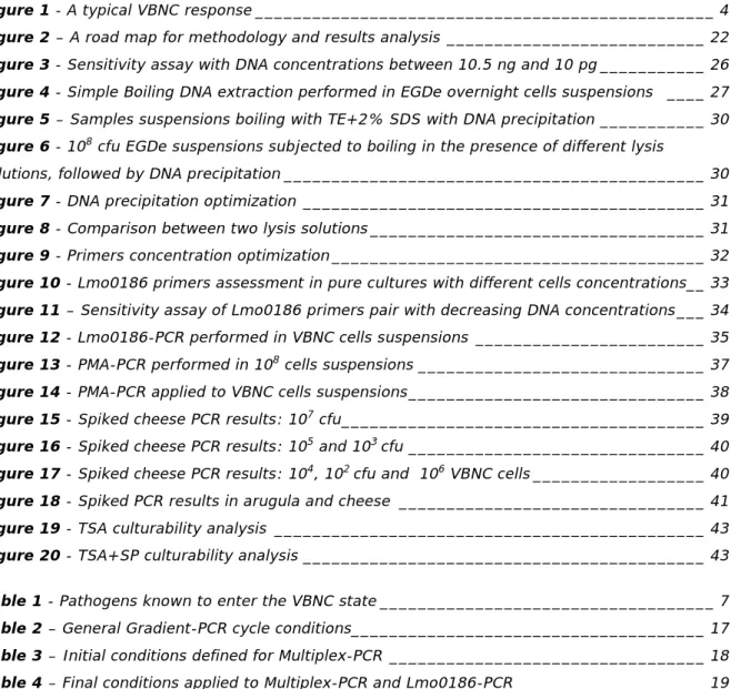
(18) However there is still not a straightforward answer to this question, once both possibilities have been reported. For instance, Cappelier et al. (2005)16 reported that, while studying the VBNC state on Listeria monocytogenes, this pathogen appear to be not virulent when in this state, but could regain virulence upon resuscitation, namely through the incubation under certain conditions5,8,16,17. Lindback et al. (2010)24, while studying the VBNC state on this pathogen also reported the absence of virulence 24. Conversely others authors have reported continued virulence in some pathogens, for example V. shiloi and V. vulnificus5,8. Furthermore, Rahman et al. (1996)33 reported that Shigella dysenteriae in VBNC state still produced shiga toxin and maintained adhesion capability towards the intestinal epithelial cells. Similarly, Vora et al. (2005)34 reported that, on the three Vibrio species tested, it was detected a continuous expression of toxins and virulence5,8,34. Nevertheless, it is important to mention that despite the virulence expression during the persistence in VBNC state, this potential can be retained and resumed upon resuscitation, leading to host infection. In fact some reports were made confirming this hypothesis5,8.. 1.2.2. ANTIBIOTIC RESISTANCE Another important question is if cells in VBNC state are more resilient to antibiotics. One could expect that the persistence in this state could promote an increased antibiotic resistance, at least for some of them, given that these target mainly actively growing cells. Reports have been made that support this information. For example, Lleò et al. (2007)13 reported a 500 times increase of the minimum inhibitory concentration of vancomycin in Ent. faecalis VBNC cells. Similarly, Bates et al. (2003)35 reported that Helicobacter pylori, responsible for ulcers, when in VBNC state was antibiotic resistant 5,8,13.. 1.3.. VIABLE BUT NONCULTURABLE STATE: DETECTION METHODS. As already mentioned, bacteria in the VBNC state fail to produce visible growth on standard bacteriological media were they would otherwise grow. Nevertheless, these bacteria are still viable and upon resuscitation become culturable 5,7,8. A typical VBNC response is shown in Fig. 1, showing a fast decline in culturable counts detected upon exposure of bacteria to one or more environmental stresses, whereas total cellular counts maintain relatively constant. Viable counts, although lower, are still higher than that of culturability assays. Viability assays are therefore crucial to determine whether bacterial cells are alive (viable) or not, as it is a fundamental characteristic for defining the VBNC state5,8. These viability assays are thus aimed to detect parameters expected to be present in the VBNC state, like respiration chain activity,. transcription,. protein. synthesis. and. additionally cellular integrity, the later being of major importance for viability. Therefore, methods that indicate subcellular activities in bacterial cells have been developed and total number of cells of a population. may. be. obtained. diamidino-2-phenylindole. using. fluorescent. the. 4',. 6-. staining. Figure 1 - A typical VBNC response. Entry of V. vulnificus into the VBNC state on incubation at 5ºC. Total cell counts (□), culturable counts (○), and viable counts●)( are represented (from Oliver, 2005)5.. 4.
(19) (DAPI) or acridine orange staining. Such methods include the detection of an intact cytoplasmic membrane, normally through the use of exclusion dyes (BacLight® Live/Dead assay or propidium iodide), which are not able to penetrate viable cells. Alternatively, viability assays can use fluorescent redox probes for detection of an active respiratory chain, like the redox dye 5-cyano-2,3-ditolyl tetrazolium chloride (CTC) used as an artificial electron acceptor that competes with molecular oxygen. The reducing power generated by the electron transport system, in viable cells, converts CTC into insoluble fluorescent crystals, detectable with UV optics and epi-illumination5,8,23,28. But since an active respiratory chain and cellular integrity does not imply that those cells are able of undergoing future growth, the direct viable count (DVC), first developed by Kogure et al. (1979)36, has also been applied to address. this. issue6,7,23,27,28.. In. this,. active. cells. are. identified. by. growth,. without. multiplication, after 6 h of incubation at 25°C, in response to the addition of nutrients (e.g. yeast extract) in the presence of an antimicrobial agent, such as nalidixic acid or ciprofloxacin. Under these conditions, active cells are unable of undergoing DNA replication and cell division but are not affected on any other metabolic activities. Thus active cells become elongated and/or flattened, hence increasing its size, whereas nonviable cells remain unchanged 5,8,27,28. Due to its simplicity, the DVC method is one of the most commonly used for assessing viability. Another’s methods concerning the detection of metabolic activity, through the use of enzymes subtracts, have also been applied as viability markers, although these cannot be considered undisputable evidence of viability or nonviability because they depend on other factors as the enzyme expression in the cells. Recent popular methods are molecular detection assays, namely the reverse transcriptase polymerase chain reaction (RTPCR), that allows gene expression detection, and relies on the relative small half-time of mRNA existence in cells, and so continuous gene expression by the VBNC cells can be detected and taken as a good indicator of their viability8,24,32.. 1.4.. VIABLE BUT NONCULTURABLE STATE: INDUCTION AND RESUSCITATION. As mentioned before, cells enter a VBNC state when exposed to stressful or adverse conditions, and so it is believed by many authors that it is indeed a survival strategy that allows bacteria to circumvent unfavorable situations5,7,8. Inducers are therefore both chemical and environmental, in which it can be included nutrient starvation, temperatures outside the normal range of growth, osmotic concentrations not adequate, presence of heavy metals or food preservatives, different pH, oxygen concentration and exposure to white light or UV radiation5,8,10,37. These should be seen as examples of factors inducing the VBNC state, since many others conditions can be a cause for the induction. Although some factors, or a conjugation of factors, are already known to lead to a VBNC state, the underlying mechanism is yet to be fully uncovered but, nevertheless, some insights have already been made. For instance it appears that hydrogen peroxide (H2 O2) has a significant role in the VBNC state induction, due to the inability of cells to deplete this metabolite (that may be present in solid media or produce upon plating cells in it)5,8. So, as reported by Kong et al. (2004)38, mutant V. vulnificus catalase-negative bacteria cannot degrade this toxic compound entering VBNC state even at room temperature, whereas incubation at lower temperature was necessary to induce the wild-type catalase-positive V. vulnificus bacteria. Additionally it has also been. 5.
(20) reported that the alternative sigma factor RpoS may be an important VBNC induction regulator and, recently, Kusumoto et al. (2012)21 described that it was necessary for delaying the entry in the VBNC state5,21. Upon induction bacteria can remain in VBNC state for a considerable amount of time, but ultimately, and in order to consider it as a survival strategy, they should be able to become culturable again. As mentioned earlier the first report of a true resuscitation was that of Whitesides and Oliver (1997)9, where a simple temperature upshift resulted in resuscitation of the V. vulnificus VBNC cells, and not due to the regrowth of residual culturable cells. Resuscitation has been most studied in V. vulnificus, since its inducing factor is temperature and the simple reversal of this is sufficient to resuscitation occurrence. However the resuscitation process is not so easy in other bacteria, and the simple reversal of the inducing factor may not be sufficient to observe recovery of the VBNC cells. There have been reports of the inability to recover VBNC cells to a culturable state and others where such recovery was only possible through the mediation of a higher organism8,17–20. A recent study of Pinto et al. (2011)39 revealed that resuscitation of E. coli strains depended on various stimuli, as the presence of specific amino acids or alteration of temperature. Even though resuscitation is not yet fully understood, a group of extracellular proteins, the resuscitation promoting factors (Rpfs), may be significantly involved, since it has been reported that these promote resuscitation in several actinobacteria and additionally in M. tuberculosis and M. smegmatis7,8,18. Furthermore, Rpf homologues have been identified in various genera8,18 . These factors were first discovered by Mukamolova et al. (1998)18 while studying resuscitation in Micrococcus luteus, and its Rpf was found to belong to a family of secreted proteins, and reports made revealed that it exists a similarity to soluble lytic transglycosylases. These factors are thought to be involved on alterations in the cell wall, namely through hydrolysis of the glycan backbone of peptidoglycan. Indeed reports revealed that this protein has a muralytic activity that is associated with its resuscitation promoting activity. Once again, biochemical modification of the cell wall appears to have an important role in the VBNC state 8,18,40. Additionally, Reissbrodt et al. (2002)41 reported the resuscitation of VBNC cells of Salmonella enterica serovar Typhimurium and two E. coli O157:H7 strains due to the presence of another kind of Rpfs, the heat-stable autoinducer of growth, which seems to be signaling molecules secreted by either gram positive and negative bacteria8,41.. 1.5.. VIABLE BUT NONCULTURABLE STATE: A PUBLIC HEALTH CONCERN?. From the characteristics already described, some can lead to the question on whether or not the existence and presence of the VBNC state can pose a threat to the public health. This is of special importance since that it has been recognized that the VBNC state occurs in more than 70 species so far, including several pathogens (human, animal or plant). A list of some of these pathogens is illustrated in Table 1, and, as it can be seen, it is quite extensive.. 6.
(21) Table 1 - Pathogens known to enter the VBNC state. From Oliver (2010)8.. The presence of pathogens persisting in VBNC state is of major importance for medicine, epidemiology, ecology, environmental quality assessment and food safety7,8,28. Indeed there is evidence that, when in VBNC state, these bacteria can persist for a long time, although remaining pathogens. undetected, 5,7,8,42. representing. therefore. an. unknown. reservoir. for. these. , that may, in some cases, be the missing link of epidemiological studies.. Concerning food microbiological safety, it should be highlighted that a foodborne pathogen is most probable to encounter several of the inducing factors mentioned before, once that these represent many of the food conservation conditions and/or composition. Furthermore, food quality assessment is still mostly based on routine culturability assays, where VBNC cells do not grow. A very recent example can be given to show the importance of VBNC cells in food products: in 2011 occurred the german outbreak of a hybrid E. coli, strain O104:H4, that involved more than 3000 cases of bloody diarrhea and hemolytic uremic syndrome. Aurass et al. (2011)43, studying this specific strain, reported that it had the ability to rapidly enter the VBNC state when in a nutrient-poor environment together with another factors such as the presence of copper ions and recover upon stress relief. Such ability would therefore allow this strain to remain undetected in the suspected sources (fenugreek sprouts and seeds), and originate the infection cases43. There is, as one can imagine, much more cases of reports of VBNC cells that have been induced in food products (for example E. coli O157:H7 on lettuce plants and L. monocytogenes on parsley leaves8,20,43,44). It becomes necessary therefore to analyze if the risk assessment in this area (food safety) of public health interest is being underestimated, for not account for the presence of VBNC cells.. 1.6.. LISTERIA MONOCYTOGENES: A FOODBORNE PATHOGEN. The Listeria genus comprises eight species, namely L. monocytogenes, L. ivanovii, L. seeligeri, L. welshimeri, L. innocua, L. grayii, L. rocourtiae and L. marthii. Bacteria belonging to this genus are ubiquitous, existing in various natural environments. Isolations of these organisms have been made from large number of domestic and wild animals, as well as from soil, silage, water, food products and other environmental sources. Listeria bacteria cells are small, having a diameter of 0.5 µm and length of 1 to 2 µm, gram positive, non-spore-forming rods. From the species mentioned above, L. monocytogenes and L. ivanovii are pathogenic to animals, but the first is commonly associated to a severe disease. 7.
(22) called human listeriosis, whereas such association is rarely attributed to the later (being most commonly responsible for listeriosis in domestic animals)45,46. The existence of listeriosis was recognized in 1924 (Murray et al., 1926)47, but only in 1929 was detected in humans. A real public health concern raised especially since the number of reports increase largely in the 1980s, with the first human listeriosis outbreak23,46,48. Indeed listeriosis, although not as frequent as other diseases of foodborne pathogens, can present severe consequences, presenting a very high death rate (of about 30%), and with a high possibility of serious squeals45,46,48. Whereas listeriosis causes flu-like symptoms in healthy people, in the established risk groups it can cause septicemia, meningitis, encephalitis, intrauterine. infection. and. immunocompromised. even adults. abortion and. and. neonatal. juveniles. (for. death. example. The. risk. patients. groups. comprise. with. acquired. immunodeficiency syndrome (AIDS) or under cancer treatment), infants including newborns, pregnant woman and the elderly. The occurrence of human listeriosis has been mostly associated to food products. Moreover from the twelve severe listeriosis outbreaks occurring in the United States between 1970 and 2002, eleven were associated to the ready-to-eat (RTE) products, which can be consumed without cooking. Several outbreaks have been reported and estimations made proclaimed an incidence in the United Stated of 0.3 cases per 100 000 inhabitants in 200745,46,48. In Portugal this disease is not of necessary notification but according to Almeida et al. (2006)49 its incidence in 2003 was estimated to be of at least 1.4 cases per million. So, despite the apparent low incidence, the severity of this disease, its high death rate and association with L. monocytogenes presence in food products, made this bacterium to be recognized as an important foodborne pathogen, and one of the most well studied. This bacterium is a facultative intracellular haemolytic pathogen that upon ingestion can penetrate the intestinal tissue and, in some extend, evade the host immune system to further multiply. It has also the ability to pass through the intestinal, blood–brain, and fetus placental barriers leading to the possible symptoms mentioned above. The infection mechanism has been already described in previous studies. All the L. monocytogenes strains have the capability of causing infection, and from the thirteen serovars associated to it, the most commonly related to infection are 4b, 1/2a and 1/2b45,46,48,50. Many countries, like Canada and Denmark, have adopted a “zero-tolerance” for the presence of this microorganism in selected food products. For example, in the United States, U.S. Food and Drug Administration (FDA) has established and maintained this policy since 1995 concerning the RTE food products45,46,48. The food safety is therefore of major importance and aims to prevent the occurrence of outbreaks of food pathogens, namely L. monocytogenes. However the microbiological analysis still relies mostly on culturability based assessments. In the case of this particular pathogen the preferred standard methodology includes two enrichments steps of the samples for Listeria spp. with plating of the cultures on two selective agar media (ALOA and other selective agar) after each step of enrichment. Enumeration of positive samples are performed by colony count on differential selective agar in combination with Most Probable Number (MPN) using the selective enrichment broth followed by plating on ALOA differential selective agar. This represents only a simple resume of the procedure defined by the International Standard ISO 11290-1 and 11290-1:1996/Amd.1:200428,45,51. 8.
(23) Nevertheless, it can be notice that besides being a time consuming procedure, it does not account for VBNC cells, since that, as already mentioned, these cells will not grow and so are not quantified. This represents a major thread to the public health given that contaminated food products can become available and lead to a new unforeseen outbreak of human listeriosis. Furthermore, evidence has been reported that this pathogen can enter the VBNC state upon adverse conditions, as those presented during food processing and storage procedures22,23. Indeed, studies performed by Besnard et al. (2000, 2002)22,23 showed that, at least, two strains of L. monocytogenes were able to enter the VBNC state under conditions as starvation, cold temperatures, nature sunlight and high NaCl concentrations22,23. Lindback et al. (2010)24 reported that the ability to enter the VBNC state was in fact present in various isolates from salmon (including processing areas) and patients, and not only on the two strains reported before, although it was apparently not virulent but an active transcription of the hlyA gene was detected24. However, the avirulence of VBNC cells of L. monocytogenes has been reported before by Cappelier et al. (2005)16, supporting these findings. Nevertheless, L. monocytogenes VBNC cells have already been shown to resuscitate in egg yolk when in the presence of an embryo, upon which they regained their virulence capability16,17. Recently Cunningham et al. (2009)37 reported that exposure of this pathogen to weak acids as potassium sorbate, commonly used as a food preservative, led to the entry in a VBNC state for several hours, although these cells became rapidly nonviable when incubated at low pH37. These studies highlight the fact that L. monocytogenes VBNC cells may pose a genuine public health concern since that, even being avirulent whenever in this state, virulence can be regained upon resuscitation. It should therefore be assessed their presence in food products to thereby guarantee the consumers safety upon consumption.. 1.7.. POLYMERASE CHAIN REACTION: A MOLECULAR APPROACH. The amplification of nucleic acids through the polymerase chain reaction (PCR) was discovered in 1983 by Kary Mullis52, being assigned to him the Chemistry Nobel Prize in 1993. No other tool had such a revolutionary impact on molecular approach and since its discovery it became a powerful diagnostic tool for the analysis of microbial infections as well as for the analysis of microorganisms in food samples. Despite its wide applications, the underlying principle is quite simple. DNA polymerase is an enzyme involved in DNA replication. In this process this enzyme synthesizes new DNA strands, in a 5’-3’ direction, from a single-stranded DNA template. This leads to an increase on DNA copies, and PCR relies on this feature. So, two oligonucleotide primers flanking a specific region are added to the reaction, therefore defining the target sequence to be amplified. Each primer hybridizes to opposite strands of the DNA functioning as reaction initiators of the DNA synthesis. With the addition of a DNA polymerase, most commonly the thermostable Taq polymerase, deoxyribonucleic acids (dATP, dCTP, dGTP, and dTTP), and the DNA template, the reaction occurs leading to the exponential increase of DNA molecules, since each newly synthesized target DNA copy will function as template in the subsequent cycles. Each reaction has three different cycles: a denaturation cycle, where DNA is single-stranded; annealing, where conditions are established towards the hybridization of the primers and extension which leads to the synthesis of the target sequence. These three cycles are repeated several times, giving thereby the amplification. 9.
Imagem


+7


Documentos relacionados
Desse modo, o artigo apresenta um estudo sobre a deficiência visual, em espe- cífico a baixa visão em relação à iluminação artificial de ambientes internos, e busca.. apresentar
Of the 128 samples that had double collection, one (microscopy and culture-positive) showed inhibition in the PCR reaction for both specimens, 14 had positive results for culture
Métodos para avaliar efetividade de gestão: o caso particular das Áreas de Proteção Ambiental (APAs)
Deste modo, o questionário foi dividido em cinco elementos (Tabela 4): contexto, planejamento, insumos, processos e resultados, distribuídos em 16 módulos temáticos: Contexto
Semiquantification by PCR, based on comparison between the intensities that correspond to the amplified products and a standard curve, plotted upon DNA amplification of samples with
Segundo Dumont et al (2010), o critério para a utilização de um jogo, prende-se muitas vezes com a preocupação de tornar a vida do professor mais fácil e não com
Todos os trabalhos de Koselleck sobre o problema dos tempos históricos publicados nas décadas de 1970 e 1980, grande parte dos quais reunidos nos volumes Futuro Passado
Utilizamos como variáveis explicativas para TRAF_DROGA o IVS e também número de jovens com idade entre 15 e 29 anos (JOVENS_1529), já que a literatura indica uma relação positiva
The results demonstrated that after 3 min of ultrasonication, 19 of the 35 compounds studied showed similar reaction yield to those obtained with the classic procedure that last
