Authors
Brenda Navarro de Souza1 Maria Brandão Tavares2
Maria Fernanda Sanches Soares3 Washington Luis Conrado dos Santos1
1 Fundação Oswaldo Cruz, Brasil,
Instituto Gonçalo Moniz, Salvador, BA, Brasil.
2 Escola Bahiana de Medicina e
Saúde Pública, Salvador, BA, Brasil. 3 John Radcliffe Hospital, OUH NHS
Foundation Trust, USA.
Submitted on: 07/10/2017. Approved on: 08/23/2017.
Correspondence to:
Washington Luis Conrado dos Santos. E-mail: [email protected]
IgA Nephropathy in Salvador, Brazil. Clinical and laboratory
presentation at diagnosis
Nefropatia por IgA em Salvador, Brasil. Apresentação clínica e
laboratorial no momento do diagnóstico
Introdução: A nefropatia por IgA (NIgA) é a glomerulopatia primária mais preva-lente no mundo, mas grande variação é relatada em diferentes países. No Brasil, a prevalência relatada é alta nos estados do Sudeste e baixa em Salvador, Bahia, Brasil. Objetivos: Este estudo investigou os pa-drões clínicos e histológicos de pacientes com NIgA em Salvador, Brasil. Métodos: Trata-se de um estudo descritivo que in-cluiu todos os pacientes com diagnóstico de NIgA, realizados em biópsias de rins nativos, coletados nos serviços de referên-cia em nefrologia dos hospitais públicos de Salvador, entre 2010 e 2015. Resultados: Foram identificados 32 casos de NIgA, correspondendo a 6% de glomerulopatias primárias. Houve uma ligeira predominân-cia do sexo masculino (56%) e a mediana da idade foi de 30 [22-40] anos. Hematúria esteve presente em 79%, proteinúria não nefrótica esteve presente em 61% e hiper-tensão esteve presente em 69% dos pacien-tes. A esclerose segmentar (lesão S1) estava presente em 81% dos casos, e lesões túb-ulo-intersticiais crônicas (lesões T1 e T2) estavam presentes em 44% dos casos. Pa-cientes com escores M1 e T2 MEST-C exi-biram maior ureia e creatinina séricas que outros pacientes. Conclusão: A prevalência de NIgA foi menor em Salvador do que em outras regiões do Brasil. Lesões histológi-cas crônihistológi-cas e marcadores laboratoriais de doença grave foram frequentes. Os escores M1 e T2 MEST-C foram correlacionados com marcadores de disfunção renal.
R
ESUMOPalavras-chave: Glomerulonefrite; Glo-merulopatia, IgA; Classificação.
Introduction: IgA nephropathy (IgAN) is the most prevalent primary glomeru-lopathy in the world, but great variation is reported in different countries. In Bra-zil, the reported prevalence is high in the Southeastern States and low in Salvador, Bahia State, Brazil. Objectives: This study investigated the clinical and histological patterns of patients with IgAN in Salva-dor, Brazil. Methods: This is a descripti-ve study that included all patients with a diagnosis of IgAN performed in native kidney biopsies collected from referral nephrology services of public hospitals in Salvador between 2010 and 2015. Re-sults: Thirty-two cases of IgAN were iden-tified, corresponding to 6% of primary glomerulopathies. There was a slight male predominance (56%) and the median age was 30 [22-40] years. Hematuria was pre-sent in 79%, non-nephrotic proteinuria was present in 61%, and hypertension was present in 69% of patients. Segmental sclerosis (S1 lesions) was present in 81% of cases, and chronic tubulo-interstitial le-sions (T1 and T2 lele-sions) were present in 44% of cases. Patients with M1 and T2 MEST-C scores exhibited higher serum urea and creatinine than other patients.
Conclusion: The prevalence of IgAN was lower in Salvador than other regions of Brazil. Chronic histological lesions and laboratory markers of severe disease were frequent. M1 and T2 MEST-C scores were correlated with markers of renal dysfunction.
A
BSTRACTKeywords: Glomerulonephritis; Glomeru-lopathy, IgA; Classification.
DOI: 10.1590/2175-8239-JBN-3851
I
NTRODUCTIONIgA nephropathy (IgAN) is the most prevalent glomerular disease
countries and in different regions of the same coun-try, such as Brazil.3,4,5 Differences in the frequency of IgAN are attributed to ethnical background or selection bias, which stems from heterogeneous bi-opsy indication policies.2 For example, the preva-lence of IgAN is high in Japan (30%), Italy (35%), and Spain (15%), and it is becoming the most prevalent glomerular disease in these areas.6,7,8 However, it is much lower in Saudi Arabia (4.7%), Africa (2.8%), India (8.1%), Colombia (11.8%), Peru (1.5%), and Mexico (7%).9,10,11,12,13,14 The prevalence of IgAN in the USA is high in Caucasian (38%) and East Asian populations (36%), and low in African-American (3%) and Hispanic popula-tions (19%).15 The estimated prevalence of IgAN varies in different Brazilian States. It is high in the Southeastern States of Minas Gerais (16.15%) and São Paulo (17.8%), and it is low in the States of Para (6.3%), Amazonas (4.3%), and Bahia (5%).16,17,18,4,19
The estimated prevalence of IgAN in Salvador, BA, Brazil is 7% of primary glomerular diseases (dos-Santos et al., in press).[1] Salvador’s ethnical background might be accountable for this low fig-ure: approximately 73% of the population self-declares as being of African heritage according to the Brazilian Institute of Geography and Statistics (IBGE). Comparatively, the estimated population self-declared of African heritage is 27.2% and 45.4% in São Paulo and Minas Gerais (IBGE), respectively, where the prevalence of IgAN are 17.8% and 16.15%, respectively.16,17 Conversely, the estimated self-declared African heritage in the State of Para is 71.9% (IBGE), whereas the prevalence of IgAN is 6.3%.18 However, it is not clear whether these differences are due to the eth-nic backgrounds or to different criteria to indicate kidney biopsy.
The present study investigated the pattern of clinical and histological presentations of patients with IgAN from Salvador, Brazil, at disease diag-nosis. We aimed to shed light on the actual disease prevalence in this highly African-decent populated area in Northeastern Brazil.
M
ETHODSin referral nephrology services of public hospitals in Salvador, State of Bahia, Brazil, and examined at the Gonçalo Moniz Institute, Fiocruz (IGM-Fiocruz) between 2010 and 2015. Only native kidney biopsies with available and sufficient his-tologic material and clinical records were included.
Renal biopsies: All renal biopsies underwent:
1) routine light microscopy processing (fixed in Bouin’s solution, embedded in paraffin, sectioned at 2-µm thickness, and stained with hematoxylin and eosin, Periodic Acid Schiff, Periodic Schiff-Methenamine Silver, Azan, and Picro Sirius red); and 2) immunofluorescence processing (embedded in cryopreservation medium and incubated with antisera anti- IgA, IgG, IgM, kappa chains, lamb-da chains, C1q, C3, and fibrinogen). All samples were fixed in 1% glutaraldehyde in cacodylate buffer, post-fixed in osmium tetroxide, and embed-ded in Poly/Bed® for ultrastructural analysis when required.
Histological analysis: Two pathologists (MFSS
and WLCS) without previous knowledge of the reported pattern of renal lesion independently reviewed the histological slides of each patient. Discrepancies in independent analyses between the two pathologists were resolved in a consensus analysis. The histological analyses are classified according to the Oxford classification of IgAN (MEST-C scores).20,21
Clinical data: The following data were obtained
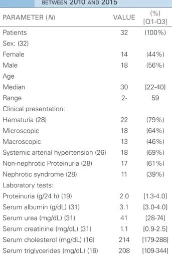
PARAMETER (N) VALUE (%) [Q1-Q3]
Patients 32 (100%)
Sex: (32)
Female 14 (44%)
Male 18 (56%)
Age
Median 30 [22-40]
Range 2- 59
Clinical presentation:
Hematuria (28) 22 (79%)
Microscopic 18 (64%)
Macroscopic 13 (46%)
Systemic arterial hypertension (26) 18 (69%)
Non-nephrotic Proteinuria (28) 17 (61%)
Nephrotic syndrome (28) 11 (39%) Laboratory tests:
Proteinuria (g/24 h) (19) 2.0 [1.3-4.0]
Serum albumin (g/dL) (31) 3.1 [3.0-4.0]
Serum urea (mg/dL) (31) 41 [28-74] Serum creatinine (mg/dL) (31) 1.1 [0.9-2.5]
Serum cholesterol (mg/dL) (16) 214 [179-288]
Serum triglycerides (mg/dL) (16) 208 [109-344] TABLE 1 GENERALCHARACTERISTICSOFPATIENTS
WITH IGA NEPHROPATHYWHOUNDERWENT RENALBIOPSYIN SALVADOR, BA, BRAZIL BETWEEN 2010 AND 2015
Data analysis: Data are reported as percentages
and absolute numbers, and summarized as means ± standard deviations or medians and the 25% and 75% percentiles. Data were summarized using Prism 5.01 (GraphPad, San Diego, CA, USA) and StataIC11 software.
Ethical considerations: The study was performed
in accordance with resolution No. 196/96 of the National Health Council, and the Ethics Committee for Research Involving Human Subjects of the Instituto Gonçalo Moniz; Fiocruz approved the pro-cedure (Protocol No. 1642146).
R
ESULTSGeneral patient characteristics: A total of 1,045
renal biopsies were examined in the IGM-Fiocruz between 2010 and 2015. However, 134 biopsies were from transplanted kidneys, 110 had under-represented renal parenchyma (mostly due to the absence of glomerulus for immunofluorescence), seven cases had an inconclusive diagnosis, and two cases were received for a second opinion. A total of 253 cases were excluded from the study, and 792 cases were included. Of the included cases, 556 were primary glomerulopathy and 236 were secondary glomerulopathy. Thirty-two cases were diagnosed as IgAN, and one case presented clinical findings suggestive of Henoch-Shoenlein vasculitis. Therefore, the prevalence of IgAN was 6% in the primary glomerulopathy cases and 4% in the renal biopsies of native kidneys.
Table 1 shows the primary clinical and demo-graphic characteristics of these patients. Age var-ied from 2 to 59 years with a median of 30 (22-40; first and third quartiles, respectively) years. Four (12.5%) patients were children, and 28 (87.5%) patients were adults, with a slight male predominance.
The primary reported clinical presentations were hematuria in 22/28 (79%), systemic hypertension in 18/26 (69%), and non-nephrotic proteinuria in 17/28 (61%) of cases.
Table 2 shows the distribution of MEST-C scores of the renal biopsies of the IgAN patients.
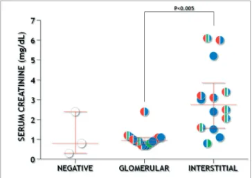
The most common lesion observed was segmental sclerosis (26; 81%). There was a predominance of chronic sclerosis over proliferative glomerular sions. As expected, chronic tubuinterstitial le-sions were associated with renal dysfunction (Fig. 1). Serum urea and creatinine concentrations were higher in patients with the combination of M1 and T2 scores (Table 2 and Fig. 2). There was a trend for increased proteinuria, serum urea, and creati-nine concentrations in individuals with S1 com-pared with patients with S0, but this difference was not statistically significant. Furthermore, all the six patients with S0 also had T0, while 14/26 (54%) patients with S1 had T1 or T2. Such asso-ciation was statistically significant (Fisher’s test p
Figure 1. Glomerular and tubule-interstitial MEST-C scores and serum creatinine concentrations. Colors represent positive MEST-C scores: M1-red, E1-green, S1-blue.
MEST N UREA CREATININE ALBLBUMIN CHOLESTEROL TRIGLYCERIDES 24 h PTU
ALL 32 (100) 41 [28-74] 1.1 [0.9-2.5] 3.1 [3.0-4.0] 213 [179-288] 208 [109-344] 2.0 [1.3-4.0]
M0 17 (53) 30 [22-59]a 1.0 [0.8-1.9]b 3.1 [2.5-4.0] 220 [198-368] 103 [71-354] 1.9 [1.2-2.0]
M1 15 (47) 67 [36-85]ª 2.1 [1.1-3.2]b 3.1 [3.0-4.0] 187 [172-259] 212 [201-335] 2.3 [2.2-4.7]
E0 23 (72) 37 [25-74] 1.1 [0.9-2.4] 3.1 [3.0-4.0] 211 [185-318] 214 [86-318] 1.7 [1.2-3.1] E1 9 (28) 42 [36-83] 1.6 [0.9-2.5] 3.6 [3.0-4.0] 216 [173-269] 202 [125-350] 2.3 [2.0-5.1]
S0 6 (19) 28 [21-30] 0.8 [0.8-1.1] 2.0 [1.5-4.1] 417 [216-513] 354 [103-578] 1.2 [0.4-2.0]
S1 26 (81) 46 [31-74] 1.4 [0.9-3.0] 3.1 [3.0-4.0] 189 [173-249] 204 [115-283] 2.2 [1.5-4.0]
T0 18 (56) 30 [23-36]c,d 0.9 [0.8-1.1]e 3.5 [2.4-4.0] 216 [189-308] 216 [100-354] 1.5 [1.2-2.2]
T1 8 (25) 60 [46-88]c 2.0 [1.3-2.8] 3.1 [2.8-3.6] 173 [170-225] 204 [201-514] 3.0 [1.9-5.1]
T2 6 (19) 74 [65-130]d 4.2 [3.1-6.0]e 3.1 [3.0-4.0] 227 [169-294] 174 [125-248] 2.2 [2.1-3.5]
C0 25 (78) 39 [79-28] 1.1 [2.8-0.8] 4.0 [4.0-3.0] 189 [249-173] 208 [335-103] 2.0 [2.8-1.3]
C1 7 (22) 41 [51-25] 1.2 [2.1-0.9] 3.1 [2.1-2.5] 269 [308-225] 250 [364-135] 3.4 [4.7-2.2] TABLE 2 MEST-C SCORESANDDISTRIBUTIONOFLABORATORYVARIABLESINPATIENTSWITH IGAN SUBJECTEDTOKIDNEY
BIOPSYIN SALVADOR, BRAZIL, 2010-2015
Mann-Whitney or Kruskall-Wallis tests were applied where applicable. a, b and c = p < 0.05; d and e = p < 0.005. (%), [Q1-Q3].
Figure 2. M1 and T1/2 scores combination and serum creatinine concentrations. Colors represent positive MEST-C scores: M1-red, E1-green, S1-blue.
D
ISCUSSIONThe demographic characteristics and clinical pre-sentation of patients in this work such as age, sex, frequency of hematuria, and non-nephrotic protein-uria, are similar to previously published studies.20,27-29 Microscopic hematuria with minimal proteinuria is the most common presentation of IgAN, and it is associated with a favorable prognosis. In contrast, the presence of significant proteinuria, hypertension, and decreased glomerular filtration rate is related to a poor prognosis. The median protein concentration in urine was slightly higher in this study than previ-ously published studies,20,25,28 and hypertension was recorded in 69% of patients. These data suggest that patients with IgAN reported here had already evolved to a late stage of progression to chronic kidney dis-ease at the time of biopsy.
Our series demonstrated a high frequency of posi-tive MEST-C scores for segmental sclerosis (81%) and tubule-interstitial lesion (44%). Other authors reported similar proportions.26,28 Together, positive T or M scores were associated with increased serum urea and creatinine concentrations in the present study. Tubulo-interstitial changes are consistently as-sociated with the clinical presentation and outcome of IgAN.28,29 Despite associations between M1 and renal dysfunction being less well established, Lee et al. demonstrated an M1 association with disease pro-gression.25,28 A model for using MEST scores for renal outcome at the time of biopsy has been proposed by Barbour and colleagues (2016).30 The authors pro-pose that a combination of MEST score with the data on blood pressure, proteinuria, and eGFR at the time of biopsy may predict the renal outcome similar to using clinical data over 2 years of follow-up. Further development of models of association between com-bined MEST-C scores and clinical presentation or outcome of IgAN are still required.
The observation of a high proportion of positive MEST-C scores for segmental sclerosis and tubule-inter-stitial lesions in this study combined with the severity of clinical presentation indicates that patients with IgAN in Salvador are in an advanced stage of the disease when subjected to renal biopsy. Further studies will be use-ful to determine factors associated with the severity of IgAN presentation and prognosis in this city.
C
ONCLUSION1) The prevalence of IgAN in patients submitted to renal biopsies in Salvador, Bahia is among the lowest reported in Brazil.
2) Patients with IgAN in this series presented high protein concentrations in urine and a high frequency of hypertension, which suggests a late stage of CKD progression.
3) A high frequency of positive MEST scores asso-ciated with progressive kidney disease was observed in these patients: segmental sclerosis (81%) and tu-bule-interstitial lesion (44%).
4) Positive T or M scores were associated with in-creased serum urea and creatinine concentrations.
L
IST OF ABBREVIATIONSIgAN - IgA nephropathy. CKD - Chronic kidney dis-ease. IBGE - Brazilian Institute of Geography and Statistics.
A
CKNOWLEDGMENTSWe thank Dr. Marilia Bahiense and Dr. Mauricio Teixeira (Hospital Ana Nery), Dr. Reinaldo Martinelli and Mrs. Rosimary (Hospital Professor Edgard Santos), Dr. Antonio Raimundo Almeida and Dr. Nadia Khouri (Hospital Geral Roberto Santos), and Dr. Daniela Braga and Dr. Marcia Bessa for their support with clinical data collection. This work was supported by the Hepatitis Reference Laboratory from FIOCRUZ. Brenda Navarro received a scholarship from State of Bahia Research Supporting Agency - FAPESB.
R
EFERENCES1. Rodrigues JC, Haas M, Reich HN. IgA Nephropathy. Clin J Am Soc Nephrol 2017;12:677-86.
2. Fiorentino M, Bolignano D, Tesar V, Pisano A, Van Biesen W, D’Arrigo G, et al.; ERA-EDTA Immunonephrology Working Group. Renal Biopsy in 2015-From Epidemiology to Evidence--Based Indications. Am J Nephrol 2016;43:1-19.
3. Queiroz MM, Silva Júnior GB, Lopes MSR, Nogueira JOL, Correia JW, Jerônimo ALC. Estudo das doenças glomerulares em pacientes internados no Hospital Geral César Cals - Forta-leza, Ceará, Brasil. J Bras Nefrol 2009;31:6-9.
4. Cardoso ACD, Mastroianni-Kirsztajn G. Padrões histopatoló-gicos das doenças glomerulares no Amazonas. J Bras Nefrol 2006;28:39-43.
5. Ferraz FHRP, Martins CGB, Cavalcanti JC, Oliveira FL, Quirino RM, Chicon R, et al. Perfil das doenças glomerula-res em um hospital público do Distrito Federal. J Bras Nefrol 2010;32:249-56.
6. Kitajima T, Murakami M, Sakai O. Clinicopathological featu-res in the Japanese patients with IgA nephropathy. Jpn J Med 1983;22:219-22.
7. Schena FP. Survey of the Italian Registry of Renal Biopsies. Fre-quency of the renal diseases for 7 consecutive years. The Italian Group of Renal Immunopathology. Nephrol Dial Transplant 1997;12:418-26.
8. Yuste C, Rivera F, Moreno JA, López-Gómez JM. Haematu-ria on the Spanish Registry of Glomerulonephritis. Sci Rep 2016;6:19732.
10. Okpechi IG, Ameh OI, Bello AK, Ronco P, Swanepoel CR, Kengne AP. Epidemiology of Histologically Proven Glomeru-lonephritis in Africa: A Systematic Review and Meta-Analysis. PLoS One 2016;11:e0152203.
11. Mittal N, Joshi K, Rane S, Nada R, Sakhuja V. Primary IgA nephropathy in north India: is it different? Postgrad Med J 2012;88:15-20.
12. Arias LF, Henao J, Giraldo RD, Carvajal N, Rodelo J, Arbeláez M. Glomerular diseases in a Hispanic population: review of a regional renal biopsy database. São Paulo Med J 2009;127:140-4. 13. Hurtado A, Escudero E, Stromquist CS, Urcia J, Hurtado ME,
Gretch D, et al. Distinct patterns of glomerular disease in Lima, Peru. Clin Nephrol 2000;53:325-32.
14. Chávez Valencia V, Orizaga de La Cruz C, Becerra Fuentes JG, Fuentes Ramírez F, Parra Michel R, Aragaki Y, et al. Epide-miology of glomerular disease in adults: a database review. Gac Med Mex 2014;150:403-8.
15. Hall YN, Fuentes EF, Chertow GM, Olson JL. Race/ethnici-ty and disease severiRace/ethnici-ty in IgA nephropathy. BMC Nephrol 2004;5:10.
16. Neves PDMM, Machado JR, Silva MV, Abate DTRS, Rodri-gues DBR, Faleiros ACG, et al. Nefropatia por IgA: análise histológica e correlação clínico-morfológica em pacientes do Estado de Minas Gerais. J Bras Nefrol 2012;34:101-8. 17. Malafronte P, Mastroianni-Kirsztajn G, Betônico GN, Romão
JE Jr, Alves MA, Carvalho MF, et al. Paulista registry of glo-merulonephritis: 5-year data report. Nephrol Dial Transplant 2006;21:3098-105.
18. Alves Júnior JM, Pantoja RKS, Barros CV, Braz MN. Estudo clínico-patológico das glomerulopatias no Hospital de Clínicas Gaspar Vianna. Rev Para Med 2008;22:39-47.
19. Sweet GMM. Glomerulopatias prevalentes na Bahia, um estu-do baseaestu-do em biópsias [Dissertação de mestraestu-do]. Salvaestu-dor: Universidade Federal da Bahia; 2011.
20. Working Group of the International IgA Nephropathy Ne-twork and the Renal Pathology Society, Cattran DC, Coppo R, Cook HT, Feehally J, Roberts IS, Troyanov S, et al. The Oxford classification of IgA nephropathy: rationale, clinicopathologi-cal correlations, and classification. Kidney Int 2009;76:534-45.
21. Trimarchi H, Barratt J, Cattran DC, Cook HT, Coppo R, Haas M, et al.; IgAN Classification Working Group of the Interna-tional IgA Nephropathy Network and the Renal Pathology Society; Conference Participants. Oxford Clasification of IgA nephropathy 2016: an update from the IgA Nephropathy Clas-sification Working Group. Kidney Int 2017;91:1014-21. 22. Martinelli R, Okumura AS, Pereira LJ, Rocha H. Primary focal
segmental glomerulosclerosis in children: prognostic factors. Pediatr Nephrol 2001;16:658-61.
23. Queiroz PF, Brito E, Martinelli R, Rocha H. Nephrotic syndro-me in patients with Schistosoma mansoni infection. Am J Trop Med Hyg 1973;22:622-8.
24. Polito MG, de Moura LA, Kirsztajn GM. An overview on fre-quency of renal biopsy diagnosis in Brazil: clinical and patholo-gical patterns based on 9,617 native kidney biopsies. Nephrol Dial Transplant 2010;25:490-6.
25. Coppo R, Troyanov S, Bellur S, Cattran D, Cook HT, Feehally J, et al.; VALIGA study of the ERA-EDTA Immunonephrology Working Group. Validation of the Oxford classification of IgA nephropathy in cohorts with different presentations and treat-ments. Kidney Int 2014;86:828-36.
26. Nasri H, Mortazavi M, Ghorbani A, Shahbazian H, Kheiri S, Ba-radaran A, et al. Oxford-MEST classification in IgA nephropathy patients: A report from Iran. J Nephropathol 2012;1:31-42. 27. Soares MF, Caldas ML, Dos-Santos WL, Sementilli A, Furtado
P, Araújo S, et al. IgA nephropathy in Brazil: apropos of 600 cases. Springerplus. 2015;4:547.
28. Lee H, Yi SH, Seo MS, Hyun JN, Jeon JS, Noh H, et al. Va-lidation of the Oxford classification of IgA nephropathy: a single-center study in Korean adults. Korean J Intern Med 2012;27:293-300.
29. Kang SH, Choi SR, Park HS, Lee JY, Sun IO, Hwang HS, et al. The Oxford classification as a predictor of prognosis in patients with IgA nephropathy. Nephrol Dial Transplant 2012;27:252-8. 30. Barbour SJ, Espino-Hernandez G, Reich HN, Coppo R,