
Marius Nicu or GRIGORE1*, Constantin TOMA1
Abstract: Salt (chalk) glands of Plumbaginaceae represent interesting structures involved in the excretion of calcium carbonate outside plants’ organs, especially on leaves surfaces. These chalk-glands, nominated by some authors as ‘Licopoli’ or ‘Mettenius’ organs are also very important from taxonomical point of view. Their structure has been a matter of debate for decades and a historical analysis reveals that there are still some inconsistencies regarding the contributions of earlier botanists in discovering and describing chalk-glands. The present work tries to provide a picture of historical progress recorded in the 19th century related to investigation of these structures, focusing especially on the two important names usually mentioned in relation to them: Mettenius and Licopoli. In this respect, several useful clarifications are made, with emphasis on the role played by the two botanists in the stimulation of research interest for these glands among the generations of botanists to come.
Keywords: chalk-glands, Licopoli, Mettenius, Plumbaginaceae, secretion. Introduction
Plumbaginaceae constitute a well-represented cosmopolitan family in the temperate zones of the Northern Hemisphere and showing preferences for arid or saline, often coastal, environments [KUBITZKI, 1993]. The Angiosperm Phylogeny Group classification of flowering plants [APG, 2003] included this family in the Caryophyllales order, together with other families adapted to extreme environments including oligotrophic soils, arid zones, and soils with high salt content. The taxonomy and taxonomical affinities of this striking family are still very problematic and controversial [CRONQUIST, 1981; LLEDO & al. 1998, 2001, 2005; REYES, 1997; SHORT & WIGHTMAN, 2011; TAKHTAJAN, 2009]. For this reason, the number of genera and species included in the Plumbaginaceae differ greatly from one author to another: from about 12 genera and 400-500 species [REYES, 1997] to 10-27 genera and about 1,000 species [SHORT & WIGHTMAN, 2011], Plumbaginaceae is a well-known halophytic family [GRIGORE, 2008, 2012; GRIGORE & TOMA, 2010; GRIGORE & al. 2014] a reality since long recognized in botanical research [ENDLICHER, 1836-1840; LINCEVSKII & CERNIAKOVSKOI, 1952; BENTHAM & HOOKER, 1876; VOLKENS, 1884; PAX, 1897; STRASBURGER & al. 1894; LINDLEY, 1846; RĂVĂRU , 1960; MOORE, 1972; TAKHTAJAN, 2009].
When referring to the Plumbaginaceae family, one should emphasize that one of the most obvious anatomical traits of its representatives is the presence of epidermal glands (chalk-glands and mucilage (chalk-glands) located on leaves and stems. Actually, these (chalk-glands were closely integrated in the taxonomical characteristics of Plumbaginaceae, as a significant anatomical
1“Alexandru Ioan Cuza” University, Faculty of Biology, Bd. Carol I, 20 A, 700505, Iaşi– Romania.
38
feature, a tendency adopted by both older [PAX, 1897; STRASBURGER & al. 1894; VOLKENS, 1884] and recent authors [KUBITZKI, 1993; TAKHTAJAN, 2009].
When describing the secreting glands of the Plumbaginaceae species, METCALFE & CHALK (1972) and, earlier, SOLEREDER (1908) classify them into two categories:
1. Chalk (chalk-secreting) glands, also known as Mettenius glands or Licopoli glands, which generally occur on or inside the cavities on the inner side of the leaves and stem, sometimes surrounded by groups of elongated epidermal cells or by simple hairs. Individual glands of this sort are made up of 4 or 8 epidermal cells arranged in palisade surrounded by 1 or 2 layers, each one made up of 4 “accessory” cells. The walls between the secreting cells of the gland and the surrounding (“accessory”) cells are cutinized. The secreting “organs” of this sort have been generally described as chalk glands, because they exude calcium salt and water; calcium salts are sometimes scattered on the leaf or stem surface by rain drops. The amount of secreted calcium salts depends on the type of soil, although, for instance, the British Limonium
species analyzed by de FRAINE (1916) do not secrete limestone-containing substances. 2. Mucilage glands occur in some representatives of the Plumbaginaceae family; those occurring in the axils of the upper side of the Limonium bellidifolium and L. binervosum basal leaf, described by de FRAINE (1916), evidence a head resting on a head borne on a base consisting of few cells with very thick cuticle-lined walls.
The present contribution will actually deal only with chalk-glands (‘Mettenius’ or ‘Licopoli’ organs), and not with mucilage glands of Plumbaginaceae [GRIGORE & TOMA, 2010], which are the other type of epidermal glands found in the species of this botanical family. It is worth mentioning that an interesting phenomenon occurs regarding the semantics of these glands. Sometimes, in older botanical papers, `Mettenian gland(s)` expression is being used [de FRAINE, 1916; JACKSON, 1928]. This word derivation may suggest that authors have attributed to Mettenius the discovering and description of these intriguing structures.
Throughout the present work, the authors maintained the nomenclature used in the papers consulted, without any intention to find and use instead updated synonyms.
Historical approach
Frequently, the structure of salt glands from Plumbaginaceae has been differently interpreted by some authors, although these controversies are related rather to details than to their basic structure. These formations drew botanists’ attention as early as the second half of the 19th century, as we will describe herein.
The chalk secretion and deposit on the surface of these organs have been noted long time before the detection and description of these glands. Thus, the French chemist BRACONNOT (1836, consulted paper) (and not from 1830, as he is erroneously quoted by MAURY, 1886) was the first who tried to analyze this mineral substance secreted by glands of different species of
Statice: S. monopetala,S. pruinosa, S. aphylla and others and of Plumbago: P. zeylanica, P.
auriculata, P. scandens and P. rosea. He investigated the ‘inorganic scales (écailles de nature inorganique) produced by species of Plumbaginaceae family’; when examined with a magnifier
glass, these white deposits on the surface of leaves appeared to Braconnot as a ‘small parasitic fungus embedded in the tissues of host plant’. He has also anticipated the existence of special secreting formations involved in the occurrence of these deposits, but he did not use a specific term to nominate them. However, he made an interesting anatomical-like observation: after washing the leaves of Staticespecies with acids, he observed on their surface ‘visible cavities indicating the
reason, even nowadays, the terms ‘Licopoli’ and ‘Mettenius’ organs are being used in parallel in botanical literature. The reason for this perception is perhaps explained by the fact that some authors knew only Mettenius’s or only Licopoli’s paper and not both of them, so that they could not have an accurate historical picture. For instance, neither MAURY nor VUILLEMIN do mention Mettenius’s work, whereas, out of the two French botanists, only MAURY (1886) mentions Braconnot’s earliest paper. One may assume that Mettenius’s paper, published in German, was inaccessible to French botanists and thus it has not been consulted; however, METTENIUS (1856) does mention Braconnot’s findings.
METTENIUS (1856) described chalk-glands in a very succinct, but quite precise manner, in the way that he did not hesitate at all in using correct terms related to the chalk secreting function of these glands: Kalksecretion (chalk secretion) and Kalkschüppchen (chalk scales). He described the chalk glands of Goniolimon tataricum (Fig. 1), Limoniastrum monopetalum (Fig. 2), Plumbago europaea (Fig. 3) and P. zeylanica (Fig. 4).
Nevertheless, Mettenius’s work (1856) represents a significant progress in the research of chalk-glands as compared to earlier Braconnot’s (1836) paper, assumed to be the first in signaling chalk-secreting process. In his brief considerations, Mettenius underlined several considerable aspects. For instance, he has correctly shown that chalk-glands belong to the epidermal complex, and that they are derived from epidermal cells divisions, and – most important – that they are not connected with stomata or the vascular system. However, the structure of gland was incorrectly described by Mettenius as consisting of a group of four cells; his mistake was maintained subsequently by LICOPOLI (1866) and MAURY (1886).
A B
Fig. 1. Chalk glands (g) in the leaf of Goniolimon tataricum
40
Fig. 2. Chalk glands (g) in the leaf of Limoniastrum monopetalum
[METTENIUS, 1856]
Fig. 3. Chalk glands (g) in the leaf of
Plumbago europaea, surface view [METTENIUS, 1856]
Fig. 4. Chalk glands (g) in the leaf of
Plumbago zeylanica, cross section [METTENIUS, 1856]
The Italian botanist LICOPOLI, still considered the first who made a description of chalk glands of Plumbaginaceae species (Statice monopetala, 1866) provided a detailed analysis of these glands and depicted them in several drawings (Figs. 5-8). Indeed, his contribution is very extended and detailed; unfortunately, it has no references included, so that it is almost impossible to assert whether he knew Mettenius’s paper or had other data in hand. Except for the fact that he did not nominate the exact types of gland-consisting cells, he was able however to distinguish them from an anatomical point of view and finally to deliver an accurate description of glands (known, as shown, as ‘Licopoli organs’). In addition, he pointed out several important details with respect to these ‘organs’; he correctly concluded that these glands are connected neither with the vascular system, nor with the stomata of plant leaf. Another important observation was that the excreted material of these glands is calcium carbonate; on his microscopical observations, he identified a chalk deposit at the top of the glands –clearly nominated as ‘glandole’.
LICOPOLI resumed his observations in a paper from 1879, where he used the term ‘glandole calcifere’. He states that: these glands have an organization (structure) based on the type discovered and described in Statice monopetala in my previous work –
After Licopoli’s findings –
After Licopoli’s findings – already known and commented by the botanists to come - the interest for the study of chalk-glands was intensified towards the end of the 19th century; the great majority of botanists recognize these glands as ‘Licopoli’ rather as ‘Mettenius’ glands.
MAURY (1886), in his extensive study on the structural organization of
Plumbaginaceae species, evidenced and described Licopoli ‘organs’ in: Plumbago europaea (Figs. 9-10), P. larpentae (Fig. 11), Statice limonium (Figs. 12-13), S. elata (Fig. 14), and S. lychnidifolia (Fig. 15).
Fig. 6. Licopoli ‘organ’ in the lamina of
Statice monopetala, front view (ap g - aperture of the gland; st – stomata) [LICOPOLI, 1866]
Fig. 5. Licopoli ‘organs’ in the lamina of
Statice monopetala (C1, C2 – different types of cells; C3 – a complex of cells – borsetta, forming the bottom of the gland, ch – chalk deposit) [LICOPOLI, 1866]
Fig. 8.Licopoli ‘organ’ in the lamina of Statice
monopetala, detail in front view (C1, C2 – different types of cells; an – angles formed on the intersection of different types of cells; ap g - aperture of the gland; b –borsetta, forming the bottom of the gland) [LICOPOLI, 1866]
Fig. 7.Licopoli ‘organ’ in the lamina of Statice
42
Fig. 9. Cross section through the lamina of
Plumbago europaea (ep – epidermis; Lc o –
Licopoli ‘organ’) [MAURY, 1886] Fig. 10.Plumbago europaeaLicopoli ‘organs’ (st – stomata) [MAURY, 1886] (Lc o) in epidermis of
Fig. 11. Cross section through the lamina of
Plumbago larpentae (s c – secretory cells of
Licopoli ‘organs’) [MAURY, 1886]
Fig. 12. Cross section through the lamina of Statice limonium (ep – epidermis; ct – cuticle; p – pore; s c –
secretory cells of Licopoli ‘organs’) [MAURY, 1886]
Fig. 13.Licopoli ‘organs’ (Lc o) at epidermis level of the lamina
of Statice limonium (st – stomata) [MAURY, 1886] Lc o
ep
Lc o
De BARY (1877) described this secreting “organ” in a different manner; he stated that it included 8 cells originating in the divisions of a single primary mother cell, which is round or square in surface section. This cell is divided in four by two cell wall divisions, perpendicularly on the surface and on each other. In its turn, each of them is divided again, so that one of the new cells is triangular and internal, and the other is rectangular and peripheral.
VOLKENS (1884) and WORONIN (1885) adopted de BARY (1877) descriptions and interpretations. It seems that they were not aware of Licopoli’s findings, since no mention is made of his interpretations. This is quite unexpectedly even for the papers of the 19th century, which are usually well documented and supported by literature, in the manner we know nowadays. Neither Volkens’ nor Woronin’s papers – written in German – have mentioned Licopoli’s findings, while the Italian paper has no references, as already shown. VOLKENS (1884) maintained the basic 8-cell structure of these glands, and pointed out their irregular layout and their role in water elimination, seeing them as “safety valves” that start working when the absorption/transpiration ratio is altered. In his opinion, any excessive calcium salt is eliminated as carbonic acid. In Statice limonium, the cells adjacent to the gland become prominent and turn into conical protrusions.
Figures 16-21 show drawings of these glands in different Plumbaginaceae species, as VOLKENS (1884) depicted them. However, Volkens uses the terms: ‘Sekretionsapparat, Kalkschuppe’, and ‘drüse’ corresponding to secretory structures.
Fig. 14. Licopoli ‘organs’ (Lc o) at epidermis
level of the lamina of Statice elata (st – stomata; t – trichome) [MAURY, 1886]
Fig. 15. Licopoli ‘organs’ (Lc o) at epidermis
44
Fig. 16. Salt-secreting ‘apparatus’ of
Statice limonium [VOLKENS, 1884] Fig. 17. Chalk gland (gl) at epidermis
level of the lamina of Statice latifolia
[VOLKENS, 1884]
Fig. 18. Chalk glands (gl) of Limoniastrum monopetalum (a – surface view; b – cross section)
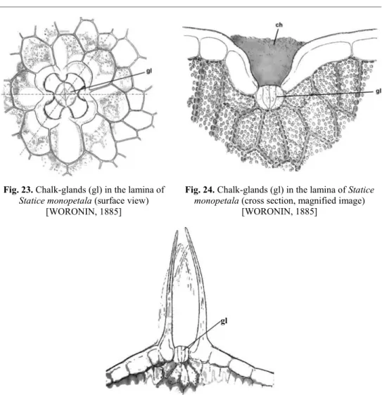
WORONIN (1885) investigated the leaf structure of Statice monopetala and evidenced the chalk glands (‘Kalkdrüse’) (Figs. 22-24); he also made a drawing of these glands in S. sareptana (Fig. 25). In addition to the anatomical description of these glands, he made an interesting ecological observation: the secretion of calcium carbonate by species of Plumbaginaceae is conditioned by soil composition, precisely by its content in calcium carbonate. Woronin correctly stated that many species of this botanical family do not show an excretory process.
Fig. 19. Salt-secreting ‘apparatus’ (gl) of
Statice pruinosa [VOLKENS, 1884]
Fig. 20. Chalk gland (gl) of Statice rhodia
[VOLKENS, 1884]
Fig. 21. Chalk gland of Statice occidentalis [VOLKENS, 1884]
Fig. 22. Chalk glands (gl) in the lamina of Statice monopetala
(cross section; ch – chalk deposit) [WORONIN, 1885] gl
gl
46
MAURY (1886) tried to elucidate the structure of the Plumbaginaceae glands, by pointing out the possible reasons for which other authors considered that these structures rely on 8 and not on 4 cells. When viewed from the top, on a small area of the epidermis, the “organ” looks like a circle divided into four sectors by two diameters perpendicular on each other. Each of these sectors seems(Maury’s emphasis in the text) divided itself in two
by a tangential line, which is more blurred than those of the other sectors. This is actually the inner wall of each secreting cell, which borders the central intercellular space; thus, it is this wall that corresponds to this line (which may be best seen on a longitudinal section of the “organ”). The secreting cells are curved, joined together at the bottom and then loosened along their whole length. Although the substance produced is mixed in this intercellular space, it expands at mid-cell height, the upper ends of which remain close to one another, so that the amount of secreted substance is not very large. The internal
Fig. 23. Chalk-glands (gl) in the lamina of
Statice monopetala (surface view) [WORONIN, 1885]
Fig. 24. Chalk-glands (gl) in the lamina of Statice monopetala (cross section, magnified image)
[WORONIN, 1885]
Fig. 25. Chalk-gland (gl) in the lamina of Statice sareptana (cross section)
fine salt filaments, occurring on the surface of Plumbago capensis and P. zeylanica organs.
These experiments also permitted several conclusions:
1. The mineral substance secreted by the Licopoli “organs” are shaped like filaments, due to the pressure put on the central cavity of the organ by the 4 secreting cells;
2. Under humid conditions or in the presence of water (rain water, irrigation), the mineral substance becomes hydrated and the filaments turn into small discs on the epidermis;
3. The role of this mineral substance is similar to that played by hairs in other plants; the author argues that it regulates transpiration.
MAURY (1886) substantiates this last aspect in the following manner: the
Plumbaginaceae living in arid or maritime environments should cope with the absence of hairs by accumulating a mineral substance on their surface. Species living in arid environments, Limoniastrum species and a specific number of Statice species are covered by a calcareous coating, which protects them against a too abundant transpiration. The data supporting his assumptions would be that the Armeria, Acantholimon species living in the uplands are less affected by these influences. The Plumbago species vegetate mostly in shady areas and, therefore, have a reduced number of Licopoli “organs”.
Whereas MAURY (1886) was positively supporting the 4-cell structure of these Licopoli “organs”, VUILLEMIN (1887) claimed that the 8-secretting-cell structure was very easy to prove. Although thin, the walls of these cells are easily dissolved in reagents; the accessory cells are persistent and their boundaries are hard and cutinized, and they are joined together at the bottom of the gland. These edges are carinated and followed by two side expansions applied directly on the connection line separating the accessory cells. The latter thus form a continuous barrier between the glandular cells, on one hand, and the parenchyma and epidermis, on the other; all substances that shift from one to the other have to pass through the accessory cells. The cutinized ridges have a rather constant layout in the various genera of the Plumbaginaceae family;each of them is made up of a lateral and a deep side. The lateral side makes up a triangle pointing towards the inside of the gland; the 4 deep sections, which form a cross, are almost parallel to the surface of the epidermis.
Unlike MAURY (1886), who claimed that the Limoniastrum monopetalum
48
Statice latifolia – Fig. 27), one may notice the completely loose and empty gland, despite the limestone covering the external side. The concretion stuck to the inner chamber (inner space) diverticula, which precedes the gland, is made up of two parts joined together by a constriction: the outer part, found on the surface of the epidermis, and the inner four-lobed part, which resembles the shape of the actual gland.
In Statice imbricata (Fig. 28), 6 cells, separated by very thin angled walls, can be noticed. There are actually 4 glandular cells flanked by two accessory cells. The thin cellulosic walls stretching between the accessory cells and the secreting components are almost always partially masked by cutinized borders. Glandular cells usually stick out from the surface of the leaf, since the accessory cells sink between the gland and the adjacent portions of the epidermis.
The parenchyma cells have an oblong shape and a palisade-like layout (with much reduced meatuses) in the gland (Fig. 28b). In the section joined to the epidermis, the accessory cells are often much thicker than in the deep section. The epidermal cells have punctuations both on their lateral sides and on their deep side. These punctuations are evenly scattered on the lateral sides and grouped on the deep one in round surfaces (corresponding to parenchyma cell insertions), whereas the opaque ones correspond to intercellular meatuses.
The cuticle is interrupted in the hypostomatic chambers (Fig. 26), and fenestrated outside these chambers.
Generally speaking, the basic structure of the glands detected and studied by VUILLEMIN in the Plumbaginaceae species remains constant. Only 4 of the 8 glandular cells are excretive. The two rows of cells are sometimes similar in terms of their dark and fine-grained content, which clearly differentiates them from the accessory cells and from epidermal or cortical elements. Exchanges occur easily among them, due to their thin walls. The external secreting cells communicate easily with the accessory cells through osmosis, along their walls, which are also thin, but separated from the latter by other leaf tissues. Cutinized ridges prevent any communication between the parenchyma and glandular cells in the interstice separating the accessory cells, as well as the formation of any meatus, by providing a proper sealing of the latter.
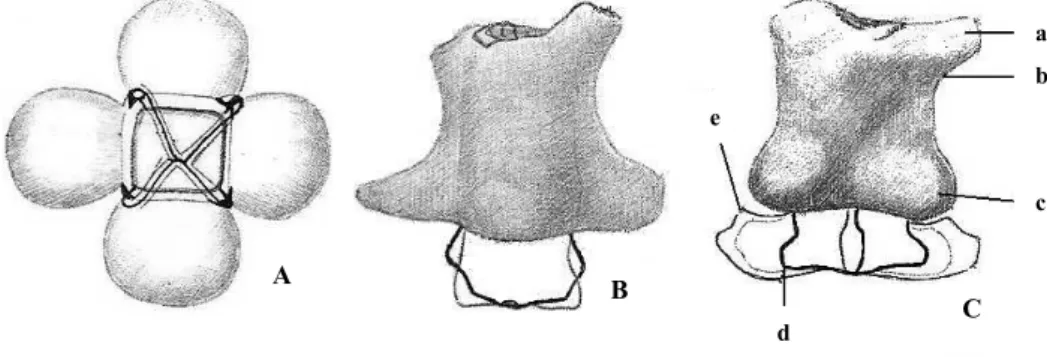
Fig. 26. Structure of chalk-gland in Limoniastrum monopetalum (A – gland observed in front view, without chalk mass; B – skeleton of gland, without accessory cells; C – a, external limit of cutinized frame that forms the edge of the internal chamber; b, orifice of the chamber in which basis gland opens; c, basis of chambers diverticula; e, extremity of free side of accessory cells) [VUILLEMIN, 1887]
A
B
C
a
b
c
In the species whose accessory cells are very well developed and partly sealed on their sides, like Limoniastrum guyonianum (Fig. 29), a cuticle sheet grows between them and bifurcates on their outer side, so that to prevent wall detachment. The accessory cells are connected with the epidermis and parenchyma cells, appearing as bridges connecting the leaf tissues with the gland; from this point of view, they behave like the basal cells of glandular hairs. The above-cited author considers the two anatomic structures, i.e. gland and hair, as homologous. The accessory cells would correspond to the foot, whereas the secreting cells to the head of a glandular hair, yet one that underwent an extreme shortening.
The surface section of glandular cells differs from that of the other walls due to its complete cutinization. The cutinized plate was best noticed on the front view of an epidermis. In Statice tatarica (Fig. 30), the depth of the chamber preceding the gland (which is almost as thick as the epidermis) and the plate are located at the level of inner side of this layer. After having treated the epidermis with a chlorine-iodine solution, the author viewed it as a violet lamella covered with yellow discs (representing glands). Each disc still leaves the impression of two dividing walls in a cross-like layout and other four walls in a rhombus-like layout. The surface is also divided into 4 triangles close to the middle and 4 neighboring trapezoids close to the borders.
Fig. 27.Statice latifolia. Epidermis, surface view (gl – chalk glands; h – hairs) [VUILLEMIN, 1887]
h gl
Fig. 28.Statice imbricata. Chalk-gland(a - cuticular network of the deep side of epidermis, continued in the proximity of a stoma; b – gland, in cross section, with 4 secretory cells and 2 accessory cells) [VUILLEMIN, 1887]
50
Fig. 29.Limoniastrum guyonianum (a – frame delimitating the
free surface of secretory cells; b – cutinized edges supporting the gland; c – projection of edges between accessory cells; d – orifice at whose basis the gland opens; e – external limit of cutinized frame constituting the limit of the chamber; f – basis of diverticula of the chamber; g – the most external segment of the accessory cells) [VUILLEMIN, 1887]
Fig. 30. Statice tatarica (a – orifice of excavation in the depth of which the gland opens; b – frame delimitating the free surface of secretory cells) [VUILLEMIN, 1887]
Conclusions
The salt glands (chalk-glands) of Plumbaginaceae represent striking structures involved in the excretion of calcium carbonate at the level of aerial organs (leaves, stems) of halophytes from arid and saline environments. According to our analysis, their secretion product has been evidenced about 20 years prior to their anatomical description. While many authors still consider that LICOPOLI (1866) was the first botanist who mentions them, it is by now obvious that, actually, METTENIUS (1856) did this prior to the Italian botanist. Indeed, Licopoli gave an extended and accurate description of them and his research could be considered as exclusively focused on the chalk-glands of Statice monopetala. As a matter of fact, all experiments developed by the plant anatomists of the 19th century in this direction were intense attempts at clarifying the structure and functions of these chalk-glands.
Acknowledgements
2
Henrietta Street, Covent Garden: 1279 pp.
CRONQUIST A. 1981. An integrated system of classification of flowering plants, second ed. New York: Columbia University Press, 1262 pp.
ENDLICHER S. 1836-1840. Genera plantarum secundum ordines naturales disposita, Vindobonae apud Fr. Beck Universitatis Bibliopolam, 1483 pp.
FRAINE E. de. 1916. The morphology and anatomy of the genus Statice as represented at Blakeney Point. I.
Statice binervosa G. E. Smith and S bellidifolia D.C. (= S reticulata). Ann. Bot. 30: 239-282. GRIGORE M. N. 2008. Introducere în halofitologie. Elemente de anatomie integrativă. Iaşi: Edit. Pim, 238 pp. GRIGORE M. N. 2012. Romanian Salt Tolerant Plants. Taxonomy and Ecology.Ia i: Edit. Tehnopress, 455 pp. GRIGORE M. N. & TOMA C. 2010. Structuri secretoare de săruri la halofite. O abordare integrativă. Bucure ti:
Edit. Academiei Române, 289 pp.
GRIGORE M. N., IVĂNESCU L. Ț TOMA C. 2014. Halophytes. An integrative anatomical study, Springer, Cham, Heidelberg, New York, Dordrecht, London: 548 pp.
JACKSON B. D. 1928. A glossary of botanic terms with their derivation and accent, fourth edition, Gerald Duckworth & CO. LTD., London, Hafner Publishing CO. INC., New York: 481 pp.
KUBITZKI K. 1993. Plumbaginaceae,In: KUBITZKI K., ROHWER J. G. & BITTRICH V.(eds.) The families and genera of vascular plants, vol. 2, Springer, Berlin, Germany: 523-530.
LLEDO M. D., CRESPO M. B., CAMERON K. M., FAY M. F. & CHASE M. W. 1998. Systematics of
Plumbaginaceae based upon cladistic analysis of rbcL sequence data. Systematic Bot. 23: 21-29. LLEDO M. D., KARIS P. O., CRESPO M. B., FAY M. F. & CHASE M. W. 2001. Phylogenetic position and
taxonomic status of the genus Aegialitis and subfamilies Staticoideae and Plumbaginoideae
(Plumbaginaceae): evidence from plastid DNA sequences and morphology. Plant Systematics and Evol. 229: 107-124.
LLEDO M. D., CRESPO M. B., FAY M. F. & CHASE M. W. 2005. Molecular phylogenetics of Limonium and related genera (Plumbaginaceae): biogeographical and systematic implications. Am. J. Bot. 92(7): 1189-1198.
LICOPOLI G. 1866. Richerche microscopiche sopra alcuni organi particolari della Statice monopetala. Sulla formazione di alcune organi nella Statice monopetaladestinati all’escrezione di sostanza minerale,
(Extract from) Ann. dell’ Acad. d. aspir. natural. di Napoli: 1-14.
LICOPOLI G. 1879. Gli stomi e le glandole delle piante. Atti dell’ R. Acad. d. Sci.Fis. e Mat. 8(5): 1-69. LINDLEY J. 1846. The vegetable kingdom; or, the structure, classification, and class of plants, illustrated upon
the natural system, London, Bradbury & Evans, Printers, Whitefriars: 908 pp.
LINCEVSKII I. A. & CERNIAKOVSKOI E. G. 1952. Plumbaginaceae, In: SHISHKIN B. K. & BOBROV E. G. (eds.) Flora SSSR,vol. 18, Izdatel’stvo Akademii Nauk SSSR, Moskva, Leningrad: 292-474. MAURY P. 1886. Etude sur l´organisation et distribution géographique des Plombaginacées. Ann. Sci. Nat., sér. 7,
Bot.4:1-134.
METCALFE C. R. & CHALK L. 1972. Anatomy of the Dicotyledons.Vol. 2,Oxford: Clarendon Press, 852–857. METTENIUS G. 1856. Filices Horti Botanici Lipsiensis. Die Farne Des Botanischen Gartens zu Leipzig, Leipzig:
von Leopold Voss Verlag, 135 pp.
MOORE D. M. 1972. Plumbaginaceae, In: TUTIN T. G., HEYWOOD V. H., BURGES N. A., MOORE D. M., VALENTINE D. H., WALTERS S. M. & WEBB D. A. (eds.), Flora Europaea, vol. 3 (Cambridge University Press: 29-51.
PAX F. 1897. Plumbaginaceae, In: ENGLER A. & PRANTL K. (eds.) Die natürlichen Pflanzenfamilien, 4(1-2), Leipzig, von Wilhelm Engelmann Verlag: 116-125.
RĂVĂRU M. 1960. Plumbaginaceae, In: SĂVULESCU T. (ed.), Flora R.P.R, vol. 7. Bucure ti: Edit. Academiei R. P. R., 21-40.
52
SHORT P. S. & WIGHTMAN G. M. 2011. Plumbaginaceae, In: SHORT P. S. & COWIE I. D. (eds.), Flora of the Darwin Region,vol. 1 Northern Territory Herbarium, Palmerston, Australia: 1-4.
SOLEREDER H. 1908. Systematic anatomy of the Dicotyledons. A handbook for laboratories of pure and applied Botany. Vol. 1, Clarendon Press Oxford: 644 pp.
STRASBURGER E., NOLL F., SCHENCK H. & SCHIMPER A. F. W. 1894. Lehrbuch der Botanik für Hochschulen, Jena, von Gustav Fischer Verlag: 710 pp.
TAKHTAJAN A. 2009. Flowering Plants (second ed.), Springer Science + Business Media B. V.: 871 pp. VOLKENS G. 1884. Die Kalkdrüsen der Plumbagineen. Ber. deutsch. bot. Ges. 2: 334-342.
VUILLEMIN P. 1887. Recherches sur quelques glandes épidermiques. Ann. Sci. Nat., sér. 7, Bot.5: 152-177. WORONIN M. 1885. Notiz über die Structur der Blätter von Statice monopetala L. Bot. Zeit. 43: 177-191.
How to cite this article:
GRIGORE M. N. & TOMA C. 2016. Structure of salt glands of Plumbaginaceae. Rediscovering old findings of the 19thCentury: ‘Mettenius’ or ‘Licopoli’ organs? J. Plant Develop. 23: 37-52.
![Fig. 1. Chalk glands (g) in the leaf of Goniolimon tataricum (A – cross section; B – surface view) [METTENIUS, 1856]](https://thumb-eu.123doks.com/thumbv2/123dok_br/18134610.325724/3.892.274.594.775.960/chalk-glands-goniolimon-tataricum-cross-section-surface-mettenius.webp)
![Fig. 2. Chalk glands (g) in the leaf of Limoniastrum monopetalum [METTENIUS, 1856]](https://thumb-eu.123doks.com/thumbv2/123dok_br/18134610.325724/4.892.183.720.212.649/fig-chalk-glands-g-leaf-limoniastrum-monopetalum-mettenius.webp)
![Fig. 6. Licopoli ‘organ’ in the lamina of Statice monopetala, front view (ap g - aperture of the gland; st – stomata) [LICOPOLI, 1866]](https://thumb-eu.123doks.com/thumbv2/123dok_br/18134610.325724/5.892.173.731.216.804/licopoli-lamina-statice-monopetala-aperture-gland-stomata-licopoli.webp)
![Fig. 13. Licopoli ‘organs’ (Lc o) at epidermis level of the lamina of Statice limonium (st – stomata) [ MAURY, 1886 ]Lc o](https://thumb-eu.123doks.com/thumbv2/123dok_br/18134610.325724/6.892.449.675.524.684/licopoli-organs-epidermis-lamina-statice-limonium-stomata-maury.webp)
![Fig. 15. Licopoli ‘organs’ (Lc o) at epidermis level of the lamina of Statice lychnidifolia (st – stomata) [ MAURY, 1886 ]](https://thumb-eu.123doks.com/thumbv2/123dok_br/18134610.325724/7.892.457.682.272.466/licopoli-organs-epidermis-lamina-statice-lychnidifolia-stomata-maury.webp)
![Fig. 19. Salt-secreting ‘apparatus’ (gl) of Statice pruinosa [VOLKENS, 1884]](https://thumb-eu.123doks.com/thumbv2/123dok_br/18134610.325724/9.892.180.715.213.628/fig-salt-secreting-apparatus-gl-statice-pruinosa-volkens.webp)
![Fig. 28. Statice imbricata. Chalk-gland (a - cuticular network of the deep side of epidermis, continued in the proximity of a stoma; b – gland, in cross section, with 4 secretory cells and 2 accessory cells) [VUILLEMIN, 1887]](https://thumb-eu.123doks.com/thumbv2/123dok_br/18134610.325724/13.892.249.627.789.968/imbricata-cuticular-epidermis-continued-proximity-secretory-accessory-vuillemin.webp)
![Fig. 30. Statice tatarica (a – orifice of excavation in the depth of which the gland opens; b – frame delimitating the free surface of secretory cells) [VUILLEMIN, 1887]](https://thumb-eu.123doks.com/thumbv2/123dok_br/18134610.325724/14.892.173.716.217.424/statice-tatarica-orifice-excavation-delimitating-surface-secretory-vuillemin.webp)
