Functional
Neuroanatomy
for Posture and
Gait Control
Kaoru Takakusaki
The Research Center for Brain Function and Medical Engineering, Asahikawa Medical University, Asahikawa, Japan
Received: December 13, 2016 Accepted: December 15, 2016
Corresponding author: Kaoru Takakusaki, MD, PhD, The Research Center for Brain Function and Medical Engineering, Asahikawa Medical University, 2-1, 1-1 Midorigaoka-Higashi, Asahikawa 078-8511, Japan
Tel: +81-166-68-2884 Fax: +81-166-68-2887 E-mail: [email protected]
ABSTRACT
Here we argue functional neuroanatomy for pos-ture-gait control. Multi-sensory information such as somatosensory, visual and vestibular sensation act on various areas of the brain so that adaptable pos-ture-gait control can be achieved. Automatic process of gait, which is steady-state stepping movements associating with postural reflexes including head-eye coordination accompanied by appropriate align-ment of body segalign-ments and optimal level of pos-tural muscle tone, is mediated by the descending pathways from the brainstem to the spinal cord. Par-ticularly, reticulospinal pathways arising from the lat-eral part of the mesopontine tegmentum and spinal locomotor network contribute to this process. On the other hand, walking in unfamiliar circumstance requires cognitive process of postural control, which depends on knowledges of self-body, such as body schema and body motion in space. The cognitive in-formation is produced at the temporoparietal asso-ciation cortex, and is fundamental to sustention of vertical posture and construction of motor programs. The programs in the motor cortical areas run to ex-ecute anticipatory postural adjustment that is opti-mal for achievement of goal-directed movements. The basal ganglia and cerebellum may affect both the automatic and cognitive processes of posture-gait control through reciprocal connections with the brainstem and cerebral cortex, respectively. Conse-quently, impairments in cognitive function by dam-ages in the cerebral cortex, basal ganglia and cere-bellum may disturb posture-gait control, resulting in falling.
Key Words
Multisensory information;
midbrain locomotor region; reticulospinal system; body schema; motor programs;
Parkinson’s disease. https://doi.org/10.14802/jmd.16062 / J Mov Disord 2017;10(1):1-17
pISSN 2005-940X / eISSN 2093-4939
J Mov Disord 2017;10(1):1-17
JMD
GENERAL SCHEMA OF
POSTURE-GAIT CONTROL
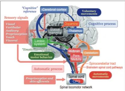
Figure 1 illustrates our recent understanding of basic signal lows involved in motor control. Senso-ry signals arising from external stimuli and/or inter-nal visceral information have various functions. For example, they are to be utilized for cognitive process-ing such as production of workprocess-ing memory which guides future behavior. Alternatively, they may afect emotional and arousal states. Sensory signals are fur-ther available to detect and correct postural instabili-ty by acting on the cerebral cortex, cerebellum, and brainstem. Accordingly, animal initiates movements depending on either a “cognitive reference” or an “emotional reference”.1,2
Voluntary movements are derived from intention-ally-elicited motor commands arising from the cere-bral cortex to the brainstem and spinal cord. On the other hand, emotional reference may contribute to emotional motor behavior which is generated by the projections from the limbic-hypothalamus to the brainstem, such as fight or flight reactions.1,3,4 Re-gardless of whether the initiation is volitional or
emotional, goal-directed behaviors are always ac-companied by automatic process of postural control including balance adjustment and muscle tone regu-lation. he subject is largely unaware of this process which is evoked by sequential activations of neurons in the brainstem and spinal cord. Basic locomotor motor pattern is generated by spinal locomotor net-works that is termed as the central pattern generators (CPG). However, in order to learn motor skills or behave in unfamiliar circumstance, the subject re-quires cognitive posture-gait control that depends on cognition of self-body information together with spatial localization of objects in extra-personal space. he cerebellum regulates the cognitive and auto-matic processes of posture-gait control by acting on the cerebral cortex via the thalamocortical projection and on the brainstem, respectively. Both the feed-forward information from the cerebral cortex via the cortico-ponto-cerebellar pathway and real-time sen-sory feedback via the spinocerebellar tract to the cer-ebellum may play major roles in these operations. he basal ganglia may also contribute to the modu-lation of each process though its gamma-aminobu-tyric acid (GABA)-ergic projections to the cerebral cortex and brainstem.2,5,6 he degree of GABAergic inluence from the basal ganglia is regulated by the midbrain dopaminergic neurons.7
BRAINSTEM AND SPINAL CORD;
CORE-STRUCTURES OF
POSTURE-GAIT CONTROL
In the absence of their forebrain, like a decere-brate cat, it can walk, trot and gallop. When the de-cerebration is made at precollicular-postmammilla-ry level, the cat initiates locomotion by electrical or chemical stimulation applied to the mesencephalic or midbrain locomotor region (MLR).1,8,9 However, if the neuraxis is transected slightly higher at the precollicular-premammillary level, cats can sponta-neously elicit locomotion with well-coordinated pos-tural control.10 herefore, a critical region exists be-tween these decerebrate levels. his area is recognized as the subthalamic locomotor region (SLR), which mostly corresponds to the lateral hypothalamic area. Stimulation of the SLR evoked locomotion after a large lesion was made in the MLR area,11 indicating that the SLR has direct connections with the brain-stem locomotor pathway beyond the MLR.
er, the walking in the decerebrate preparations is machine-like and is neither goal-directed nor adap-tive to the environment. Hence, the SLR connec-tions to the MLR are likely important for normal control of posture and gait.
So far three locomotor regions have been identi-fied in animals: the MLR in the mesopontine teg-mentum, the SLR and the cerebellar locomotor region (CLR) in the mid-part of the cerebellum.12 Human imaging study demonstrated that the organization of these supraspinal locomotor centers was preserved during the transition to bipedal locomotion human.13 he clinical relevance of these centers has so far been largely neglected.
Role of the mesencephalic area in the control
of posture and locomotion
The MLR appears to be present in all classes of vertebrates.14 It likely includes the cuneiform nucle-us (CNF) and the pedunculopontine tegmental nu-cleus (PPN), although the precise location of the lo-comotor regulation still remains a matter of debate. The PPN is located in the ventrolateral part of the caudal mesencephalic reticular formation, composed of a heterogeneous population of neurons, containing GABA and glutamate in addition to acetylcholine.15 Different neuronal types within the PPN area may have different functions with their own inter-nections to multiple parts of the brain. here are con-nections to cerebral cortex, multiple basal ganglia and limbic areas, the thalamus, the brainstem, the spinal cord and the cerebellum.16 This key location including multiple segregate functions, renders igur-ing out the precise function of these regions quite complicated.17
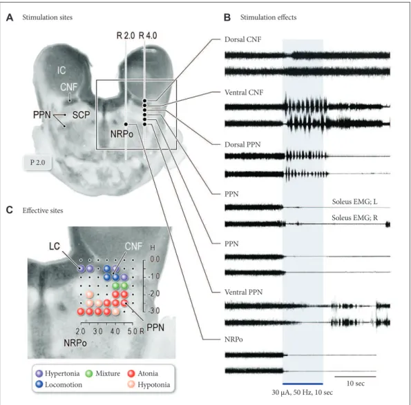
In the experiments using decerebrate cats, activa-tion of neurons in the PPN suppress muscle tone via its cholinergic projections to the pontine reticular formation (PRF), while activation of neurons in the CNF mostly elicits locomotion.5,15,18-20 Microstimu-lation of the transition zone between the two areas induced a mixture of locomotor rhythm with asso-ciated muscle tone suppression (Figure 2). More-over, blocking the PRF by injecting atropine sulfate, blocked the PPN-induced atonia but facilitated MLR-induced locomotion, indicating that cholinergic PPN neurons not only control the level of muscle tone but also modulate the locomotor pattern, and do this by efects at the pontine level.15,18 Studies in
rats by Sherman et al.21 show non-cholinergic neu-rons just medial to the PPN have projections to the spinal cord, while the cholinergic neurons of the PPN do not. his area at the mesopontine junction may be the true MLR. he dorsal neurons of this MLR area (laterodorsal tegmental nucleus) with spinal projec-tions are active in locomotion, while the ventral neu-rons are active in standing and do not have spinal pro-jections.
Functional organization of the reticular
formation in the control of posture
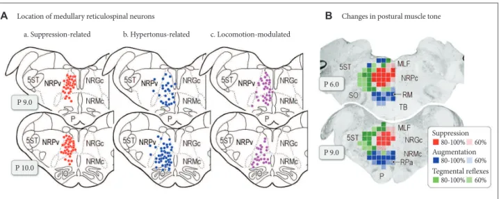
It is generally agreed that the reticulospinal tract (RST) contributes to regulation of the level of mus-cle tone. here may exist functional organization in the pontomedullary reticular formation (PMRF) in relation to the control of postural muscle tone (Fig-ure 3).15 Direct recording of reticulospinal neurons (RSNs) revealed that those located in the dorso-me-dial part of the PMRF are active during the period of muscle tone suppression or muscular atonia (Fig-ure 3Aa), and those in the ventromedial part are ac-tive during relex standing due to decerebrate rigidity or hypertonus (Figure 3Ab). Accordingly, functional topographical organization may exist in the PMRF in the control of postural muscle tone. On the other hand, during MLR-elicited locomotion or sponta-neously evoked locomotion in high-decerebrated preparation, RSNs located in both the dorsomedial and ventromedial PMRF were modulated in response to step cycles (Figure 3Ac), indicating that muscle tone-related RSNs participate in the execution of lo-comotion so that locomotor rhythm and muscle tone can be simultaneously regulated by the reticulospinal system during locomotion.
J Mov Disord 2017;10(1):1-17
JMD
characterized by extension of the unilateral limb and lexion of the contralateral limb, was evoked from the area between the inhibitory and excitatory areas and the lateral part of the PMRF where few RSNs arise in the cat (Figure 3B, green areas).
In addition to the RST, vestibulospinal tract (VST) plays an important role in the control of postural equi-librium by its similar architectonic organization of
de-scending ibers within the spinal cord with the RST.22,23 Matsuyama and Drew24,25 examined firing property of neurons in the RST and VST in the cat during loco-motion on an inclined surface. Specifically, the VST controls primarily the overall level of postural muscle tone, while the RST has an additional role in deter-mining the relative level of diferent muscles, particu-larly when the pattern is asymmetric.
Figure 2. Effects of midbrain stimulation on posture and locomotion in decerebrate cat preparation. A: Stimulation sites in the right mesopontine tegmentum. Stimulation consists of 30 μA in intensity and 50 Hz in frequency with a du -ration of 10 seconds. B: Effects of stimulation applied to each site in (A) on left and right soleus muscle electromyo-grams. Stimulation of the dorsal part of the CNF induced muscle tone augmentation. While stimulation of the ventral CNF and the dorsal PPN induced locomotor rhythm, the latter was accompanied by a decrease in muscle tone. Stim-ulation of the PPN and PRF corresponding to the nucleus reticularis pontis oralis (NRPo) immediately suppressed soleus muscle activities. C: Topography of stimulus effects in the mesopontine tegmentum. Locomotion was evoked by stimulating the CNF (blue). Stimulation of the locus coeruleus (LC) and dorsolateral CNF induced hypertonia (violet; muscle tone augmentation). Ventrolateral part of the PPN and NRPo, induced muscular atonia (red) and hypotonia (orange). Stimuli applied to the locomotion-evoking sites and atonia-evoking sites elicited a mixture of rhythmic limb movements and muscle tone suppression (green). Modified from Takakusaki et al. J Neural Transm (Vienna) 2016;123:695-729, with permission of Springer.15 CNF: cuneiform nucleus, PPN: pedunculopontine tegmental nucleus, IC: inferior colliculus, SCP: superior cerebellar peduncle.
A
C
B
Stimulation sites
Efective sites
Hypertonia Mixture Atonia Locomotion Hypotonia
Stimulation efects
Dorsal CNF
Ventral CNF
Dorsal PPN
PPN
PPN
Ventral PPN
NRPo
10 sec 30 μA, 50 Hz, 10 sec
Soleus EMG; L
Locomotor pathway and its control by the
forebrain structures
In the “locomotor pathway”, signals from the MLR also activate medullary RSNs, in turn commanding the spinal locomotor network to generate the oscil-latory pattern of locomotion.8,26 However, the SLR and CLR may also activate this reticulospinal loco-motor pathway through distinct and direct projec-tions.27 Signals from the MLR may also activate mono-aminergic descending pathways including the coerulo-spinal and raphecoerulo-spinal tracts, acting as a muscle tone excitatory system.28-30 hen we focus on the inputs of the forebrain structures to the midbrain MLR/PPN area. In decerebrate cats, the basal ganglia control locomotion and posture using diferent GABAergic output pathways of the substantia nigra reticulata (SNr); the lateral part of the SNr blocks the PPN-induced muscle tone suppression, whereas the me-dial part of the SNr suppresses the MLR-induced lo-comotion.18,20,31 Recent rat studies conirm that inhibi-tory input from the SNc (GABAergic and dopami-nergic) to ventral MLR regulate posture, while
in-hibitory projection from the GABAergic SNr to the dorsal MLR regulates locomotion.21 In Parkinson’s disease (PD), GABAergic outputs of the basal gan-glia are abnormally increased,7 so excessive SNr-in-hibition of the MLR may cause gait disturbance and muscle rigidity.5,18
However, it is unknown whether these mecha-nisms are the same for bipedal humans and quadru-pedal animals. It is also unclear what drives or dic-tates the SNr-induced control of movements. here are sub-compartments in the basal ganglia; neostri-atum-dorsal pallidal pathway (“dorsal pathway”, Figure 4A) and ventral striatum-ventral pallidal path-way (“ventral pathpath-way”, Figure 4B). he nucleus ac-cumbens, as a component of the limbic system, may be important in releasing locomotion, via GABAer-gic projections disinhibiting the MLR via the ventral pallidum32,33 and via ibers to the SNr (Figure 4B).34 Because the nucleus accumbens also receives inputs from the hippocampus and amygdala, the ventral pathway may be implicated in reward-oriented loco-motor behaviors, as it receives inputs from ventral
Figure 3. Functional organization of medullary reticulospinal systems in decerebrate cats. A: Locations of the medullary reticulospinal neu-rons relating to muscle tone suppression (a), muscle tone augmentation (hypertonus) (b), and locomotion (c). During relex standing of the decerebrate cats, reticulospinal neurons with a iring frequency more than 10 Hz during relex standing of decerebrate cats are judged as hypertonus-related reticulospinal neurons (b; n = 76). When carbachol (long-acting cholinomimetic agents) was injected into the pontine re-ticular formation muscle tone of decerebrate cats was abolished. Reticulospinal neurons of which iring frequency was increased to more than 10 Hz during carbachol-induced atonia are judged as atonia-related reticulospinal neurons (a; n = 75). During relex standing (decere -brate rigidity) these cells usually had no spontaneous iring. Locomotion-related neurons (n = 59) were judged as those displaying rhythmic iring relating to step cycles of locomotion. Recording was made in both high decerebrated cats which displayed spontaneous locomotion and normal decerebrated cats with stimulation of the MLR. B: Results obtained from ive animals are superimposed on representative coro -nal planes of the caudal pons and medulla. Sites from which either suppression (red), augmentation (blue), or tegmental relexes (green) was elicited in more than three out of ive animals are marked. Sites from which the stimulation induced postural changes in more than four ani -mals are indicated by darker colored squares; conversely, light colored squares indicate that the postural changes were induced in three ani -mals. Modiied from Takakusaki et al. J Neural Transm (Vienna) 2016;123:695-729, with permission of Springer.15 P: pyramidal tract, MLF: medi-al longitudinmedi-al fasciculus, 5ST: spinmedi-al trigeminmedi-al tract, NRPc: nucleus reticularis pontis caudmedi-alis, TB: trapezoid body, RM: nucleus raphe magnus, SO: superior olive, NRGc: nucleus reticularis gigantocellularis, NRMc: nucleus reticularis magnocellularis, RPa: nucleus raphe pallidus, NRPv: nucleus reticulars parvocellularis.
A Location of medullary reticulospinal neurons B Changes in postural muscle tone
a. Suppression-related b. Hypertonus-related c. Locomotion-modulated
P 9.0
P 10.0
P 9.0 P 6.0
Suppression
Augmentation
Tegmental relexes 80-100%
80-100%
80-100% 60%
60%
J Mov Disord 2017;10(1):1-17
JMD
tegmental area, hippocampus and amygdala. On the other hand, the more recently evolved parts of the basal ganglia make up the dorsal system (Figure 4A).35 hese parts may achieve locomotor control depend-ing on cognitive behavioral context, such as sensory-guided locomotor control.
Spinal control of posture and gait
Spinal circuitry involved in the stretch relex, re-ciprocal inhibition, non-rere-ciprocal inhibition (or au-togenic inhibition) and lexion relexes are involved in the control of posture. Particularly, stretch relex and non-reciprocal inhibition (Ib inhibition) play major role in static control of posture. On the other hand, interneuronal networks involved in reciprocal Ia
in-Figure 4. Neuronal mechanisms of cognitive (A) and emotional (B) control of locomotion in the cat. A: Dorsal sys-tem for cognitive locomotor control. A visuo-motor pathway from the visual cortex to motor cortex via the parietal cortex contributes to this control. Corticofugal projections act on to the basal ganglia nuclei, brainstem and spinal cord. Dopaminergic projection from the substantia nigra pars compacta (SNc) to the caudate-putamen (CPu) may be involved in learning the locomotor behaviors. GABAergic output from the basal ganglia nuclei (internal segment of the globus pallidus and substantia nigra pars reticulata; GPi/SNr) acts on MLR/PPN area may control locomo-tion and posture. Efferents from the midbrain locomotor region (MLR) recruit excitatory system, inhibitory system and locomotor pathway. The excitatory system arises from the LC and the raphe nuclei. The inhibitory system which arises from cholinergic neurons in the PPN sequentially activates PRF neurons, medullary reticulospinal neurons in the nucleus reticularis gigantocellularis (NRGc) and spinal inhibitory interneurons. The inhibitory interneurons may inhibit both motoneurons and interneurons. The locomotor pathway consists of reticulospinal neurons arising from the ventromedial medulla corresponding to the nucleus reticularis magnocellularis (NRMc). Cholinergic and gluta-matergic projections from the PPN excite SNc-DA neurons. These descending tracts act on CPGs in spinal cord so that muscle tone and locomotion are regulated. Efferents from the (CLR may excite locomotor pathway. B: Ventral system for emotional locomotor control. Efferents from the amygdala (AMD) and hippocampus (Hipp) project to the nucleus accumbens (NAc). GABAergic NAc neurons project to ventral pallidum (VP) and the SNr, which control activity of the MLR/PPN neurons. Efferents from the AMD and the Hipp also act on lateral hypothalamic area, which corresponds to the SLR. DA projections from the ventral tegmental area (VTA) may contribute to the reward-oriented locomotor behaviors. Modiied from Snijders et al. Ann Neurol 2016;80:644-659, with permission of Wi -ley.164 E: extensor motoneurons, F: lexor motoneurons, PRF: pontine reticular formation, PPN: pedunculopontine tegmental nucleus, LC: locus coeruleus, RN: raphe nuclei, DA: dopamine, CLR: cerebellar locomotor region, SLR: subthalamic locomotor region, CNF: cuneiform nucleus, CTX: cortex, GPe: external segment of the globus pallidus.
A
B
Inhibitory interneurons Inhibitory interneurons
Spinal locomotor network Spinal locomotor network
Excitatory system
Excitatory system
Inhibitory system
Inhibitory system Locomotor pathway
Locomotor pathway
Motoneurons Motoneurons
Muscles Muscles
hibition and lexion relexes including crossed-exten-sion relex and are critical to produce postural igures with extension-lexion movements of let-right leg alternation during walking. Integration of all spinal relex networks therefore can be essential to full exe-cution of muscle tone regulation during movement. While spinal relex networks generate rhythm and pattern of locomotor movements through the activa-tion of the CPG, they play crucial role in supporting body during stance phase of locomotion as well. Be-cause spinal preparations in quadruped animals do not express postural reflex described above, neural networks within the spinal cord alone does not enable to control postural equilibrium,36,37 and integration of descending supraspinal signals and peripheral senso-ry aferents at the level of spinal cord is necessasenso-ry for full execution of postural control.
Organization of the spinal locomotor network
Once animals start locomotion, muscle tone must be regulated depending on locomotor cycles. A par-ticular group of spinal interneuronal networks that generates rhythmic activity in the absence of rhyth-mic inputs is termed CPG.1,36,37 he rhythmic neuronal activity is sent to the second-order inter-neurons in the intermediate region (lamina IV-VII of Rexed), which shape “locomotor patterns” of each limb’s movements.26,38 he signals are then transmit-ted to the target motoneurons innervating ipsilateral limb muscles through their excitatory and inhibitory actions.26,36 Reciprocal Ia interneurons, classical Ib in-terneurons and Renshaw cells are likely included in this group.36 On the other hand, lamina VIII interneu-rons projecting to the contralateral side contribute to the let-right alternations of limb movements.39,40 he rhythm and pattern are transmitted back to the supra-spinal structures via the spinothalamic, spinoreticular and spinocerebellar tracts so that the supraspinal structures monitor all events in the spinal cord.36
Spinal control of muscle tone during locomotion
Activity of the spinal locomotor networks is mod-ulated by sensory afferents in a phase dependent manner.36,38,41,42 For example, proprioceptors in mus-cles at the hip joint are primarily responsible for reg-ulating the stance phase. Aferents from propriocep-tors in extensor muscles regulate transition from stance to swing phase. It should be critically noted that
signals in Ib aferents from tendon organ in ankle ex-tensor muscles inhibit homonymous motoneurons at rest, while they excite extensor motoneurons dur-ing stance phase.36,41 he functional consequence of this “relex reversal” is that the swing phase is not ini-tiated until the extensor muscles are unloaded and the forces exerted by these muscles are low.
Skin afferents also exert a powerful influence on the CPG.26,36,41 Skin receptors are important to detect obstacles and adjust stepping to avoid them such as the “stumble-corrective reaction.43 Importantly, the corrective flexion movements are produced only if the paw is stimulated during the swing phase. An identical stimulus applied during stance phase elicits the opposite response, an excitation of extensor mus-cles that reinforces the ongoing extensor activity. his is another example of the relex reversal. he relex reversal phenomenon is critically involved in pos-tural control during locomotion. However, its mech-anisms have not been elucidated.
HIGHER-ORDER REGULATION OF
POSTURE-GAIT CONTROL
Classical lesion studies in the cat
Even cerebral cortex was removed, the kitten can alive more than several years.44 hey could eat and exhibit periods of rest, become active, search for fool, and were able to remember the location of food. hey utilized the visual and haptic senses with respect to external space. However, in the adult cats, skilled locomotor performance was disturbed when lesions were made in the motor-related cortical regions.45 If the caudate nucleus of the cat is selectively removed both sides,46 a remarkable behavior develops referred to as the “compulsory approaching syndrome”. The cat faithfully followed any moving object that catches its attention, seemingly unable to terminate the loco-motor behavior. his was referred to as “visually-de-termined cortical automatism”.47 he main manifesta-tions consisted of loss of drive (apathy), obsessive-compulsive behavior, cognitive deficits, stimulus-bound perseverative behavior, and hyperactivity.46 On the other hand, removal of both the cerebral cortex and the striatum (diencephalic cat; the thalamus and hypothalamus were preserved) resulted in the cats walking incessantly, even though they did not attend to any environmental stimuli.48
J Mov Disord 2017;10(1):1-17
JMD
the limbic-hypothalamic structures, and the basal ganglia as well as the cerebellum control posture and gait largely by acting on the reticulospinal system through their direct and indirect connections via the MLR/PPN area (Figure 4). hese cortical and sub-cortical projections may enable animals to express volitional and emotional motor behaviors depend-ing on the context.2,14
Control by the cerebral cortex
While basic locomotor synergy was not largely disturbed if pyramidal tracts were bilaterally discon-nected,49 skilled locomotor task was severely impaired. Liddell and Phillips50 found ater unilateral or bilat-eral pyramidal lesions that the cats became ‘helplessly immobile’, unable to take a step without slipping or falling, when they were required to walk along a
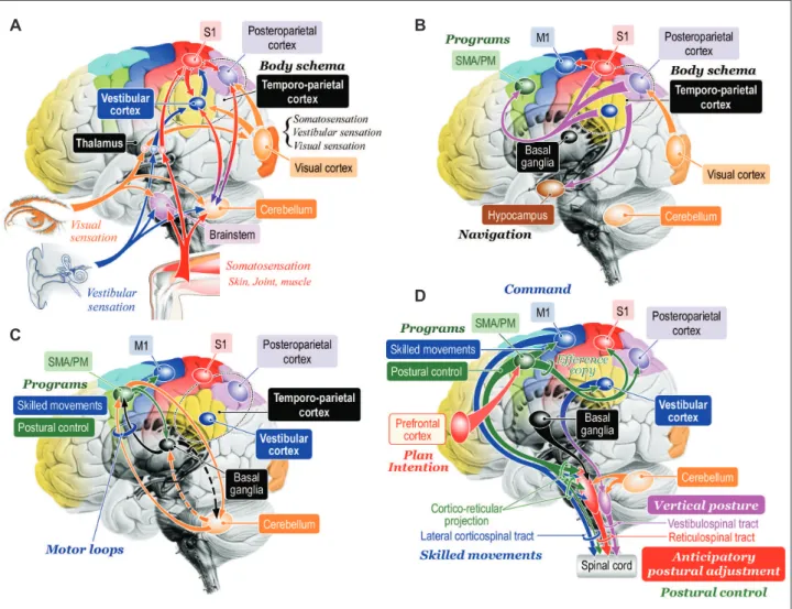
nar-Figure 5. Hypotheses of cognitive process of posture-gait control. A: Cognition of bodily information. Sensory signals lowing into the cen -tral nervous system converge to the brainstem, cerebellum, thalamus, and cerebral cortex. At the level of cerebral cortex, signals from the visual cortex, vestibular cortex and primary sensory cortex (S1) is integrated and internal model of self-body, such as body schema and verticality can be constructed at the temporoparietal cortex including the vestibular cortex and posteroparietal cortex. Reciprocal connection between the temporoparietal cortex and cerebellum may contribute to this process. B: Transmission of the bodily information. The bodil y information is then transmitted to the supplementary motor area (SMA) and premotor area (PM) where the information can be utilized as materials to produce motor programs. Similarly, the information is transferred to hippocampus and is used to navigate further behaviors. C: Motor programming. The motor cortical areas closely cooperate with the basal ganglia and cerebellum so that appropriate motor programs are constructed. D: Postural control by corticofugal projections to the brainstem and spinal cord. The bodily information generated at the ves-tibular cortex may be utilized for sustention of vertical posture via cortico-vesves-tibular and vestibulospinal tract. Signals from the prefrontal cor-tex including plans and intentions may trigger to run motor programs in the SMA/PM, which may include those for purposeful movements and associating postural control. The postural control program may be utilized to generate anticipatory postural adjustment via cortico-reticu-lar and reticulospinal tract. Then motor programs are sent to the M1 so that goal-directed purposeful skilled movements can be achieved. Modiied from http://dx.doi.org/10.1080/01691864.2016.1252690, with permisson of Taylor & Francis.165
A
C
B
row beam or horizontal ladder. Such a skilled perfor-mance became more severely damaged by postcruci-ate than by precrucipostcruci-ate lesions. After postcrucipostcruci-ate lesions including both the somatosensory and parietal cortices, the cat refused to walk on narrow trucks.45 he precruciate area, which corresponds to supple-mentary motor area (SMA) and premotor area (PM) of the primates, may be involved in movement initia-tion. On the other hand, the postcruciate cortices may utilize speciic somatosensory inputs to fulil a role in the regulation of ongoing movements51 in the manner of anticipatory or feed-forward adjustments.52 Skilled posture-gait control, therefore, can be achieved on the basis of knowledges of the orientation and motion of the body in space as well as motion perception and spatial localization of objects in extra-personal space.53-55 Such a knowledge is provided by integration of ves-tibular, somatosensory and visual sensory signals which occurs at both the cerebral cortex and cerebel-lum (Figure 5A).56
Precise visuomotor coordination occurred at the cerebral cortex plays critical roles in the execution of precise arm-hand movements such as reaching and grasping.57 Similarly, when a walking subject encoun-ters obstacles, each leg must be placed with a high de-gree of accuracy through the visuo-parieto-frontal cortical projection, as in the subject has to modify the leg trajectory in each step in order to achieve appro-priate foot placement.58 Such a visuomotor coordi-nation is particularly necessary in quadrupeds be-cause an obstacle is no longer within the visual ield by the time the hindlimbs are stepping over it. When the posterior parietal cortex was bilaterally removed, the cat’s hindlimbs did not step over the obstacles if their forelimbs cleared them.59 herefore, the poste-rior parietal cortex must be engaged to register and store the temporospatial relationship between the ob-stacle and one’s bodily information, such as body sche-ma, in short-term memory that is utilized to produce motor programs in the motor cortices (Figure 5B) so that the cat can precisely modify the limb trajecto-ry.59,60 To successfully achieve such an accurate control of limb movements during walking, posture must be optimized in advance to the purposeful action so that bodily equilibrium can be maintained. herefore, the visuo-parieto-frontal cortical projection (Figure 5B) is critically involved in the fulilment of ongoing pur-poseful control via anticipatory adjustments of pos-ture.61 It follows that both the intentional limb
move-ments and anticipatory postural adjustment are programmed at motor cortical areas (Figure 5B and D).
Anticipatory postural adjustment
hen, what part of the motor cortex contributes to the programming of the anticipatory postural ad-justments? One of the most candidate areas is the SMA and PM (Figure 5B and C). In bipedal walk-ing monkey, an inactivation of the leg area of the primary motor cortex (M1) by injecting muscimol (GABAA agonist) partly paralyzed the contralateral leg.62 On the other hand, muscimol injections into the trunk/leg regions of the bilateral SMA largely disturbed postural control without inducing paraly-sis.63 When it was injected into the dorsal PM, spon-taneous walking was maintained; however, the mon-key could not start walking using sensory cue. hese indings indicate the SMA and PM may contribute to postural control during bipedal walking and ini-tiation of gait, respectively.
Studies using neural tracers have demonstrated abundant cortico-fugal projections to the PMRF from the premotor cortices (SMA/PM) in quadruped64 and biped65 animals. Recent studies have focused onto the importance of cortico-pedunculopontine projection in terms of motor control. Probabilistic dif-fusion tractography in rhesus monkey as well as hu-mans, shows that the SMA is strongly connected to the lateral PPN, while the dorsal PM is connected to the medial PPN.66 he RST innervates whole spinal segments22 so that it controls postural muscle tone and symmetric postural igures (Figure 3).15 herefore, the cortico-reticular and RST may achieve anticipatory postural adjustment (Figure 5D) Possibly, the SMA contributes to the anticipatory postural adjustment for step initiation, which is impaired in PD patients.67 On the other hand, the PM/SMA may forward programs of precise leg-foot movement to the M1,68 which, in turn, sends motor command via the corticospinal tract.
J Mov Disord 2017;10(1):1-17
JMD
are at higher risk of falling particularly when more cognitive tasks are required.69,70
Maintenance of vertical posture
Next critical question as to the cortical control of posture is “how does the brain acquire access to an internal estimate of body motion and postural ver-ticality?” Postural vertical is supported by a sense of verticality which is synthesized by visual, somato-sensory and vestibular information.71-75 Among them, the vestibular sensation is superior to others in terms of absoluteness of sensation because it always refers the gravity,76 and the vestibular system provides the brain with sensory signals about three-dimensional head rotations and translations. Vestibulothalamic projections are bilateral and mainly involve the pos-terior thalamus.77-79 While there was no cortical area that receives inputs exclusively from vestibular afer-ents, there are multiple presentations of vestibular in-formation in the cerebral cortex,80 such as the frontal eye ield, PM, somatosensory cortex, ventral intra-parietal cortex, medial superior temporal area, and parieto-insular vestibular cortex (PIVC). he PIVC has particularly dense connections with other vestib-ular-relating cortical areas, and receives information from other sensory modalities.81-83 Now, both the posterior thalamus and PIVC are areas of interest for the internal model of postural verticality.84-88 Because the PIVC has descending projections to the contralat-eral vestibular nuclei,79,89-91 the vestibular cortical sys-tems possibly contribute to upright standing by acting to the vestibulospinal system based on the internal model of postural verticality (Figure 5B).
Postural verticality is oten disturbed in pathological conditions such as “pusher syndrome” ater stroke and “Pisa syndrome” in advanced PD. Stroke patients with pusher syndrome actively push away from the ipsile-sional side and have a tendency to fall towards their paretic, contralesional side (the let side for right-hemi-sphere patients). hey had lesions including the pari-etal insular cortex or posterior thalamus.84,87,88,92 his phenomenon is more prominent when patients are upright compared to when lying down. Now the pusher phenomenon can be arising from a conlict or mismatch between visual and postural vertical.84,87,88 However, it is still uncertain whether the same patho-physiological mechanism is operating in Pisa syn-drome.
Posture-gait control by the cerebellum
Postural control by the cerebellum highly depends on sensory aferents. Signals from the labyrinth as-cend the vestibular nerve to the loccules and ver-mis of the cerebellum in addition to the vestibular nuclei. he fastigial nuclei (FN) receive a copy of the output of the spinal cord in addition to peripheral sensory information via spinocerebellar tracts.93 he FN also receive visual94 and vestibular95 information. These multisensory features may provide “an error-correction mechanism”, which permits FN neurons to afect motor functions such as coordinating postural responses during walking which entail changes in limb position. he FN may therefore send highly in-tegrated bodily information to the posture-gait relat-ed areas in the brainstem and motor cortical areas.96Action on the brainstem structures
Output from the FN to the brainstem contributes to the control of postural muscle tone. Electrical stimula-tion applied to the mid-part of the cerebellar white matter in decerebrate cats either increased97 or reduced the level of muscle tone.98 he cerebellar stimulation possibly activated the excitatory RST and VST of both sides so that extensor muscle tone was bilater-ally increased. On the other hand, the decrease in the level of muscle tone is considered to be due to with-drawal of excitatory inluence upon motoneurons.98 Because, postural muscle tone is generally reduced by the damage of the medial part of the cerebellum, the cerebellum contributes to the activation of antigravi-ty muscles. Possibly, the FN regulates posture-gait subprograms in the brainstem and spinal cord by in-parallel activation of fastigio-spinal, fastigio-reticular, and fastigio-vestibular pathways.27,99,100 herefore, the deiciency in these pathways may reduce the degree of α–γ linkage in patients with cerebellar diseases, resulting in hypotonia. he hypotonia state reduces the accuracy of the sensory feedback so that posture-gait control can be seriously disturbed.
Cerebellar actions on the cerebral cortex
The FN in the cerebellum, as well as the vestibular cortex (PIVC), is critically involved in encoding inter-nal postural model in space and self-motion.56 Some studies have suggested the presence of FN projec-tions to the parietal cortex,102,103 motor cortex and multimodal visual areas.104 Reciprocal connection between parietal cortex and the cerebellum may be involved in perception of body motion in space (Fig-ure 5B). Such a bodily information can be utilized to maintain upright posture during standing and to achieve anticipatory postural adjustment. he latter may involve reciprocal connections between the mo-tor cortical area (SMA/PM) and cerebellum (momo-tor loop) in order to construct motor programs (Figure 5C).105,106 Accordingly, cerebellar disease patients may have problems in cognitive process of postural con-trol. However, the perception of verticality in patients with cerebellar ataxia may only deteriorate in a more advanced stage of the disease.107 In addition, only few abnormalities of anticipatory postural adjust-ment were found in the cerebellar disease patients compared to controls, while the patients appeared to be less able to use predictive information.108 Because the cerebellum is reciprocally connected with the basal ganglia,109 it is possible that basal ganglia in addition to the cerebral cortex may complement the cerebellar role of cognitive process of postural control.
Posture-gait control by the basal ganglia in
relation to PD
Because posture-gait control is seriously impaired in PD, the basal ganglia has long been functionally regarded to be predominantly involved in motor con-trol but is increasingly recognized to play additional roles in sensory processing, cognition, and behav-ior.110-112 Here, emphasis has been placed on the me-chanisms of posture-gait impairment in PD so that understanding the role of basal ganglia to the pos-tural control is facilitated. Based on our recent un-derstanding, postural disturbances in PD attribute to following mechanisms: 1) disturbances in the dopa-minergic and cholinergic systems, 2) impairment of cognitive functions due to failure of integrative sen-sory processing that allows to produce internal pos-tural model (body schema), 3) failure in motor pro-gramming due to reduced activity in the motor cor-tical areas, and 4) disturbances in posture-gait areas in the brainstem.15,18,113
Disturbances in the dopaminergic and cholinergic systems
Recent clinical studies suggest that the postural impairments in PD is based on dysfunction of both the dopaminergic and cholinergic systems. Distur-bances in these chemospeciic systems may critically contribute to posture-gait failure in this disease. For example, a damage of dopamine (DA) neurons in the substantia nigra pars compacta (SNc), which project to the basal ganglia nuclei, is considered to increase in the GABAergic inhibitory output from the basal gan-glia.7,114,115 his may strongly inhibit thalamocortical systems and brainstem structures (Figure 1).
In addition, cholinergic neurons in the PPN (brain-stem) and basal forebrain (BF) are seriously dam-aged in PD.116-119 Indeed, a reduction of the thalamic cholinergic innervation in patients with PD has no cognitive and motor impairments but exhibits an in-crease in postural sway speed.120 Cholinergic PPN neurons project to the non-speciic thalamocortical system,121-123 basal ganglia nuclei including DA neu-rons in the SNc and PMRF.124-126 On the other hand, cholinergic BF projections to the cerebral cortex are necessary for attentional performance and cognitive processing.127
herefore, disturbances in attention, sensori-mo-tor integration and cognitive processing in PD can be largely attributed to the damage of the cholinergic systems. Accordingly, both of the excessive inhibi-tion from the basal ganglia and the damage of cho-linergic systems may impair both the cortical and subcortical, particularly the brainstem, functions.
Impairments in sensory processing and cognition
J Mov Disord 2017;10(1):1-17
JMD
and vestibular graviception,133 may result in deicits of internal model of postural verticality which is pos-sibly constructed at the temporoparietal association area including the vestibular cortex. herefore, asym-metry in the activity of the let and right vestibular cortices may induce leaning upright posture, which is oten called as Pisa syndrome. Pisa syndrome is a dystonic laterolexion of the trunk with a postural dis-turbance resembling the leaning tower of Pisa, and is more often observed in patients with advanced PD.134 he marked laterolexion become worsen dur-ing walkdur-ing but is almost completely alleviated by passive mobilization or supine positioning.135 Be-cause PD without Pisa syndrome also had deicien-cies in postural verticality compared to healthy con-trols,132,136-138 mismatch between proprioception and vestibular gravitation in PD may alter subjective postural verticality, resulting in Pisa syndrome. Al-ternatively, asymmetry of the basal ganglia output, which is due to cholinergic-dopaminergic imbalance in the striatum134,139,140 or disturbance of the pallidal output,141,142 may also elicit let-right disproportion of the thalamocortical processing of vestibular infor-mation.
Failure in motor programming
he motor cortical areas including the M1, SMA, and PM have connections with the basal ganglia and cerebellum, constituting motor loop that contributes to execution and motor programming of voluntary movements (Figure 5C).105,106 Because of increasing inhibitory output from the basal ganglia to the thal-amocortical projections in addition to reduced cog-nitive information processing, the capability of pro-ducing motor programs in response to changes in circumstance can be deteriorated. In fact, the SMA contributes to the anticipatory postural adjustment for step initiation via corticofugal projections to the PPN and PMRF, and this process is seriously im-paired in PD patients.67 Also, the dorsal part of the PM is involved in sensory-guided gait control as suggested in bipedally walking monkey.62 Because the activity of dorsal part of the PM was increased during visually-guided paradoxical gait in PD, pos-ture-gait programs in SMA/PM became reusable by the activation of visuo-motor pathway.129 According-ly, failure in motor programming in PD can be due to the decrease in excitability of the motor cortical ar-eas in addition to impairment of sensory processing
in the temporoparietal cortices. Recently, role of the cerebellum in the pathophysiology of PD is highly recognized.143,144 Because the cerebellum has recip-rocal connections with the basal ganglia (Figure 5C) in addition to the cerebral cortex and brainstem, there is a need to elucidate whether the cerebellum partici-pates in compensatory mechanisms associated with the disease or contributes to the pathophysiology of PD.
Reduced activity in posture-gait area in the midbrain
We propose that reduced excitability in the meso-pontine tegmentum including the PPN/MLR can be also involved in posture-gait failure in PD.6,15,18,20,113 In decerebrate cats, the basal ganglia control locomo-tion and posture using diferent GABAergic output pathways of the SNr; the lateral part of the SNr blocks the PPN-induced muscle tone suppression, whereas the medial part of the SNr suppresses the MLR-in-duced locomotion.18 Recent studies in rodents con-irm that inhibitory input from the SNr to the glutama-tergic neurons in the MLR regulates locomotion.21,145 In PD, GABAergic outputs of the basal ganglia are abnormally increased,7,114 so excessive SNr-inhibition of the MLR may cause gait disturbance and muscle rigidity acting on MLR and muscle tone inhibitory region in the PPN.18 Muscle tone rigidity in PD is called as “lead-pipe like rigidity” which is character-ized by co-contraction of extensor and lexor muscles. Such a co-contraction is observed in neck, back and leg muscles, resulting in lexion posture including cam-ptocormia.146
PD patients with cholinergic cell loss in the PPN showed more severe motor disabilities with gait and posture, which were associated with L-3,4-dihy-droxyphenylalanine (L-DOPA)-resistant akinesia.147,148 Subsequent post-mortem study in PD patients estab-lished a correlation between the occurrence of falls and freezing and the loss of cholinergic PPN neurons. However, the degree of neuronal loss in the CNF was not signiicantly diferent between fallers and non-fallers in PD patients.147 In PD patients, individual neurons in the dorsal PPN increased their firing rates with increased stepping frequency.149 Moreover, gait speed in PD patients was correlated with a power of alpha-oscillations (7–10 Hz) of field potentials re-corded from the PPN area.150
gait-posture deficiency in PD, deep brain stimulation (DBS) targeting the PPN with the aim of stimulating remaining cholinergic neurons.151-155 he irst studies using DBS in advanced PD patients concluded that low-frequency stimulation of the PPN could be ef-fective to control freezing of gait and falls. However, further clinical studies concluded that freezing of gait were mildly improved by PPN-DBS but some results were rather disappointing.156,157 hese results empha-size the need to determine the optimal surgical tar-get.158,159 Ferraye et al.156 suggest that the most suit-able targets are located slightly posterior to the PPN pars compacta, probably in the ventral part of the CNF where stimulation-induced locomotion has been re-ported in animals.18 his area possibly corresponds to the subcuneiform nucleus as described by Alam et al.160 and Karachi et al.161 also suggest that it may be the case that treating PD patients sufering from failure of gait initiation versus falling may require speciically targeting the CNF and the dorsal part of the PPN, respectively.
Reorganization of cortico-cerebello-brainstem pathways in PD
In human, Fling et al.162 used functional neuroimag-ing approach and revealed strong functional connec-tivity between the SMA and PPN/MLR area, which was positively correlated with freezing severity in pa-tients of PD. In contrast, connectivity between the STN and SMA was lost. hey suggested that the for-mer connectivity may potentially due to a maladap-tive compensation, and the latter may relect the re-duced automatic control of gait by the basal ganglia. A study using difusion tensor imaging revealed the connection between the cerebellum and the PPN in PD patients without freezing of gait. However, freezers of patients in PD showed the absence of cerebelloteg-mental connectivity and increased visibility of the de-cussation of corticopontine fibers in the anterior pons.163 These findings highlight the importance of corticopontine-cerebellar pathways in the pathophysi-ology of gait when the cerebellotegmental connec-tion that may contribute to automatic execuconnec-tion of gait control is damaged in freezers of PD.
Conflicts of Interest
he author has no inancial conlicts of interest.
Acknowledgments
his work is partially supported by grants from JSPS
KAKEN-HI (Grant Numbers 26120004 and 25290001) and from Japan Agency for Medical Research and Development (AMED) for K.T. KT is also supported by granting foundations from the QOLER Medical Group and Sasson Hospital.
REFERENCES
1. Grillner S. Control of locomotion in bipeds, tetrapods, and fish. In: Brookhart JM, Mountcastle VB, editors. Handbook of physiology. he nervous system II. Bethes-da, MD: American Physiological Society, 1981;1179-1236. 2. Takakusaki K. Forebrain control of locomotor behaviors.
Brain Res Rev 2008;57:192-198.
3. Sinnamon HM. Preoptic and hypothalamic neurons and the initiation of locomotion in the anesthetized rat. Prog Neurobiol 1993;41:323-344.
4. Takakusaki K, Takahashi K, Saitoh K, Harada H, Okumu-ra T, Kayama Y, et al. Orexinergic projections to the cat midbrain mediate alternation of emotional behavioural states from locomotion to cataplexy. J Physiol 2005;568(Pt 3):1003-1020.
5. Takakusaki K, Tomita N, Yano M. Substrates for normal gait and pathophysiology of gait disturbances with respect to the basal ganglia dysfunction. J Neurol 2008;255 Suppl 4:19-29.
6. Takakusaki K. Neurophysiology of gait: from the spinal cord to the frontal lobe. Mov Disord 2013;28:1483-1491. 7. DeLong MR, Wichmann T. Circuits and circuit disorders
of the basal ganglia. Arch Neurol 2007;64:20-24. 8. Armstrong DM. Supraspinal contributions to the
initia-tion and control of locomoinitia-tion in the cat. Prog Neurobiol 1986;26:273-361.
9. Mori S. Integration of posture and locomotion in acute decerebrate cats and in awake, freely moving cats. Prog Neurobiol 1987;28:161-195.
10. Hinsey JC, Ranson SW, McNattin RF. he role of the hy-pothalamus and mesencephalon in locomotion. Arch Neur-Psych 1930;23:1-43.
11. Shik ML, Orlovsky GN. Neurophysiology of locomotor automatism. Physiol Rev 1976;56:465-501.
12. Mori S, Matsui T, Kuze B, Asanome M, Nakajima K, Mat-suyama K. Stimulation of a restricted region in the mid-line cerebellar white matter evokes coordinated quadru-pedal locomotion in the decerebrate cat. J Neurophysiol 1999;82:290-300.
13. Jahn K, Deutschländer A, Stephan T, Kalla R, Wiesmann M, Strupp M, et al. Imaging human supraspinal locomotor centers in brainstem and cerebellum. Neuroimage 2008; 39:786-792.
14. Grillner S, Georgopoulos AP, Jordan LM. Selection and initiation of motor behavior. In: Stein PSG, Grillner S, Selverston AI, Stuart DG, editors. Neurons, networks, and motor behavior. Cambridge, MA: he MIT Press, 1997;3-19.
15. Takakusaki K, Chiba R, Nozu T, Okumura T. Brainstem control of locomotion and muscle tone with special refer-ence to the role of the mesopontine tegmentum and med-ullary reticulospinal systems. J Neural Transm (Vienna) 2016;123:695-729.
16. Mena-Segovia J. Structural and functional considerations of the cholinergic brainstem. J Neural Transm (Vienna) 2016;123:731-736.
J Mov Disord 2017;10(1):1-17
JMD
Circuits 2015;9:68.
18. Takakusaki K, Habaguchi T, Ohtinata-Sugimoto J, Saitoh K, Sakamoto T. Basal ganglia efferents to the brainstem centers controlling postural muscle tone and locomotion: a new concept for understanding motor disorders in basal ganglia dysfunction. Neuroscience 2003;119:293-308. 19. Takakusaki K, Habaguchi T, Saitoh K, Kohyama J.
Chang-es in the excitability of hindlimb motoneurons during muscular atonia induced by stimulating the pedunculo-pontine tegmental nucleus in cats. Neuroscience 2004;124: 467-480.
20. Takakusaki K, Obara K, Nozu T, Okumura T. Modulatory effects of the GABAergic basal ganglia neurons on the PPN and the muscle tone inhibitory system in cats. Arch Ital Biol 2011;149:385-405.
21. Sherman D, Fuller PM, Marcus J, Yu J, Zhang P, Cham-berlin NL, et al. Anatomical location of the mesencephalic locomotor region and its possible role in locomotion, pos-ture, cataplexy, and parkinsonism. Front Neurol 2015;6: 140.
22. Kuze B, Matsuyama K, Matsui T, Miyata H, Mori S. Seg-ment-speciic branching patterns of single vestibulospinal tract axons arising from the lateral vestibular nucleus in the cat: a PHA-L tracing study. J Comp Neurol 1999;414: 80-96.
23. Matsuyama K, Takakusaki K, Nakajima K, Mori S. Multi-segmental innervation of single pontine reticulospinal ax-ons in the cervico-thoracic region of the cat: anterograde PHA-L tracing study. J Comp Neurol 1997;377:234-250. 24. Matsuyama K, Drew T. Vestibulospinal and reticulospinal
neuronal activity during locomotion in the intact cat. I. Walking on a level surface. J Neurophysiol 2000;84:2237-2256.
25. Matsuyama K, Drew T. Vestibulospinal and reticulospinal neuronal activity during locomotion in the intact cat. II. Walking on an inclined plane. J Neurophysiol 2000;84: 2257-2276.
26. Rossignol S. Neural control of stereotypic limb move-ments. In: Rowell LB, Sheperd JT, editors. Handbook of physiology, section 12. Exercise: regulation and integra-tion of multiple systems. New York, NY: Oxford Universi-ty Press, 1996;173-216.
27. Mori S, Matsui T, Kuze B, Asanome M, Nakajima K, Mat-suyama K. Cerebellar-induced locomotion: reticulospinal control of spinal rhythm generating mechanism in cats. Ann N Y Acad Sci 1998;860:94-105.
28. Fung SJ, Barnes CD. Evidence of facilitatory coerulospinal action in lumbar motoneurons of cats. Brain Res 1981;216: 299-311.
29. Mori S, Kawahara K, Sakamoto T, Aoki M, Tomiyama T. Setting and resetting of level of postural muscle tone in decerebrate cat by stimulation of brain stem. J Neuro-physiol 1982;48:737-748.
30. Sakai M, Matsunaga M, Kubota A, Yamanishi Y, Nishiza-wa Y. Reduction in excessive muscle tone by selective de-pletion of serotonin in intercollicularly decerebrated rats. Brain Res 2000;860:104-111.
31. Takakusaki K, Saitoh K, Harada H, Okumura T, Sakamoto T. Evidence for a role of basal ganglia in the regulation of rapid eye movement sleep by electrical and chemical stim-ulation for the pedunculopontine tegmental nucleus and the substantia nigra pars reticulata in decerebrate cats. Neuroscience 2004;124:207-220.
32. Sławińska U, Kasicki S. heta-like rhythm in depth EEG activity of hypothalamic areas during spontaneous or
elec-trically induced locomotion in the rat. Brain Res 1995;678: 117-126.
33. Swanson LW, Mogenson GJ. Neural mechanisms for the functional coupling of autonomic, endocrine and somato-motor responses in adaptive behavior. Brain Res 1981;228: 1-34.
34. Lynd-Balta E, Haber SN. Primate striatonigral projections: a comparison of the sensorimotor-related striatum and the ventral striatum. J Comp Neurol 1994;345:562-578. 35. Robertson B, Kardamakis A, Capantini L,
Pérez-Fernán-dez J, Suryanarayana SM, Wallén P, et al. he lamprey blue-print of the mammalian nervous system. Prog Brain Res 2014;212:337-349.
36. Rossignol S, Dubuc R, Gossard JP. Dynamic sensorimotor interactions in locomotion. Physiol Rev 2006;86:89-154. 37. Rossignol S, Barrière G, Frigon A, Barthélemy D, Bouyer
L, Provencher J, et al. Plasticity of locomotor sensorimotor interactions ater peripheral and/or spinal lesions. Brain Res Rev 2008;57:228-240.
38. McCrea DA, Rybak IA. Organization of mammalian loco-motor rhythm and pattern generation. Brain Res Rev 2008; 57:134-46.
39. Jankowska E. Interneuronal relay in spinal pathways from proprioceptors. Prog Neurobiol 1992;38:335-378. 40. Matsuyama K, Takakusaki K. Organizing principles of
ax-onal projections of the long descending reticulospinal pathway and its target spinal lamina VIII commissural neurons: with special reference to the locomotor function. In: Westland TB, Calton RN, editors. Handbook on White Matter: Structure, Function and Changes. New York, NY: Nova Science Publishing, 2009:335-356.
41. Pearson KG. Generating the walking gait: role of sensory feedback. Prog Brain Res 2004;143:123-129.
42. Frigon A, Sirois J, Gossard JP. Effects of ankle and hip muscle aferent inputs on rhythm generation during ic-tive locomotion. J Neurophysiol 2010;103:1591-1605. 43. Forssberg H. Stumbling corrective reaction: a
phase-de-pendent compensatory reaction during locomotion. J Neu-rophysiol 1979;42:936-953.
44. Bjursten LM, Norrsell K, Norrsell U. Behavioural reperto-ry of cats without cerebral cortex from infancy. Exp Brain Res 1976;25:115-130.
45. Adkins RJ, Cegnar MR, Rafuse DD. Diferential efects of lesions of the anterior and posterior sigmoid gyri in cats. Brain Res 1971;30:411-414.
46. Villablanca JR. Why do we have a caudate nucleus? Acta Neurobiol Exp (Wars) 2010;70:95-105.
47. Denny-Brown D. The midbrain and motor integration. Proc R Soc Med 1962;55:527-538.
48. Villablanca J, Marcus R. Sleep-wakefulness, EEG and be-havioral studies of chronic cats without neocortex and stri-atum: the ‘diencephalic’ cat. Arch Ital Biol 1972;110:348-382.
49. Eidelberg E, Yu J. Effects of corticospinal lesions upon treadmill locomotion by cats. Exp Brain Res 1981;43:101-103.
50. Liddell EGT, Phillips CG. Pyramidal section in the cat. Brain 1944;67:1-9.
51. Brooks VB, Stoney SD Jr. Motor mechanisms: the role of the pyramidal system in motor control. Annu Rev Physiol 1971;33:337-392.
52. Vicario DS, Martin JH, Ghez C. Specialized subregions in the cat motor cortex: a single unit analysis in the behaving animal. Exp Brain Res 1983;51:351-367.
human upright stance. Exp Brain Res 2006;171:231-250. 54. Mergner T, Becker W. A modeling approach to the human
spatial orientation system. Ann N Y Acad Sci 2003;1004: 303-315.
55. Horak FB, Shupert CL, Dietz V, Horstmann G. Vestibular and somatosensory contributions to responses to head and body displacements in stance. Exp Brain Res 1994; 100:93-106.
56. Shaikh AG, Meng H, Angelaki DE. Multiple reference frames for motion in the primate cerebellum. J Neurosci 2004;24:4491-4497.
57. Kravitz DJ, Saleem KS, Baker CI, Mishkin M. A new neu-ral framework for visuospatial processing. Nat Rev Neu-rosci 2011;12:217-230.
58. Georgopoulos AP, Grillner S. Visuomotor coordination in reaching and locomotion. Science 1989;245:1209-1210. 59. Lajoie K, Andujar JE, Pearson K, Drew T. Neurons in area
5 of the posterior parietal cortex in the cat contribute to in-terlimb coordination during visually guided locomotion: a role in working memory. J Neurophysiol 2010;103:2234-2254.
60. Marigold DS, Drew T. Contribution of cells in the posteri-or parietal cposteri-ortex to the planning of visually guided loco-motion in the cat: efects of temporary visual interruption. J Neurophysiol 2011;105:2457-2470.
61. Massion J. Movement, posture and equilibrium: interac-tion and coordinainterac-tion. Prog Neurobiol 1992;38:35-56. 62. Nakajima K, Mori F, Tachibana A, Nambu A, Mori S.
Cortical mechanisms for the control of bipedal locomo-tion in Japanese monkeys: I. Local inactivalocomo-tion of the pri-mary motor cortex (M1). Neurosci Res 2003;46(Suppl 1):S156.
63. Mori F, Nakajima K, Tachibana A, Nambu A, Mori S. Cortical mechanisms for the control of bipedal locomo-tion in Japanese monkeys: II. Local inactivalocomo-tion of the sup-plementary motor area (SMA). Neurosci Res 2003;46(Sup-pl 1):S157.
64. Matsuyama K, Drew T. Organization of the projections from the pericruciate cortex to the pontomedullary brain-stem of the cat: a study using the anterograde tracer Phaseolus vulgaris-leucoagglutinin. J Comp Neurol 1997; 389:617-641.
65. Keizer K, Kuypers HG. Distribution of corticospinal neu-rons with collaterals to the lower brain stem reticular forma-tion in monkey (Macaca fascicularis). Exp Brain Res 1989; 74:311-318.
66. Aravamuthan BR, McNab JA, Miller KL, Rushworth M, Jenkinson N, Stein JF, et al. Cortical and subcortical con-nections within the pedunculopontine nucleus of the pri-mate Macaca mulatta determined using probabilistic dif-fusion tractography. J Clin Neurosci 2009;16:413-420. 67. Jacobs JV, Lou JS, Kraakevik JA, Horak FB. The
supple-mentary motor area contributes to the timing of the antic-ipatory postural adjustment during step initiation in par-ticipants with and without Parkinson’s disease. Neuroscience 2009;164:877-885.
68. Hoshi E, Tanji J. Distinctions between dorsal and ventral premotor areas: anatomical connectivity and functional properties. Curr Opin Neurobiol 2007;17:234-242. 69. Snijders AH, van de Warrenburg BP, Giladi N, Bloem BR.
Neurological gait disorders in elderly people: clinical ap-proach and classiication. Lancet Neurol 2007;6:63-74. 70. Cohen RG, Nutt JG, Horak FB. Errors in postural
prepara-tion lead to increased choice reacprepara-tion times for step initia-tion in older adults. J Gerontol A Biol Sci Med Sci 2011;66:
705-713.
71. Barbieri G, Gissot AS, Fouque F, Casillas JM, Pozzo T, Péren-nou D. Does proprioception contribute to the sense of verticality? Exp Brain Res 2008;185:545-552.
72. Bisdorff AR, Wolsley CJ, Anastasopoulos D, Bronstein AM, Gresty MA. he perception of body verticality (sub-jective postural vertical) in peripheral and central vestibu-lar disorders. Brain 1996;119(Pt 5):1523-1534.
73. Merfeld DM, Zupan L, Peterka RJ. Humans use internal models to estimate gravity and linear acceleration. Nature 1999;398:615-618.
74. Van Beuzekom AD, Van Gisbergen JA. Properties of the internal representation of gravity inferred from spatial-di-rection and body-tilt estimates. J Neurophysiol 2000;84: 11-27.
75. Pérennou DA, Mazibrada G, Chauvineau V, Greenwood R, Rothwell J, Gresty MA, et al. Lateropulsion, pushing and verticality perception in hemisphere stroke: a causal relationship? Brain 2008;131(Pt 9):2401-2413.
76. Lopez C, Falconer CJ, Deroualle D, Mast FW. In the pres-ence of others: self-location, balance control and vestibu-lar processing. Neurophysiol Clin 2015;45:241-254. 77. Akbarian S, Grüsser OJ, Guldin WO. halamic
connec-tions of the vestibular cortical ields in the squirrel mon-key (Saimiri sciureus). J Comp Neurol 1992;326:423-441. 78. Lopez C, Blanke O. he thalamocortical vestibular system
in animals and humans. Brain Res Rev 2011;67:119-146. 79. Fukushima K. Corticovestibular interactions: anatomy,
electrophysiology, and functional considerations. Exp Brain Res 1997;117:1-16.
80. Faugier-Grimaud S, Ventre J. Anatomic connections of in-ferior parietal cortex (area 7) with subcortical structures related to vestibulo-ocular function in a monkey (Macaca fascicularis). J Comp Neurol 1989;280:1-14.
81. Guldin WO, Grüsser OJ. Is there a vestibular cortex? Trends Neurosci 1998;21:254-259.
82. Brandt T, Dieterich M. he vestibular cortex. Its locations, functions, and disorders. Ann N Y Acad Sci 1999;871:293-312.
83. Sugiuchi Y, Izawa Y, Ebata S, Shinoda Y. Vestibular corti-cal area in the periarcuate cortex: its aferent and eferent projections. Ann N Y Acad Sci 2005;1039:111-123. 84. Barra J, Marquer A, Joassin R, Reymond C, Metge L,
Chauvineau V, et al. Humans use internal models to con-struct and update a sense of verticality. Brain 2010;133(Pt 12):3552-3563.
85. Mafei V, Mazzarella E, Piras F, Spalletta G, Caltagirone C, Lacquaniti F, et al. Processing of visual gravitational mo-tion in the peri-sylvian cortex: evidence from brain-dam-aged patients. Cortex 2016;78:55-69.
86. Zhang LL, Wang JQ, Qi RR, Pan LL, Li M, Cai YL. Motion sickness: current knowledge and recent advance. CNS Neu-rosci her 2016;22:15-24.
87. Pérennou D, Piscicelli C, Barbieri G, Jaeger M, Marquer A, Barra J. Measuring verticality perception ater stroke: why and how? Neurophysiol Clin 2014;44:25-32.
88. Karnath HO, Dieterich M. Spatial neglect--a vestibular disorder? Brain 2006;129(Pt 2):293-305.
89. Wilson VJ, Zarzecki P, Schor RH, Isu N, Rose PK, Sato H, et al. Cortical inluences on the vestibular nuclei of the cat. Exp Brain Res 1999;125:1-13.
J Mov Disord 2017;10(1):1-17
JMD
91. Akbarian S, Grüsser OJ, Guldin WO. Corticofugal projec-tions to the vestibular nuclei in squirrel monkeys: further evidence of multiple cortical vestibular fields. J Comp Neurol 1993;332:89-104.
92. Ticini LF, Klose U, Nägele T, Karnath HO. Perfusion im-aging in pusher syndrome to investigate the neural sub-strates involved in controlling upright body position. PLoS One 2009;4:e5737.
93. Stecina K, Fedirchuk B, Hultborn H. Information to cere-bellum on spinal motor networks mediated by the dorsal spinocerebellar tract. J Physiol 2013;591:5433-5443. 94. Büttner U, Glasauer S, Glonti L, Guan Y, Kipiani E, Kleine
J, et al. Multimodal signal integration in vestibular neu-rons of the primate fastigial nucleus. Ann N Y Acad Sci 2003;1004:241-251.
95. McCall AA, Miller DJ, Catanzaro MF, Cotter LA, Yates BJ. Hindlimb movement modulates the activity of rostral fas-tigial nucleus neurons that process vestibular input. Exp Brain Res 2015;233:2411-2419.
96. Cavdar S, Onat FY, Yananli HR, Sehirli US, Tulay C, Saka E, et al. Cerebellar connections to the rostral reticular nu-cleus of the thalamus in the rat. J Anat 2002;201:485-491. 97. Asanome M, Matsuyama K, Mori S. Augmentation of
postural muscle tone induced by the stimulation of the descending ibers in the midline area of the cerebellar white matter in the acute decerebrate cat. Neurosci Res 1998; 30:257-269.
98. Llinas R. Mechanisms of supraspinal actions upon spinal cord activities. Diferences between reticular and cerebel-lar inhibitory actions upon alpha extensor motoneurons. J Neurophysiol 1964;27:1117-1126.
99. Eccles JC, Nicoll RA, Schwarz WF, Táboriková H, Willey TJ. Reticulospinal neurons with and without monosynap-tic inputs from cerebellar nuclei. J Neurophysiol 1975;38: 513-530.
100. Fukushima K, Peterson BW, Uchino Y, Coulter JD, Wilson VJ. Direct fastigiospinal ibers in the cat. Brain Res 1977; 126:538-542.
101. Bostan AC, Dum RP, Strick PL. Cerebellar networks with the cerebral cortex and basal ganglia. Trends Cogn Sci 2013; 17:241-254.
102. Sasaki K, Kawaguchi S, Oka H, Sakai M, Mizuno N. Elec-trophysiological studies on the cerebellocerebral projec-tions in monkeys. Exp Brain Res 1976;24:495-507. 103. Amino Y, Kyuhou S, Matsuzaki R, Gemba H.
Cerebello-thalamo-cortical projections to the posterior parietal cor-tex in the macaque monkey. Neurosci Lett 2001;309:29-32.
104. Kyuhou S, Kawaguchi S. Cerebellocerebral projection from the fastigial nucleus onto the frontal eye ield and anterior ectosylvian visual area in the cat. J Comp Neurol 1987;259: 571-590.
105. Middleton FA, Strick PL. Basal ganglia and cerebellar loops: motor and cognitive circuits. Brain Res Rev 2000;31: 236-250.
106. Hikosaka O. GABAergic output of the basal ganglia. Prog Brain Res 2007;160:209-226.
107. Tarnutzer AA, Marti S, Straumann D. Gravity perception in cerebellar patients. Prog Brain Res 2008;171:369-372. 108. Timmann D, Horak FB. Perturbed step initiation in
cere-bellar subjects: 2. Modification of anticipatory postural adjustments. Exp Brain Res 2001;141:110-120.
109. Bostan AC, Dum RP, Strick PL. The basal ganglia com-municate with the cerebellum. Proc Natl Acad Sci U S A 2010;107:8452-8456.
110. Brown LL, Schneider JS, Lidsky TI. Sensory and cognitive
functions of the basal ganglia. Curr Opin Neurobiol 1997; 7:157-163.
111. Bloem BR, Valkenburg VV, Slabbekoorn M, van Dijk JG. he multiple tasks test. Strategies in Parkinson’s disease. Exp Brain Res 2001;137:478-486.
112. Bhatia KP, Marsden CD. he behavioural and motor conse-quences of focal lesions of the basal ganglia in man. Brain 1994;117(Pt 4):859-876.
113. Takakusaki K, Saitoh K, Harada H, Kashiwayanagi M. Role of basal ganglia-brainstem pathways in the control of motor behaviors. Neurosci Res 2004;50:137-151. 114. Filion M, Tremblay L. Abnormal spontaneous activity of
globus pallidus neurons in monkeys with MPTP-induced parkinsonism. Brain Res 1991;547:142-151.
115. Nambu A. Seven problems on the basal ganglia. Curr Opin Neurobiol 2008;18:595-604.
116. Bohnen NI, Albin RL. he cholinergic system and Parkin-son disease. Behav Brain Res 2011;221:564-573. 117. Hirsch EC, Graybiel AM, Duyckaerts C, Javoy-Agid F.
Neuronal loss in the pedunculopontine tegmental nucleus in Parkinson disease and in progressive supranuclear pal-sy. Proc Natl Acad Sci U S A 1987;84:5976-5980. 118. Jellinger K. he pedunculopontine nucleus in Parkinson’s
disease, progressive supranuclear palsy and Alzheimer’s disease. J Neurol Neurosurg Psychiatry 1988;51:540-543. 119. Zweig RM, Jankel WR, Hedreen JC, Mayeux R, Price DL.
The pedunculopontine nucleus in Parkinson’s disease. Ann Neurol 1989;26:41-46.
120. Müller ML, Albin RL, Kotagal V, Koeppe RA, Scott PJ, Frey KA, et al. halamic cholinergic innervation and pos-tural sensory integration function in Parkinson’s disease. Brain. 2013;136(Pt 11):3282-3289.
121. Jones BE. From waking to sleeping: neuronal and chemi-cal substrates. Trends Pharmacol Sci 2005;26:578-586. 122. Steriade M, McCormick DA, Sejnowski TJ.
halamocorti-cal oscillations in the sleeping and aroused brain. Science 1993;262:679-685.
123. Winn P. Experimental studies of pedunculopontine func-tions: are they motor, sensory or integrative? Parkinson-ism Relat Disord 2008;14 Suppl 2:S194-S198.
124. Dautan D, Huerta-Ocampo I, Witten IB, Deisseroth K, Bolam JP, Gerdjikov T, et al. A major external source of cholinergic innervation of the striatum and nucleus ac-cumbens originates in the brainstem. J Neurosci 2014;34: 4509-4518.
125. Mena-Segovia J, Winn P, Bolam JP. Cholinergic modula-tion of midbrain dopaminergic systems. Brain Res Rev 2008;58:265-271.
126. Takakusaki K, Shiroyama T, Yamamoto T, Kitai ST. Cho-linergic and nonchoCho-linergic tegmental pedunculopontine projection neurons in rats revealed by intracellular label-ing. J Comp Neurol 1996;371:345-361.
127. Hasselmo ME, Sarter M. Modes and models of forebrain cholinergic neuromodulation of cognition. Neuropsycho-pharmacology 2011;36:52-73.
128. Hanakawa T, Fukuyama H, Katsumi Y, Honda M, Shiba-saki H. Enhanced lateral premotor activity during para-doxical gait in Parkinson’s disease. Ann Neurol 1999;45: 329-336.
129. Hanakawa T, Katsumi Y, Fukuyama H, Honda M, Hayashi T, Kimura J, et al. Mechanisms underlying gait distur-bance in Parkinson’s disease: a single photon emission computed tomography study. Brain 1999;122(Pt 7):1271-1282.