Copyright © 2013 The Korean Society of Plastic and Reconstructive Surgeons
This is an Open Access article distributed under the terms of the Creative Commons Attribution Non-Commercial License (http://creativecommons.org/
licenses/by-nc/3.0/) which permits unrestricted non-commercial use, distribution, and reproduction in any medium, provided the original work is properly cited. www.e-aps.org
INTRODUCTION
Xanthelasma palpebrarum is a common benign disorder mani-festing as yellowish plaques on the eyelids, resulting from the deposition of cholesterol laden histiocytes. Xanthelasma palpa-brarum is the most common cutaneous xanthoma. Histologi-cally, xanthelasmas are composed of xanthoma cells, or foam cells, which are typically found in the middle and supericial lay-ers of the dermis in perivascular and periadnexal locations, with
evidence of concurring ibrosis and inlammation [1].
Xanthelasma often indicates an underlying plasma lipid dis-order and has been identified as a marker for increased risk of atherosclerosis inferred from associated lipoprotein and apolipo-protein abnormalities [2]. Hyperlipidemia is reported to occur in approximately 50 percent of patients with xanthelasma [1]. However, xanthelasma can occur in patients with a normolipid-emic proile [2].
These lesions rarely cause functional problems such as
ob-Outcomes of Surgical Management of Xanthelasma
Palpebrarum
Hoon Young Lee
1, Ung Sik Jin
1, Kyung Won Minn
1, Young-Oh Park
21Department of Plastic and Reconstructive Surgery, Seoul National University College of Medicine, Seoul; 2Ever Plastic Surgery Clinic, Seoul, Korea
Correspondence: Ung Sik Jin Department of Plastic and Reconstructive Surgery, Seoul National University College of Medicine, 101 Daehang-ro, Jongno-gu, Seoul 110-744, Korea Tel: +82-2-2072-2375 Fax: +82-2-3675-7792 E-mail: usj1011@snu.ac.kr Background Xanthelasma palpebrarum (XP) is a benign disorder manifesting as yellowish
cholesterol-laden plaques on the eyelids. This paper presents the outcomes in patients with XP who have undergone surgical excision as the main modality of treatment.
Methods A retrospective review of patients who received surgery for xanthelasma palpebrarum from March 2007 to March 2011 was conducted. Patients were classified into four grades according to the location and extent of the lesion, with grade I being the mildest and grade IV being the most diffuse. Simple excision was performed in grade I and II lesions, while local laps and skin grafts were performed in the more advanced grades.
Results Ninety-five cases from March 2007 to March 2011 were included in this study. 66 cases (70%), were treated by simple excision. Twenty-four cases (25%) and 5 cases (5%) were treated by simple excision in combination with or without local laps and skin grafts. In approximately 1/4 of the patients, orbicularis oris muscle involvement was observed. 4 patients (4.2%) developed scar contracture postoperatively, which required a secondary procedure. Recurrence was reported in 3 patients (3.1%). Otherwise. There were no other reports of major complications or disigurement.
Conclusions We found that for lesions involving the deep dermis and/or muscle, surgical excision was the most appropriate therapeutic option.
Keywords Xanthomatosis / Foam cells / Reconstructive surgical procedures / Eyelids
Received: 7 Feb 2013 • Revised: 17 Mar 2013 • Accepted: 27 May 2013
pISSN: 2234-6163 • eISSN: 2234-6171 • http://dx.doi.org/10.5999/aps.2013.40.4.380 • Arch Plast Surg 2013;40:380-386
This article was presented at the 69th Congress of the Korean Society of Plastic and Reconstructive Surgeons on November 11-13, 2011 in Seoul, Korea.
No potential conlict of interest relevant to this article was reported.
structed vision, and treatment is usually sought due to aesthetic compromises of the periorbital area caused by ibrotic plaques. Numerous treatment options, both local and systemic, have been reviewed in the literature, and include surgical resection [3]; abla-tion with various lasers such as carbon dioxide [4], erbium-YAG [5], pulsed dye [6], and Nd-YAG [7]; tricholoracetic acid peels [6]; and treatment of the underlying medical condition [1,2,8].
his paper presents the outcomes in patients presenting with xanthelasma palpebrarum who underwent surgical excision as the main modality of treatment. We performed a retrospective review of patient records from 2007 to 2011 of xanthelasma pal-pebrarum at our clinic. he purpose of our study was to further deine the clinicopathologic factors of xanthelasma palpebrarum and the role of surgery in the treatment of xanthelasma by evalu-ating recurrence and complications.
METHODS
A retrospective review of 95 patients (male:female, 2:3), who underwent surgery for xanthelasma palpebrarum from March 2007 to March 2011, was conducted. he diagnosis of
xanthelas-ma palpebrarum was based on physical presentation and clinical examination. A lipid proile was obtained for all patients through standard laboratory blood tests. Preoperative biopsies were ob-tained to determine the lesion depth.
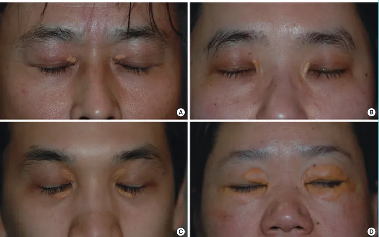
The patients were classified into four grades according to the location and extent of the lesion. Grade I comprised patients who presented with lesions on the upper eyelids only. Grade II comprised patients whose lesions extended to the medial canthal area. Grade III comprised patients with lesions on the medial side of the upper and lower eyelids. Grade IV comprised patients whose lesions were difuse, with involvement of the medial and lateral sides of the upper and lower eyelids (Fig. 1). he height of the lesions was also noted.
Surgical excision was performed in conjunction with blepha-roplasty, medial epicanthoplasty, “uncapping surgery” where the lesion is “uncapped” and the cholesterol deposit is removed in one piece followed by closure1, local skin advancement laps or orbicularis oculi muscle laps, and skin grats. he results of the surgical modality used in each aforementioned classiication were assessed for recurrence and complications. Patient charac-teristics according to the lesion grade are summarized in Table 1.
Fig. 1. Clinical presentation of xanthelasma palpebrarum according to grade
(A) Grade I lesion, involving only the upper eyelids. (B) Grade II lesion, extending to the medial canthal area. (C) Grade III lesion, involving the medial side of the upper and lower eyelids. (D) Grade IV lesion, diffuse involvement of the medial and lateral side of the upper and lower eyelids.
A B
RESULTS
A total of 95 cases from March 2007 to March 2011 were in-cluded in this study. he mean age of the patients was 56 years, and the male-to-female ratio of the patient population was ap-proximately 2:3. he patient population who presented with an abnormal lipid proile was 52%. Preoperative biopsies were per-formed in all of the cases.
Simple excision was performed for the grade I and II lesions, while local flaps and skin grafts were performed in the more advanced grades of the disease. Sixty-six cases (70%), generally
grade I or II lesions, were treated by simple excision with blepha-roplasty design. In 33 patients (35%) who presented with grade I lesions, limited to the supericial dermis, simple excision was performed. In 33 patients (35%) who presented with grade II le-sions extending to the medial canthal area, concomitant medial epicanthoplasty was necessary.
he surgical modality for more advanced stages of the disease with involvement of the medial side of the upper and lower eyelids (grade III), and with difuse involvement of the medial and lateral sides of both upper and lower eyelids (grade IV) was simple excision by blepharoplasty in combination with or
with-Table 1. Patient characteristics and surgical methods
Lesion grade No. (%) Patient Surgical methods Accompanying surgery Minor complications Recurrence
Grade I 33 (35) Simple excision Blepharoplasty None 1 (1%)
Grade II 33 (35) Simple excision Blepharoplasty, medial epicanthoplasty None None Grade III 24 (25) Simple excision, uncapping surgery,
skin lap advancement, orbicularis muscle lap, skin graft
Blepharoplasty, medial epicanthoplasty Scar contracture 2 (2.1%) 2 (2.1%)
Grade IV 5 (5) Blepharoplasty, medial epicanthoplasty Scar contracture 2 (2.1%) None Total 95 (100)
A B
C D
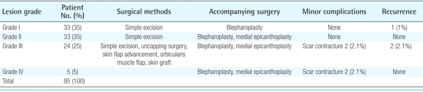
(A, B) Preoperative photo of a grade IV diffuse lesion involving the medial and lateral sides of both upper and lower eyelids. (C, D) Postoperative photo at 1 year after surgical excision by upper blepharoplasty and lower lid orbicularis myocutaneous advancement lap.
out local laps and skin grats. Skin grats were performed using the excised normal skin from the blepharoplasty. Twenty-four cases (25%) were grade III lesions, and 5 cases (5%) were grade IV lesions (Figs. 2, 3).
In our study, 7 cases of grade I lesions and 6 cases of grade II lesions showed involvement of the orbicularis oculi muscle, and the affected musculature was completely resected. Grade III and grade IV lesions generally showed muscle involvement, and to provide tension-free closure ater excision of these difuse lesions, concomitant muscle excision was compulsory. When the size of the lesions exceeded 5 mm in height, surgical manage-ment, regardless of the lesion depth, was preferred.
Four patients (4.2%) developed scar contracture postoperative-ly, which required a secondary procedure such as scar revision or medial epicanthoplasty. During 1 year of postoperative follow-up, lesion recurrence was reported in only 3 patients (3.1%). Other-wise, there were no other major complications or disigurement reported in this study.
DISCUSSION
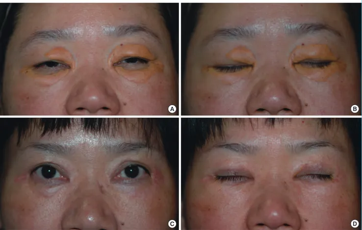
Xanthelasma palpebrarum manifests as yellowish fibrotic plaques on the eyelids, resulting from the deposition of choles-terol laden histiocytes in the dermis. Histologically, xanthelas-mas are composed of xanthoma cells, or foam cells, which are typically found within the middle and supericial layers of the dermis in perivascular and periadnexal locations, with evidence of concurrent fibrosis and inflammation [1,2]. In some cases, xanthelasma can iniltrate the muscle layer and muscle resection is required for complete excision of the lesion [9]. In this study, muscular iniltration was seen in approximately 25% of patients (Fig. 4). Incomplete excision of the lesion leads to a higher inci-dence of recurrence.
Numerous treatment options, both local and systemic, have been reviewed in the literature, including surgical resection [3]; ablation with various lasers such as carbon dioxide [4], erbium-YAG [5], pulsed dye [6], and Nd-erbium-YAG [7]; tricholoracetic acid peels [6]; and treatment of the underlying medical condition [1,2,8]. However, each method of treatment is associated with (A, B) Preoperative photo of a grade IV diffuse lesion involving the medial and lateral sides of both upper and lower eyelids. (C, D) Postoperative photo at 1 year after surgical excision by upper blepharoplasty, lower lid orbicularis myocutaneous advancement lap, and full-thickness skin graft with excised skin from blepharoplasty. Postoperative photos show complete excision of the lesions with relatively minimal scarring and no sign of recurrence. The patient expressed satisfaction with the overall aesthetic result.
A B
C D
Fig. 4. Histopathologic analysis of xanthelasma palpebrarum
limitations and complications such as persistent erythema, re-currence, ectropion, scarring, and hypo- or hyperpigmentation [10]. Mendelson and Masson [3] found that 40% of patients had recurrence after primary surgical excision, 60% after sec-ondary excision, and 80% when all four eyelids were involved. Mendelson and Masson did not mention lesion extension in regard to depth, and may not have acquired an adequate resec-tion depth margin in the surgical treatment of xanthelasma palpebrarum, accounting for their relatively high recurrence rate. However, this was not discussed in their paper, and it is diicult to draw conclusions.
Treatment with various lasers for xanthelasma palpebrarum has been reviewed in the literature with extensive variations in outcome [4,5,7,10-12]. At our center, laser therapy as well as surgical excision has been utilized as a therapeutic module with satisfactory results. Primary laser therapy with an ablative carbon dioxide laser or erbium-YAG was reserved for treatment only in small aberrant lesions and lesions involving the supericial der-mis. However, because the focus of our paper is on the surgical results of xanthelasma palpebrarum, the results analysis of laser therapy for supericial lesions has not been included in this study. Fusade [7] suggested that a 1,064-nm Q-switched Nd:YAG la-ser is a valuable treatment option for xanthelasma, while Karsai et al. [13] reported that Q-switched Nd:YAG laser treatment of xanthelasma is not recommended. Argon laser photocoagula-tion treatment was found by Hintschich1 to have a high recur-rence rate [1]. Raulin et al. [4] reviewed their experiences with ultrapulsed carbon dioxide laser and reported that treatment in the early stages of xanthelasma development was crucial in order to prevent recurrence. Lieb et al. [14] revealed that wound healing with carbon dioxide laser was signiicantly slower due to a larger thermal necrosis zone. Tricholoracetic acid in the
man-agement of xanthelasma was found to be efective for treatment of smaller lesions, but repeat procedures were common and pig-mentation and scarring were common cosmetic concerns [6]. Mitelviehaus et al. [9] found through histologic specimens that in a large number of cases, the depth of tissue invasion in xanthe-lasma iniltrates through the entire dermis, reaching the stratum muscular, and even invading into this layer; these lesions, they proposed, should not be treated by lasers, but should be surgi-cally excised [8].
Recurrence of xanthelasma palpebrarum is a common prob-lem [1]. here are numerous treatment options, both local and systemic, for this condition. In lesions presented at our clinic that were conined to the supericial dermis, less than 5 mm in height, and with an onset of less than one year, laser ablation was a treat-ment option, and in some cases this treattreat-ment alone sufficed. However, when lesions involved deeper layers, recurrence rates were higher, and this is atributable to the fact that lasers may not provide a clean resection margin due to a lack of technical skill or penetration. Furthermore, supericial ablation of lesions may require multiple therapy sessions. When lesions involve the deep dermis or iniltrate the underlying musculature, surgical excision is the mainstay of treatment. hrough our experience, we have found that for longstanding lesions with an onset exceeding a year, or large lesions extending beyond 5 mm in height, local laps and skin grats may be necessary to preserve the aesthetic continuity of the eyelids, regardless of the lesion depth. Also, simple excision by blepharoplasty may provide a more aestheti-cally pleasing result ater surgery. It is generally known that only small eyelid defects that are less than 5 mm are amenable to healing by secondary intention [15]. Lasers and other nonsurgi-cal treatment options ultimately result in a partial skin defect of the eyelid. Thus, to prevent eyelid deformation and to achieve a more tolerable scar, surgical management is recommended. A clinically large lesion (i.e., lesions greater than 5 mm in height), regardless of depth, when treated with lasers, resulted in a more blatant secondary deformity and was more susceptible to skin discoloration, scarring, and recurrence.
Our study presents only the outcomes of surgical cases of xan-thelasma palpebrarum. In patients with lesions involving only the supericial dermis, a multitude of less invasive treatment op-tions exists that may provide satisfactory outcomes. In patients who were hesitant to undergo invasive approaches who present-ed with supericial lesions at our clinic, lasers have also providpresent-ed satisfactory outcomes. We recommended surgical treatment to patients who we predicted would benefit aesthetically from a surgical approach, such as those with difuse lesions, dermato-chalasia, baggy lower eyelids, or skin laxity. hus, in the lesions that are confined to the superficial dermis, treatment should Lipid laden histiocytes (foam cells) observed infiltrating into the
be individualized for patient preference and characteristics. We acknowledge that this is a limitation of our study and a compara-tive study of diferent modalities of treatment with results analy-sis will provide further credibility to our conclusion.
Observational studies have linked the presence of xanthelasma palpebrarum with conditions such as hyperlipidemia, dyslipo-proteinemia, hypertension, and underlying cardiovascular dis-ease such as atherosclerosis. Underlying medical conditions such as hypothyroidism, renal syndromes, and cirrhosis have also been reported in the differential diagnosis for these cutaneous lesions. Histological analysis has revealed similarities in the ul-trastructure of xanthelasma and atheroma formation and several data suggest that there may be an association with low high den-sity lipoproteinqq cholesterol levels and other lipoprotein and apolipoprotein abnormalities [2]. Furthermore, in the patho-genesis of xanthelasma, abnormal lipid metabolism seems to play a crucial role. However, the appearance of xanthelasmas in
patients with a normolipidemic proile and studies that conclude no relationship between plasma lipoprotein abnormalities and xanthelasma suggest that the formation of xanthelasmas may be inluenced by, but not indicative of, underlying disease [2,3,8]. In our study, only 52% of patients presented with an abnormal lipid profile. Whether or not restriction of fat intake may cor-relate with regression of xanthelasma remains relatively obscure. Although xanthelasmas are not definitely determinate cutane-ous markers of atherosclerosis or other underlying conditions, inconsistency in the results in the pathogenesis of xanthelasma cannot be ignored [1,2,8]. At our clinic, it was recommended that patients with underlying medical conditions or those who presented with an abnormal lipid proile see an internal medicine specialist for a further work-up and treatment, if necessary.
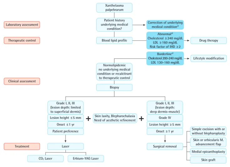
We have developed a treatment algorithm for xanthelasma, tak-ing into account patient characteristics and the size and location of the lesion. Xanthelasma palpebrarum has been correlated in
Fig. 5. Proposed algorithm for the treatment of xanthelasma palpebrarum
The size and location of the lesion should be taken into account in the treatment of xanthelasma palpebrarum. In lesions involving the deep dermis and/or muscle, surgical excision is the most appropriate therapeutic option. LDL, low density lipoprotein; IHD, ischemic heart disease. a)refer
to internal medicine specialist.
Skin laxity, Blepharochalasia Need of aesthetic reinement Laboratory assessment
Therapeutic control
Clinical assessment
Treatment
Patient history underlying medical
condition?
Normolipidemic no underlying medical condition or recalcitrant
to therapeutic control Xanthelasma palpebrarum
Simple excision with or without blepharoplasty
Skin or orbicularis M. advancement lap Blood lipid proile
Biopsy
Grade I, II, III (lesion depth: limited to supericial dermis)
Lesion height ≤ 5 mm
Onset ≤ 1 yr
Patient preference
Grade I, II, III (lesion depth: deep dermis-muscle)
Grade IV
Lesion height ≥ 5 mm
Onset ≥ 1 yr
Laser Surgical removal
CO2 Laser Erbium-YAG Laser
Medial epicanthoplasty
Skin graft Drug therapy
Lifestyle modiication Correction of underlying
medical conditiona)
Abnormala)
Cholesterol ≥ 240 mg/dL LDL ≥ 160 mg/dL Risk factor of IHD ≥ 2
Borderlinea)
some cases with the presence of an underlying medical condi-tion, and therefore, systemic treatment of the underlying medical condition with lifestyle modiication or pharmaceutical therapy can be attempted before or in conjunction with surgical exci-sion. In lesions with only a supericial extent, laser therapy may suice, but in some cases, surgery may provide a more aesthetic result. In such cases, treatment should be individualized in each case. In conclusion, among the numerous treatment options for xanthelasma, we have found that for lesions involving the deep dermis and/or muscle, surgical excision was the most appropri-ate therapeutic option (Fig. 5). In difuse lesions (grades III, IV), excision in conjunction with local laps or skin grats provided aesthetic reconstruction with a low incidence of recurrence.
REFERENCES
1. Rohrich RJ, Janis JE, Pownell PH. Xanthelasma palpebrarum: a review and current management principles. Plast Reconstr Surg 2002;110:1310-4.
2. Bergman R. The pathogenesis and clinical significance of xanthelasma palpebrarum. J Am Acad Dermatol 1994;30: 236-42.
3. Mendelson BC, Masson JK. Xanthelasma: follow-up on results ater surgical excision. Plast Reconstr Surg 1976;58: 535-8.
4. Raulin C, Schoenermark MP, Werner S, et al. Xanthelasma palpebrarum: treatment with the ultrapulsed CO2 laser. La-sers Surg Med 1999;24:122-7.
5. Borelli C, Kaudewitz P. Xanthelasma palpebrarum: treat-ment with the erbium:YAG laser. Lasers Surg Med 2001;29: 260-4.
6. Cannon PS, Ajit R, Leatherbarrow B. Eicacy of trichloro-acetic acid (95%) in the management of xanthelasma palpe-brarum. Clin Exp Dermatol 2010;35:845-8.
7. Fusade T. Treatment of xanthelasma palpebrarum by 1064-nm Q-switched Nd:YAG laser: a study of 11 cases. Br J Der-matol 2008;158:84-7.
8. Ozdol S, Sahin S, Tokgozoglu L. Xanthelasma palpebrarum and its relation to atherosclerotic risk factors and lipoprotein (a). Int J Dermatol 2008;47:785-9.
9. Mitelviehaus H, Kreusser C, Bohringer D, et al. he under-estimated depth of tissue invasion of xanthelasma: a histo-logical study. Klin Monbl Augenheilkd 2011;228:14-8. 10. Park EJ, Youn SH, Cho EB, et al. Xanthelasma palpebrarum
treatment with a 1,450-nm-diode laser. Dermatol Surg 2011; 37:791-6.
11. Hintschich C. Argon laser coagulation of xanthelasmas. Oph-thalmologe 1995;92:858-61.
12. Katz TM, Goldberg LH, Friedman PM. Fractional photo-thermolysis: a new therapeutic modality for xanthelasma. Arch Dermatol 2009;145:1091-4.
13. Karsai S, Schmit L, Raulin C. Is Q-switched neodymium-doped ytrium aluminium garnet laser an efective approach to treat xanthelasma palpebrarum? Results from a clinical study of 76 cases. Dermatol Surg 2009;35:1962-9.
14. Lieb WE, Klink T, Munnich S. CO2 and erbium YAG laser in eyelid surgery. A comparison. Ophthalmologe 2000;97: 835-41.