Analyse titanium surface irradiated with laser,
with and without deposited of durapatite
1Análise da superfície de titânio sob efeito do laser
com e sem deposição de hidroxiapatita
1Karin Ellen Sisti2, Idelmo Rangel Garcia JR.3, Antonio Carlos Guastaldi4, Andréia C. M. B. Antoniolli5, Rafael De Rossi6 , Álvaro de L. Brochado Neto7
1. Department of Odontology, University Odontology of Araçatuba - State University of São Paulo (UNESP), Brazil. 2. Master, Fellow PhD degree in Post Graduate of Health and Development in the Central West Region, Federal University
of Mato Grosso do Sul (UFMS), Brazil.
3. PhD, Associate Professor of the Department of Surgery and Integral Clinic of University of Odontology of Araçatuba UNESP, Brazil.
4. PhD, Associate Professor of the Department of Physic Chemistry of Institute de Chemistry of Araraquara – UNESP, Brazil.
5. PhD, Associate Professor of the Department of Surgery, UFMS, Brazil. 6. PhD, Associate Professor of the Department of Veterinary, UFMS, Brazil. 7. Physiotherapist of Catholic Universit Dom Bosco, Brazil.
ABSTRACT
Purpose: The aim of this study was to analyse the surface of titanium implants using disc irradiated with lasers. Methods:
Titanium discs were irradiated with laser high insensitive (Nd-YAG), deposited durapatite and used thermal treatment. Sample received qualitative morphological analyse trough micrographics with many size in SEM (Scanning Electron
Microscopy). Results: Surface laser irradiation shows roughness and isomorphic characteristic. The durapatite amplified
the titanium surface area by method biomimetic. Conclusion: The surface treatment presented more deposition of durapatite,
roughness on the surface, better isomorphic characteristic and increase quantitative in titanium surface area, samples shows rugous, roughness and homogeneity there is not found in the implants available at the market.
Key word: Lasers. Titanium . Durapatite.
RESUMO
Objetivo: Estudar a superfície de implantes osseointegráveis utilizando discos de titânio irradiados com feixe de laser.
Métodos: A amostra foi irradiada com feixes de laser de alta intensidade (Nd-YAG), posteriormente depositado hidróxiapatita e submetido a tratamento térmico. Foi analisada sob MEV (Microscópio Eletrônico de Varredura) e realizada análise
morfológica qualitativa com microfotografias em vários aumentos. Resultados: A superfície irradiada com laser apresentou
deformidade superficial e característica isomórfica; a aplicação de hidroxiapatita pelo método de biomimético aumentou
quantitativamente a área da superfície de titânio. Conclusão: Adeposição de hidroxiapatita apresentou melhor característica
isomórfica e aumento quantitativo da área superficial estudada, a amostra demonstrou características não encontradas nos implantes disposto no mercado.
Descritores: Lasers. Titânio. Durapatita.
13 - ORIGINAL ARTICLE
Introduction
The introduction of osseointegration endosseous implants make possible a new rehabilitation in odontology with more comfort to the patients, resulting in better esthetic, phonetic and mastication, providing a better quality of life as
well. Since then, a new era in odontology1 has started.
Branemark, in 1960, introduced the concept of osseointegration in odontology using titanium as implant material and since this date the implantology is the area in odontology that has presented more technological studies, with different types of implants, techniques and biomaterials, leading to a gretaer
success in the rehabilitation of edentulous patients2. The
osseointegration is the direct contact, structural and functional between order bone and the surface implant, in the optical microscopic, showing stable and supporting the masticatory force.2 In the biological vision there is no evidence of the
an aloplastic material in the bone is got and kept during the function”.3 With the objective of improving or even accelerating
this osseointegration, especially in areas with poor quality bone, various studies about titanium implant surfaces were realized in the last decades, and have shown that the recovering with durapatite improve the rigidity of bone and implant interface.4 There are various types of treatment of titanium
implant surfaces, as titanium plasma-spray, blast of a lot of particles types (sand, glass, aluminum oxide), absorptive biomaterials as micro particles of durapatite, besides, modification with acids or anodization (chemic), and physics, as irradiation with laser high intensity - Nd-YAG3,5. In 1960,
Theodore Maiman presented the first laser that had the ruby crystal as the active principle. The laser has characteristics that differs it from regular light as monochromaticity, coherence, direction, focalization in little areas and emission of high density energy.6 The oxidation or nitretation of titanium surface with
laser irradiation is very important because it increases the surface area, and as titanium oxide and titanium nitrate are biocompatible, it becomes possible to use them in the organism. The titanium nitretation is considered bioinert for Food and Drugs Administration (FDA), it increases the capacity of flow off of the implants. The high intensity laser Nd:YAG is used in
studies to modify surfaces in titanium implants.5 In 2002,
titanium implant surfaces were irradiated with laser Nd:YAG in an overheating temperature which changed the original
morphologic surface.7 Laser Nd:YAG was used over titanium
samples and indicated that the potential parameters of laser influence in the melting surface.8 In studies of oxidation by
laser in titanium surfaces, Lavisse9 concluded that the thickness
melting layer depends on the amount of emission in same local surface. However, the studies don’t kwon which real alteration of surfaces implants with irradiation lasers high intensity and association with durapatite. Based on this, the study was realized using experimental methodology, about titanium discs irradiated with lasers Nd:YAG and deposited durapatite.
Methods
Sample: 02 discs of titanium, with 4mm of diameter and 1,5mm of thickness, from Institute of chemistry – UNESP – Araraquara. The discs were denominate disc nº1 and disc nº2.
Procedure: In this study titanium discs were used from Biomaterial Group of Chemistry Institute – UNESP-Araraquara. The discs were irradiated with high intensity Nd-YAG laser, and analysed with Scanning Electron Microscopy (SEM).
Disc n°1 received irradiation of lasers Nd-Yag, with energy
of 100W/m2, frequency pulse of 35.000 Hz, scanning velocity
of 80mm/s, pace 1, matrix space of +/- 0,01cm and time almost 2minutes . Disc nº2 received irradiation of lasers the same type that n°1. The parameters of energy, frequency pulse, scanning velocity and pace were the same used in disc nº1. There was variation of matrix and time of application, that disc n°2 was almost 0,02cm and 1 minute 10 seconds. Deposition of durapatite over titanium: after the laser irradiation the discs were analysed with SEM, and immersed in the solution of SBF (solution body fluid) to durapatite deposition using the biomimetic method. Then the substrates were immersed during
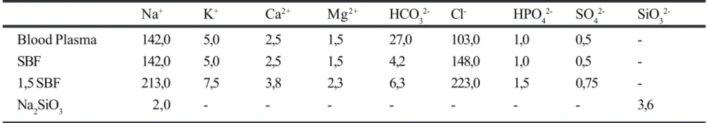
7 days at 37°C in the solution 1,5 SBF to grow up. Table 1 displays the ionic concentration of blood plasma and each solution used in this process. For easier interpretation the results of the discs were denominate 1a and 2a when received only laser irradiation, 1b and 2b after deposited durapatite. Morphologic analysis: a qualitative analysis was made with
micrographic using SEM, model JSM-T330A, Jeol, from
Chemistry Institute, Araraquara - UNESP, with zoom 500, 5.000 and 10.000 X, before and after deposited durapatite. The defects presents and the homogeneity were the question analysed.
Results
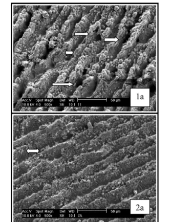
The figures 1, 3 and 5 (disc 1a and disc 2a) present micrographics in SEM of surface sample after laser irradiation. The micrographic with zoom 500X (figure 1) shows melt zone and quick solidification. The figure 2 (discs 1b e 2b) presents sample micrographics after laser irradiation and deposited durepatite, and show up the presence of durapatite in the zoom 500X, the porous and homogeneity also can be seen in this figure. As disc 1a and disc 2a show up linear defects with diameter 25µ and homogenates irregularity, however 1a presents bigger porous and melt zone and quick solidification deeper, and 2a presents a more homogenous surface.
FIGURE 2 - 1b micrographic disc 1(lasers condition I and durapatite deposition) zoom 500X in SEM, observe the durapatite (single dart), roughness surface and homogeneity. 2b micrographic disc 2 (laser condition II and durepatite deposition) zoom 500X in SEM, durapatite (double dart) on surface proportioned irregular and roughness surface.
FIGURE 3 - 1a micrographic disc 1 (laser condition I), zoom 5000X in SEM, increase on surface area because irregularity, and formation of spheres (darts). 2a micrographic disc 2 (lasers condition II) zoom 5.000X in SEM, observe increase surface for irregularity and presence of spheres (darts).
FIGURE 4 - 1b micrographic disc 1 (laser condition I and with durapatite) zoom 5.000X in SEM, there are durapatite (darts), and roughness. 2b micrographic disc 2 (laser condition II and with durapatite) zoom 5.000X in SEM, there are increase surface and durapatite (dart).
Discussion
Delay between the surgery and the prosthesis installation over implants trouble the patients. Nevertheless, the surfaces implants always caused concern and interest in the scientific communities, because the surface has close relationship whit the time, which can alternate from four until six months in machinade surfaces implants, however studies about treatment surface have proved that the time can be decreased. Orsini et al10 made analysis with blast surface
implants, machinade and with acid, proved irregular morphologic blast surface, and with acid, which can possibilities the best beginning adhesion cellular, and better osseointegration. Tests about citotoxicity were realized too, and didn’t show same toxicity cellular and
biocompatibility. Li et al11 wrote about interface bone
and endousseous implants with bland and aluminum oxide blast, then concluded that roughness surface can induce the connect bone/fibre perpendicular on surface, during the osseointegration, can improve the interfacial biomechanic and increase the osteoblast function. However study compare, in human, between machinade
surface and surface altered with acid, Trisi, at al12
presented the biggest contact in implants that made modification surface with acid, the study was made with SEM and delay with 6 months over 11 patients.
Nevertheless, Sykaras13 showed as surface as diameter,
length, design, material, surface topographic, time and
FIGURE 6 - 1b micrographic show the disc 1 (laser condition I and with durapatite), zoom 10.000X in SEM, observe durapatite on spheres (single dart). 2b micrographic disc 2 (laser condition and durepatite), zoom 10.000X in SEM, observe the durapatite with characteristic of roughness surface (double dart).
TABLE 1 - Ionic concentration of solution used to cover surface with durapatite (mmol.dm-3).
Na+ K+ Ca2+ Mg2+ HCO
3
2- Cl- HPO
4
2- SO
4
2- SiO
3
2-Blood Plasma 142,0 5,0 2,5 1,5 27,0 103,0 1,0 0,5
-SBF 142,0 5,0 2,5 1,5 4,2 148,0 1,0 0,5
-1,5 SBF 213,0 7,5 3,8 2,3 6,3 223,0 1,5 0,75
-Na2SiO3 2,0 - - - 3,6
implantation local, and a lot of factories interferer in the osseointegration process. The surface modification also can be realized for electrochemic, with anodic oxidation or for application high intensives lasers. There are same studies using the lasers to prepare the surface implants.
In 2002, Gyorgy, et al8 used high intensity lasers to
change the titanium surface, and analysed with SEM, that displayed modification in the last layer of titanium.
Yue14 studied the alloy Ti6Al4V surface irradiation with
lasers to decrease the corrosion and increase the resistance, presented reduction of corrosion on alloy and there were alteration surface without fractures and with
relative malleability. Petö and col7 irradiated the surface
titanium implants only machinade and blast surface with
Al2O3 using pulse lasers Nd:glass, that altered the
original morphologic implants surface, was used rabbit tibias and compared with machinade surface with SEM and XPS (photoelectron spectroscopic excited by X-ray), showed that treatment with lasers made the isomorphic topographic and the new bone around presented a good resistance with torch, 20% more when compare to
implants with machinade surface. Hallgren15 showed with
histomorphmetric that after 12 weeks the quantity of calcificated tissue put on surface irradiated implants was
bigger than control group. Cho and Jung16 analysed
Ncm on machinade implant, after 8 weeks of surgery, in rabbit tibias. The process of modification titanium surface by irradiation lasers is viable, clean and reproductively, the melt and quick solidification proportionate irregularity in the metal and is controlled for various factors, as parameters lasers sheaf and atmosphere during the irradiation. However in this study can see different surface because lasers Nd:YAG provide homogeneity and irregularity that can improve osseointegration of material. In micrographic 1a (figure 1) display a typical morphologic with melt and quick solidification zones, that are proven to lasers process. This treatment yields porous formation and homogenized surface, that make an increase area and volume, but in the 1a presents linear defects biggest and deeper that proportionate the increase of contact surface when
compare if 2a. However in 2a(figure 5) display a bigger
roughness than 1a(figure 5). There are homogeneous
defect on surface, it is important characteristic in endosseous implants, because optimize the osseointegration (bone tissue without interposition fibre/
tissue).2 The sheaf lasers in environment atmosphere alter
surface that provide physical and chemical propriety desirable. The durapatite merit eminence because is the principal component mineral of calcificated tissue. The durapatite is the ceramic phosphate or bioceramic, with composition and structure similar to mineral phase of
bone and tooph.17 Proussaef18 in analysis with implants
and durapatite after periods of function between 3.5 and 11 years, concluded that durapatite didn’t presents reabsorption or dissolution after few ages in function, and probably this occur only when there is contact with
soft tissue. Geurs, et al19 analysed characteristics of
surface implants in osseointegration process with 3 types of implants: plasma-spray of titanium, durapatite and cylinder with durapatite. Concluded that durapatite pressed the initial tax of osseointegration and presented a decrease faster in the micro-movement when compare
with plasma-spray of titanium. Ramires20 in study about
grown cellular with osteoblast and biomaterials showed that there are precocious aggregation, differentiation and
mineralization cellular on TiO2 surface with durapatite. In
this study after deposit durapatite on irradiated surface both groups continued presented homogeneity and irregularity, but in the sample 1b observed more durapatite deposition (figure 2), roughness surface (figure 4) and more isomorphic characteristic (figure 2) when compare with 2b (figure 4), as in 1b as in 2b comply clearly the presence of durapatite on spheres of surface. The modification of surface in this study provides an increase contact surface and improves the first stability in the endosseous implants and can decrease the osseointegration time. The characteristics phisicochemical of durapatite was evidenced in every groups proportioning more facility in keep the liquid, which improve the quality and accelerate the osseointegration process. Nevertheless, a study with animals could be considerate, to observe the repair around material in the organism.
Conclusion
The surface treatment presented more deposition of durapatite, roughness, and better isomorphic characteristics; The durapatite application through biomimetic method increased the titanium surface area. The sample showed rugous, roughness and homogeneity there is not found in the implants available at the market.
References
1. Saadoun AP, Legall ML. Clinical results and guidelines on Steri-Oss Endosseous Implants. Int J Periodont Rest Dent. 1992;12:87-99.
2. Bränemark PI, Zarb GA, Albrektsson T. Tissue Integrated Protheses: osseointegration in clinical
dentistry. Chicago: Quintessence; 1985.
3. Zarb GA, Albrektsson T. Osseointegration: a require for the periodontal ligament? Periodont Rest Dent. 1991;11:88-91.
4. Sanz A, Oyarzun A, Farias D, Diaz I. Estudo experimental da resposta óssea a um novo tipo de tratamento de superfície de implantes de titânio. Int J Oral Implantol. 2001;10:20-1.
5. György E, Pérez Del Pino A, Serra P, Morenza JL. Chemical composition of dome-shaped structures grown on titanium by multi-pulse Nd:YAG laser irradiation. Appl Surface Sci. 2003;189:1-8.
6. Cecchini SCM. Estudo in vitro das aplicações do laser
de Holmio:YLF em esmalte e dentina, visando a realização de cirurgias de acesso endodôntico e preparo cavitário. [Dissertação Mestrado] Universidade de São Paulo; 1995.
7. Petö G, Karacs A, Pászti Z, Guczi L, Divinyi T, Joób A. Surface treatment of screw shaped titanium dental implants by high intensity laser pulses. Appl Surface Sci. 2002;186:7-13.
8. György E, Mihailescu IN, Serra P, Pérez Del Pino A, Morenza JL. Single Pulse Nd:YAG laser irradiation of titanium: influence of laser intensity on surface morphology. Surface Coating Technol. 2002;154:63-7. 9. Lavisse L, Grevey D, Langlade C, Vannes B. The early stage of the laser-induced oxidation of titanium
substrates. Appl Surface Sci.2002;186:150-5.
10. Orsini G, Assenza B, Scarano A, Piattelli M, Piattelli A. Surface Analysis of Machined Versus Sandblasted and Acid-Etched Titanium Implants. J Oral Maxillofac Implants. 2000; 15:779-784.
11. Li D, Liu B, Wu J. Bone interface of dental implants cytologically influenced by a modified sandblasted surface: a preliminary in vitro study. Implant Dent. 2001;10(2):132-8.
Correspondence:
Rua José Antônio, 2200 - sala 06 79010-190 - Campo Grande - MS - Brazil e-mail: [email protected]
Conflict of interest: none Financial source: none
How to cite this article:
Sisti KE, Garcia Jr. IR, Guastaldi AC, Antoniolli ACMB, De Rossi R, Brochado Neto ÁL. Analyse titanium surface irradiated with laser, with and without deposited of durapatite. Acta Cir Bras. [serial on the Internet] 2006;21 Suppl 4. Available from URL: http://www.scielo.br/acb.
13. Sykaras N, Lacopino A, Marker VA, Triplett RG. Woody, RD. Implant Materials, Designs, and surface Topographies: Their Effect on Osseointegration. A Literature Review. Int J Oral Maxillofac Implants. 2000;15:675-690.
14. Yue TM, Yu JK, Mei Z, Man HC. Excimer laser surface treatment of Ti-6Al-4V alloy for corrosion resistance enhancement. Mat Letters. 2002;52:206-12.
15. Hallgren C, Reimers H, Chakarov D, Gold J, Wennerberg A. An in vivo study of bone response to implants topographically modified by laser micromachining. Biomaterials. 2003; 24:701.
16. Cho SA, Jung SK. A removal torque of the laser-treated titanium implants in rabbit tibia. Biomaterials. 2003;24:4859-63.
17. Kawachi EY, Bertran CA, Dos Reis RR. Biocerâmicas: tendências e perspectivas de uma área interdisciplinar. Química Nova. 2000; 23(4):518-22.
18. Proussaef P, Lozada J, Ojano M. Histologic Evoluation of Threaded HA-Coated Root-Form Implants After 3.5 to 11 Years of Function: A Report of Three Cases. Int J Periodont Rest Dent. 2001;21(1):21-9.
19. Geurs NC, Jeffcoat RL, Mcglumphy EA, Reddy MS, Jeffcoat MK. Influence of Implant Geometry and Surface Characteristcs on Progressive Osseointegration. J Oral Maxillofac Implants. 2002;17: 811-15.