O
BJECTIVEAnalysis of critical end diastolic left ventricular volume (EDLVV), defined as the lowest acceptable volume to keep cardiac output, in the selection of patients with post-valvotomy aortic stenosis, candidates to univentricular correction.
M
ETHODSA retrospective study in 21 patients with aortic stenosis, during the first year of life, and 232 patients compiled from literature. Values of end diastolic volume (EDLVV), from 20 to 60 ml/m2 were determined as normal. The EDLVV of deceased patients was compared to that from survival patients. A correlation between the age and EDLVV was carried out at the time of valvotomy, between the groups. Finally, the critical EDLVV through the theoretical relationship between the EDLVV and heart rate for different ejection fractions and designated cardiac indexes (CI): 2,000 and 2,500 20-60 ml/min/m2 was determined.
R
ESULTSFor EDLVV values <20ml/m2 and >60 ml/m2, there was statistical significance between deceased and survival patients (p< 0.0001). However, in the range between 20 and 60 ml/m2, that significance was lower (p=0.0309). A greater incidence of death took place among those who showed it in the first week of life. For a CI of 2,500 ml/min/m2 and a heart rate (HR) of 140 bpm, critical EDLVV will be 26 ml/m2 if left ventricular ejection fraction (LVEF) is 0.70, and 44.5 ml/m2, if LVFE is 0.40.
Mailing address: Marco Aurélio Santos • Rua Bulhões de Carvalho, 245/301 • 22081-000 • Rio de Janeiro, RJ - Brazil
E-mail: [email protected] Received on 12/28/04 • Accepted on 03/04/05
Critical Left Ventricular Volume in Aortic
Stenosis in First Year of Life - Its Importance in
Patients Selection Candidates to Univentricular
Surgical Correction Pos-valvotomy
Marco Aurélio Santos, Vitor Manuel Pereira Azevedo, Maria Ourinda Mesquita da Cunha
Instituto Nacional de Cardiologia Laranjeiras e Instituto Nacional do Câncer - Rio de Janeiro, RJ - Brazil
C
ONCLUSIONThe analysis of critical end diastolic left ventricular volume (EDLVV) can add another parameter in the indication of univentricular correction in patients with post-valvotomy aortic stenosis, during the first year of life.
K
EY WORDSAortic stenosis in the first year of life has high post-valvotomy mortality, either through balloon catheter or surgery1,2. That high mortality can be partly justified
by reduced left ventricular volume of those patients, which are insufficient to keep a proper post-valvotomy cardiac output. Both empiric studies and creation of physiological models suggest that, even in the absence of associate lesions, survival is little probable for those patients with ventricular volumes lower than 20 ml/ m2,3-10. So, those patients should be candidates to a
Norwood-type univentricular correction and, then, to a Fontan-type procedure.
One of determining factors in that decision will be the size of small left ventricle11-14. Although that small ventricle
may develop, due to its growth capacity, it should necessarily have an acceptable minimum volume in order to keep a proper cardiac output, immediately after a valvotomy. Then, the big questioning will be, “Which is the lowest acceptable volume the left ventricle should have and still have conditions to keep a proper post-valvotomy cardiac output?”.
M
ETHODS
A retrospective analysis of end diastolic left ventricular volume (EDLVV) was carried out in 21 patients with neonatal aortic stenosis, and compared to 232 cases compiled from literature. Values of EDLVV within a range from 20 to 60 ml/m2 were arbitrarily regarded as normal.
EDLVV from deceased patients was compared with those from survivor patients. A relationship of EDLVV with the age at the time of valvotomy was established between groups. Finally, a theoretical relationship between EDLVV and the heart rate (HR) for different left ventricular ejection fractions and determined cardiac indexes was graphically represented, from the following equation: cardiac output (CO) = EDLVV x left ventricular ejection fraction (LVEF) x HR, which means, EDLVV = CO/LVEF x HR. From that relation, it was possible to obtain critical EDLVV, that is, the lowest acceptable EDLVV for cardiac output maintenance.
R
ESULTS
Table I shows EDLVV 18 patients with neonatal aortic stenosis. Ten of them survived balloon valvuloplasty and eight patients died. Those groups of patients showed EDLVV of 51.5±25.1 and 38.4 ±2.5 ml/m2 (p=0.28).
Table II analysis demonstrates values of EDLVV from 177 patients (including 18 from our series), who were divided in three groups, according to EDLVV values (<20; 20-60 and >60ml/m2). For EDLVV values <20 ml/m2
and >60 ml/m2, there was statistic significance between
deceased and survivor patients (p<0.0001). However, within the range between 20 and 60 ml/m2, that statistic
significance was low (p=0.0309).
Table II - Relation of EDLVV with post-valvotomy survival or death
Bühlmeyer, Ziemer, Burch, Parsons, Hoffman, Huhta, Mocellin, Santos, Zeevi, Latson, Lofland. EDLVV - end diastolic left ventricular volume.
n EDLVV (ml/m2) p (lines)
<20 20-60 >60 Survivors 109 3 63 43 <0.001 Deceased 68 17 47 4 <0.001 Total 177 20 110 47 <0.001 p (columns) <0.0001 <0.0001 0.0309 <0.0001
Table I - Relation of EDLVV with post-aortic valvotomy survival or death
n EDLVV (ml/m2) Literature p
Survivors 14 42.6±26.8 Koevakin 1998 0.11 Deceased 5 21.8±7.2
Survivors 19 50.0±8.0 Hammon 1988 <0.05 Deceased 14 20.0±4.0
Survivors 19 37.0±17.0 Grundi 1986 NS Deceased 5 36.0±7.0
Survivors 10 51.5±25.1 Santos/Azevedo 2002 0.28 Deceased 8 38.4±2.5
EDLVV - end diastolic left ventricular volume.
When age was included at the time of aortic valvulotomy (tab. III), and it was correlated with EDLVV value, it was observed that, from 0 to 10 days, 15 from 27 patients survived and 12 died, and 18 had EDLVV in the range of 20-60 ml/m2. From 10 days of life, survival possibility
increased, and almost the totality of them were within the range of A EDLVV values between 20 and 60 ml/m2.
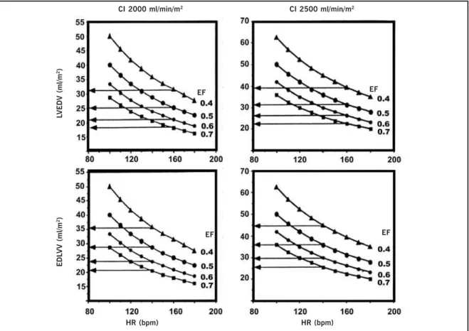
Figure 1 graphically represents a family of theoretical curves, which were obtained through the relations between EDLVV and HR for different ejection fractions and cardiac indexes. The horizontal arrow points at the critical EDLVV. So, a neonate individual with critical EDLVV of 26 ml/m2
can survive, temporarily, if he/she shows a HR of 140 bpm, LVEF of 0.70, obtaining a cardiac index of 2,500 ml/m2.
For those same cardiac index and HR, a LVFE of 0.40 is sufficient, since the critical EDLVV increases to 44.5 ml/m2.
D
ISCUSSION
Early post-valvotomy mortality among neonates and infants with aortic stenosis is high, within a range from zero to 100%, with a median of 31%, a lower quartile of zero and an upper quartile of 50%. Oppositely, aortic valvotomy, after the first year of life, has an early mortality rate in the range from zero to 18%, with a median of 3% and upper quartile of 7%15. It is possible that such mortality
would be even higher if all patients were submitted to that procedure. On the other hand, it is possible that with the development of techniques less invasive than surgeries, earlier diagnosis and better understanding and handling of intensive care, those results can be more promising.
supravalvar stenosis, or large shunts on atrial, ventricular or ductus arteriosus level, substantially add the possibility of early post-valvotomy mortality13,14. Literature also directs
place, as a single measurement in and improper place would produce an assessment mistake and that would be raised to the power of 2; second, in neonatal aortic stenosis, the left ventricle tends to be more globular than under normal situations. So, the measurement of cross-section area must be done by using the longer axel, for instance,
π
ab, in which a is the long axle radius. So, twomeasurements are incorporated to the gauging, and not the squared number of one (small axle), which minimizes the estimated mistake. Third, the presence of myocardial sinusoids in newborns and infants with aortic stenosis is not rare. Bühlmeyr et al18, in a cardiac angiographic study,
determined ventricular volume in 18 patients with critical valve aortic stenosis. In 7 from those patients, myocardial sinusoids were present. When myocardial sinusoids were included in that determination, EDLVV was 120% greater than the normal in 7 patients, normal, in 6, and 80% lower than the normal in 5 patients. When myocardial sinusoids were excluded, there was an average reaction of 6% in ventricular volume in those 7 patients. For the total of the group, EDLVV was still increased over 120% in 6 patients, but reduced under 80% in 8 patients.
For some time, literature has presenting publications trying to correlate end diastolic left ventricular volume with post-valvotomy survival (tables I and II).
Normal left ventricular volume values up to two years of life, obtained through angiographic study determined by Graham et al3, were 42 ml/m2±10 ml/m2.
Another observation demonstrated by literature is that lower volume ventricles are found in younger patients and, consequently, those who are critically ill, soon after birth, and that have high post-valvotomy mortality (tab. III). In our series, that mortality rate reached 100% of the nine patients who were submitted to balloon valvuloplasty, in the first week of life9.
In terms of ventricular configuration distortions and possible mistakes in determining diastolic left ventricular volume, two questions must be asked: 1) how could the true determination and importance of end diastolic left ventricular volume be valuated? 2) What determines when a volume is very little?
In order to try to answer those questions, we considered the basic physiology of cardiac output: cardiac output = EDLVV x LVEF x HR.
That formula allows us for relating EDLVV to cardiac output and HR for any LVEF. If we decide for a minimum cardiac output that a newborn or infant needs to survive in the first post-valvotomy weeks, which is the period that the left ventricle is under adjustment, and we determine which maximum HR is acceptable. So, we are able to build a family of curves that will define those relationships. Those are demonstrated in figure 1. In that figure, arrows point at the critical EDLVV, which will be the lowest acceptable volume to maintain a certain cardiac index for any LVEF and HR of 140 and 160 bpm. Although selected cardiac indexes are low, those patients can
Table III - Relation of EDLVV with age, at the time of valvotomy, between survivors and deceased patients
Bühlmeyer, Karl, Lakier, Messina, Zeevi, Hoffman, Santos, Rhodes, Shink, Latson, Mocellin. EDLVV - end diastolic left ventricular volume.
Age (days) n EDLVV (ml/m2) Survivors Deceased
<20 20-60 >60
00-10 27 6 18 3 15 12 11-20 12 0 10 2 12 0
21-30 3 0 2 1 3 0
31-40 5 0 5 0 4 1
the attention to the fact that many patients die immediately after the procedure, in terms of complications inherent to the procedure, maintenance of left ventricular dysfunction due to fibroelastosis or subendocardial ischemia6,9,15, or
even in consequence of repetition of the procedure. Aortic stenosis patients, who showed up in neonatal period, have higher mortality in terms of a lower left ventricular ejection fraction (LVEF)9, as well as a reduction
of EDLVV (tab. II). So, just an increase of EDLVV or LVEF is necessary to improve clinical features.
There are many possibilities of infer, indirectly, left ventricle dimensions. One of them would be gauging mitral or aortic ring diameters, obtained at angiographic or echocardiographic study. Literature has been showing works demonstrating a significant correlation between aortic ring dimensions and survival possibility: smaller than 4 mm7, 5 mm4, 6 mm11 or 6.5 mm12. In the study
carried out by Rhodes et al7, survivors among patients
with aortic ring diameters smaller than 7.5 mm were not found. In their turn, Latson et al5 did not also observed
patients with critical dimension mitral valve diameter that showed, independently, increased left ventricular dimensions. For a more accurate assessment, the ideal would be the direct determination of left ventricular volume, which is often a difficult task, due to its method. Schiller et al16 used a complex method. However, they
found similar results to those obtained through a simpler method, described by Wyatt et al17 (both investigators
used mass measurement, but volume gauging was obtained through the difference between internal and external ventricular volumes).
Left ventricular volume assessment can be obtained through the following formula17: volume=4.12b2
(b=measurement of smaller axle diameter).
For the calculation of end diastolic volume, all measurements must be carried out in diastole.
survive for some time if a proper support therapy is used. Through those charts we can see that a 20 ml/m2 EDLVV
can keep the life of the infant only if LVEF is 0.60 or higher. A cardiac index of 2,500 ml/min/m2 (a lower limit
than the normal one) and a HR of 140 bpm, acceptable for the first month of life, will need an EDLVV of 26 ml/ m2 if LVEF is 0.70; 36 ml/m2 for a 0.50 LVEF and 44.5
ml/m2 for a LVEF of 0.40.
In the example of a neonate with EDLVV of 20 ml/m2
and LVEF of 0.40 and HR of 160 bpm, the cardiac index will be 1,280 ml/min/m2, which is very low for life
maintenance. In that situation, it seems to us more physiological that the most suitable post-valvotomy procedure be the assignment for a univentricular correction. However, if, due to valve clearance and improvement of clinical conditions with inotropic measures, LVEF raises to 0.60, cardiac output will increase to 1,920 ml/min/m2,
which would allow for survival, since there was a progressive ventricular volume increase. Moreover, if such increase does not happen, univentricular correction seems to us the best choice of procedure.
A high number of neonates and infants already show proper EDLVV and LVEF, even before valvotomy. However, with the success of that procedure there may be an improvement of such parameters. Unfortunately, it is very
difficult to predict how much of LVEF and EDLVV increase will take place on the first post-valvotomy days. The limiting factor of such process is given by the level of endocardial fibroelastosis and myocardial fibrosis that already existed in intrauterine life9,15,18. As those processes
are progressive, the procedures of early delivery19-21 are
justified, or even valvular clearance in the fetus22-25.
In conclusion, our works does not aim at determining critical ventricular volume as an exact magnitude, as it is usually done by using mathematical formulas, as those by Simpson, Dodge and other formulas. Therefore, critical ventricular volume does not represents an anatomical parameter, expressed in ml/m2 of body surface, but a
physiological one, dependent on variables, such as heart rate, ejection fraction, cardiac index, etc, therefore varying in the same patient.
It is evident that many factors must be taken into consideration in the decision on which type of procedure must be the ideal one, for neonate and infant patients of post-valvotomy critical aortic stenosis. Very often, the presence itself of associated defects may contribute for the selection of patients. However, when the matter is univentricular correction versus biventricular correction, critical EDLVV parameter adds the possibility of a more coherent decision for that group of critically ill patients.
Fig. 1 - Representative charts to demonstrate relationships between end diastolic left ventricular volume (ordinate axis) and heart rate (abscissa axis) for different ejection frac-tions and determined cardiac indexes: 2,000 and 2,500 ml/min/m2. The horizontal arrow points at end diastolic left ventricular volumes (critical volume), at each ejection frac-tion, and cardiac index for heart rates of 160 bpm (upper panel) and 140 bpm (lower panel). EDLVV - end diastolic left ventricular volume; HR - heart rate; CI - cardiac output; EF - ejection fraction. BPM - beats per minute.
CI 2000 ml/min/m2
L
VEDV (ml/m
2)
EF
E
D
L
VV (ml/m
2)
EF
HR (bpm) EF
HR (bpm)
CI 2500 ml/min/m2
1. Zeevi B, Keane JF, Castaneda AR et al. Neonatal critical valvar aortic stenosis. A comparison of surgical and balloon dilatation therapy. Circulation 1989; 80: 831-9.
2. McCrindle BW, Blackstone EH, Willians WG et al. Are outcomes of surgical versus transcatheter balloon valvotomy equivalent in neonatal critical aortic stenosis? Circulation 2001; 104(Suppl I): 152-8.
3. Grahan TJ, Jarmakani JM, Canent RV et al. Left heart volume estimation in infancy and childhood: re-evaluation of methodology and normal values. Circulation 1971; 43: 895-904.
4. Edmunds LH, Wagner HR, Heymann MA. Aortic valvotomy in neonates. Circulation 1980; 61: 421-7.
5. Latson L A, Cheatham JP, Gutgesell HP. Relation of the echocardiographic estimate of left ventricular size to mortality in infants with severe left ventricular outflow obstruction. Am J Cardiol 1981; 48: 887-91.
6. Mocellin R, Sauer U, Simon B et al. Reduced left ventricular size and endocardial fibroelastosis as correlates of mortality in newborns and young infants with severe valve stenosis. Pediatr Cardiol 1983; 4: 265-72.
7. Rhodes LA, Colan SD, Sanders SP. Predictors of survival in neonates with critical aortic stenosis. Circulation 1991; 84: 2325-35.
8. Hammon JW, Lupinetti FM, Maples S et al. Predictors of operative mortality in critical valvular aortic stenosis presenting in infancy. Ann Thorac Surg 1988; 45: 537-40.
9. Santos MA, Azevedo VM. Neonate aortic stenosis. Importance of myocardial perfusion in prognosis. Arq Bras Cardiol 2002; 79: 251-5.
10. Kovalchin JP, Brook MN, Rosental EL et al. Echocardiographic, hemodinamic and morphometric predictors of survival after two-ventricle repair in infants with critical aortic stenosis. J Am Coll Cardiol 1998; 32: 337-44.
11. Pelech AN, Dyck JD, Trusler GA et al. Critical aortic stenosis survival and management. J Thorac Cardiovasc Surg 1987; 94: 510-7.
12. Lakier JB, Lewis AB, Heymann MA et al. Isolated aortic stenosis in the neonate: natural history and hemodynamic consideration. Circulation 1974; 50: 801-8.
13. Karl TR, Sano S, Brawn WJ et al. Critical aortic stenosis in the first
R
EFERENCES
month of life: surgical results in 26 infants. Ann Thorac Cardiovasc Surg 1990; 50: 105-9.
14. Messina LM, Turley K, Stanger P et al. Successful aortic valvotomy for severe congenital valvular aortic stenosis in the newborn infant. J Thorac Cardiovasc Surg 1984; 88: 92-6.
15. Hoffman JIE. Aortic stenosis. In: Moller JH, Neal WA. Fetal, Neonatal and Infant Cardiac Disease. Norwalk, CT: Apple & Lange, 1990: 451-74.
16. Schiller NB, Skiôldebrand CG, Schiller EJ et al. Canine left ventricular mass estimation by two-dimensional echocardiography. Circulation 1983; 68: 210-6.
17. Wyatt HL, Heng MK, Meerbaun S et al. Cross-sectional echocardiography I. Analysis of mathematic models for quantifying mass of the left ventricle in dogs. Circulation 1979; 60: 1104-13.
18. Bühlmeyer K, Simon B, Mocellin R, Saner U. Clinical angiocardiographic and functional studies in the assessment of critical valvular aortic stenosis. Paediatric Cardiology 2, pg 220-36. In: Godman MJ, Marques RM (Eds). Paediatric Cardiology. Edin-burgh: Churchill Livingstone, 1979: 220-36.
19. Burch M, Redington AN, Carvalho JS et al. Open valvotomy for critical aortic stenosis in infancy. Br Heart J 1991; 65: 156-8.
20. Parsons MK, Moreau GA, Graham TP et al. Echocardiographic estimation of critical ventricular size in infants with isolated aortic valve stenosis. J Am Coll Cardiol 1991; 18: 1049-55.
21. Lofland GK, Mc Crindle BW, Willians WG et al. Critical aortic stenosis in the neonate: a multi-institutional study of management, outcomes and risk factors. J Thorac Cardiovasc Surg 2001; 121: 10-27.
22. Maxweel D, Allan L, Tynan M. Balloon dilatation of the aortic valve in the fetus. Br Heart J 1991; 65: 156-8.
23. Lopes LM, Cha SC, Kajita LJ et al. Balloon dilatation of the aortic valve in the fetus. A case report. Fetal Diagn Ther 1996; 11: 296-300.
24. Kohl T, Starland G, Allan LD et al. World experience of percutaneous ultrasound-guided balloon valvuloplasty in human fetuses with severe aortic valve obstruction. Am J Cardiol 2000; 85: 1230-3.