Transactions of the VŠB – Technical University of Ostrava, Mechanical Series No. 1, 2012, vol. LVIII
article No. 1900
Martin JANEČKA*, Karel FRYDRÝŠEK**, Leopold PLEVA***, Jaroslav ROJÍČEK****
REPORT ABOUT THE DESIGN OF EXTERNAL FIXATOR FOR TREATMENT OF PELVIS AND ACETABULUM FRACTURES
INFORMOVÁNÍ O KONSTRUKCI EXTERNÍHO FIXÁTORU PRO LÉČENÍ ZLOMENIN PÁNVE A ACETABULA
Abstract
Main point in this contribution is the design of external fixators applied in traumatology and orthopaedics. These fixators can be used in the treatment of open and unstable (i.e. complicated) fractures of pelvis and its acetabulum. Numerical modelling (i.e. Finite Element Method), together with CAD modelling, experiments, material engineering, and nanotechnology are presented as a support for developing of a new design of external fixators.
Abstrakt
Hlavním bodem článku je návrhexterních fixátorů aplikovaných v traumatologii a ortopedii. Tyto fixátory mohou být používány pro léčení otevřených a nestabilních (komplikovaných) zlomenin pánve a acetabula. Obory numerické modelování (Metoda konečných prvků), společně s modelováním v CAD, experimenty, materiálové inženýrství a nanotechnologie vytváří v dnešní době podporu pro vývoj nových směrů v konstrukci externích fixátorů.
1 INTRODUCTION
This paper reports about the designing of external fixators applied in traumatology and orthopaedics. These fixators can be applied in the treatment of open and unstable (i.e. complicated) fractures of pelvis and its acetabulum, for example see an radiograph in Fig. 1.
Acetabular fractures either occur with high-energy trauma (e.g. auto collisions, falls, etc.) or as an insufficiency fracture. In younger patients, there is almost always significant trauma, and commonly associated injuries, when an acetabular fracture occurs. In elderly patients, acetabular fractures can occur due to bone weakened from osteoporosis.
*
Ing. Martin JANEČKA, VŠB – Technical University of Ostrava, Faculty of Mechanical Engineering, Department of Production Machines and Design, 17. listopadu 15/2172, 708 33 Ostrava, Czech Republic, tel. (+420) 59 732 4509, e-mail [email protected]
**
Assoc. prof., M.Sc., Karel FRYDRÝŠEK, Ph.D., ING-PAED IGIP; VŠB - Technical University of Ostrava, Faculty of Mechanical Engineering, Department of Mechanics of Materials; 17. listopadu 15/2172, 708 33 Ostrava, Czech Republic, tel. (+420) 59 732 3495, e-mail: [email protected].
***
prim. Assoc. Prof. MUDr. Leopold PLEVA, CSc., University Hospital in Ostrava, Traumatology centrum, 17.listopadu 1790, 708 52 Ostrava – Poruba, Czech Republic, e-mail: [email protected].
****
Fig. 1 Fracture of pelvis and acetabulum and its treatment (radiograph, external fixator - way of treatment).
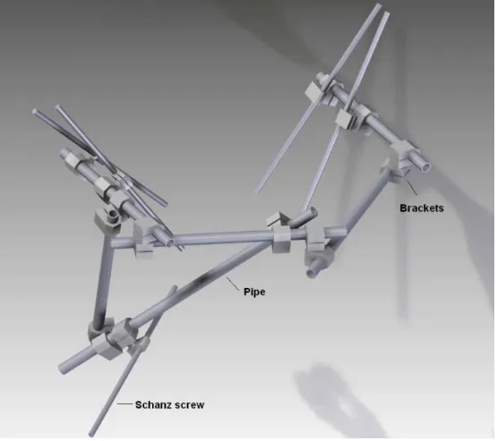
External fixation, see Fig. 1 and 2, is a surgical treatment usually used to set bone fractures in which a cast (plaster) would not allow proper alignment of the fracture. In this kind of treatment, holes are drilled into uninjured areas of bones around the fracture and special bolts or wires are screwed into the holes. Outside the body, rods and curved pieces of metal with special joints (bracket) connect the bolts to make a stiff support. The complicated fracture can be set in the proper anatomical configuration.
Fig. 2 Application of external fixator for pelvis and its acetabulum (experiments in our laboratory).
2 CAD AND FE MODEL
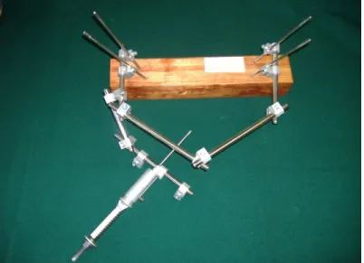
Fig. 3 CAD model of external fixator for pelvis and its acetabulum.
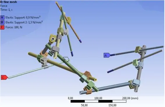
The CAD model, see Fig. 3, was imported into the Finite Element (FE) software Ansys Workbench in which the FE mesh was created, see Fig. 4 and 5.
The basic information about the boundary conditions is presented in Fig. 4.
There are defined mechanical contacts with friction between the brackets and pipes and between brackets and Schanz screws, see also Fig. 4.
Schanz screws are embed in pelvis and its acetabulum in drilled holes. Their attachments are modelled by elastic supports (i.e. by Winkler's foundation, see point “A” and “B” in the legend of Fig. 6 and 7). The elastic support is applied in the radial and axial direction on a parts of Schanz screws. This is quite good and popular simplification of the real complicated interaction between screw and bone. For more information about the elastic foundation and its applications, see references [6], [7], [8] and [9]. Tensile force is explained in the chapter 4 too.
Material behaviour of the external fixator is isotropic and elastic. However, the model is nonlinear because of mechanical contacts between the brackets and pipes and between brackets and Schanz screws.
According to the previous text, the numerical model of external fixator with pelvis interaction was solved via FEM, for example see Fig. 5 (total displacement).
Fig. 4 FE model (boundary conditions) of external fixator for pelvis and its acetabulum.
3 EXPERIMENT IN THE LABORATORY
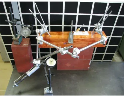
The new types of external fixators for treatment of fractures of pelvis and its acetabulum were tested in the Laboratory of Biomechanics at the VŠB – Technical University of Ostrava (Ostrava, Czech Republic), see reference [4] and for example Fig. 6, 7.
Fig. 6 Prototype of the external fixator for pelvis and acetabulum and its measurement.
Fig. 7 Measuring in the laboratory.
4 CONCLUSION
Report about the new ways to design of external fixator for the treatment of fractures of pelvis and its acetabulum, based on the results of previous research, was presented. Hence, the new designs and materials of fixators will satisfy the ambitious demands of modern traumatology, surgery and economics.
VŠB - Technical University of Ostrava together with University Hospital of Ostrava and Trauma Hospital of Brno are now in the middle of a process creating a new design for external fixators. Hence, they are in cooperation with the Czech producers MEDIN Nové Město na Moravě (Czech Republic) and ProSpon Kladno (Czech Republic).
For more information about external fixators for pelvis and its acetabulum, see references [1], [2], [3], [4] and [10].
Therefore, all results could not be published in this paper due to confidentiality reasons.
Acknowledgements
The work has been supported by the grant project MPO FR-TI3/818 sponsored by Ministry of Industry and Trade of the Czech Republic.
References
[1] Frydrýšek, K., Koštial, P., Barabaszova, K., Kukutschová, J., New ways for Designing External Fixators Applied in Treatment of Open and Unstable Fractures, in: j. World Academy of Science, Engineering and Technology, ISSN 2010-376X (print version) ISSN 2010-3778 (electronic version), vol. 7, issue 76, 2011, pp. 639–644.
[2] Pleva, L., External Fixator for Treatment of Acetabulum Fractures, final report of the project IGA MZ ČR, reg. č. 3522-4, written in Czech language, FNsP – Ostrava-Poruba, 1999, Czech Republic, pp.77.
[3] Rozum, K., External Fixators for the Treatment Open Unstable Fractures, inaugural work written in Czech language, FME, VŠB – Technical University of Ostrava, Czech Republic, 2008, ISBN 978-80-248-1670-8, pp.43.
[4] Janečka, M., Výzkum a vývoj zevního fixátoru na pánev (acetabulum), včetně experimentálního měření a počítačového modelování, thesis of Ph.D. work, VŠB – Technical University of Ostrava, Ostrava, Czech Republic, pp. 1-17, written in Czech language.
[5] Marek, P., Brozzetti, J., Guštar, M., Tikalsky, P., Probabilistic Assessment of Structures Using Monte Carlo Simulation Background, Exercises and Software, (2nd extended edition), ISBN 80-86246-19-1, ITAM CAS, Prague, Czech Republic, 2003.
[6] Frydrýšek K., Pravděpodobnostní výpočty v mechanice 1 (Probabilistic Calculations in Mechanics 1), Faculty of Mechanical Engineering, VŠB - Technical University of Ostrava, Ostrava, ISBN 978-80-248-2314-0, Ostrava, Czech Republic, 2010, pp. 1-149, written in Czech language.
[7] Frydrýšek, K..: Nosníky a rámy na pružném podkladu 1 (Beams and Frames on Elastic Foundation 1), Faculty of Mechanical Engineering, VŠB - Technical University of Ostrava, ISBN 80-248-1244-4, Ostrava, 2006, Czech Republic, pp.463, written in Czech language. [8] Frydrýšek, K., Jančo, R. at al, Nosníky a rámy na pružném podkladu 2 (Beams and Frames on
Elastic Foundation 2), Faculty of Mechanical Engineering, VŠB - Technical University of Ostrava, ISBN 978-80-248-1743-9, Ostrava, Czech Republic, pp.516, 2008, written in Czech language.