O R I G I N A L A R T I C L E UDC: 616-073.75:618.19-006-07 DOI: 10.2298/VSP140508001D
Diagnostic value of breast ultrasound in mammography BI-RADS 0
and clinically indeterminate or suspicious of malignancy breast lesions
Dijagnosti
č
ka vrednost ultrazvu
č
nog pregleda dojke kod žena sa
mamografskom kategorijom BI-RADS 0 i lezijom koja je klini
č
ki nejasna ili
sumnjiva na malignitet
Aleksandar Dobrosavljević*, Snežana Rakić*†, Branka Nikolić*†, Svetlana Janković Ražnatović*†, Svetlana Dragojević Dikić*†, Zorica Miloševi憇,
Aleksandar Jurišić*†, Milica Skrobić*
*Clinic of Obstetrics and Gynecology “Narodni Front”, Belgrade, Serbia; †Faculty of Medicine, University of Belgrade, Belgrade, Serbia; ‡Institute for Oncology and
Radiology of Serbia, Belgrade, Serbia
Abstract
Background/Aim. Not only that ultrasound makes the dif-ference between cystic and solid changes in breast tissue, as it was the case at the beginning of its use, but it also makes the differential diagnosis in terms of benign-malignant. The aim of this study was to assess the role of sonography in the diagnosis of palpable breast masses according to the American College of Radiology Ultrasonographic Breast Imaging Reporting and Data System (BI-RADS) and to correlate the BI-RADS 4 and BI-RADS 5 category with pathohistological findings. Methods. A retrospective study was conducted with the breast sonograms of 30 women presented with palpable breast masses found to be mammography category BI-RADS 0 and ultrasonographic BI-RADS categories 4 and 5. The sonographic categories were correlated with pathohistological findings. Results. Surgical biopsy in 30 masses revealed: malignancy (56.7%), fibroadenoma (26.7%), fibrocystic dysplasia with/without atypia (10%), lipoma (3.3%) and intramammary lymph node (3.3%). Correlation between BI-RADS categories and pathohistological findings was found (p < 0.05). All BI-RADS 5 masses were malignant, while in BI-RADS 4A category fibroadenomas dominated. A total of 53.8% of all benign le-sions were found in women 49 years of age or younger as compared with 35.3% of all malignancies in this group (p < 0.05). Conclusion. Ultrasonography BI-RADS improved classification of breast masses. The ultrasound BI-RADS 4 (A, B, C) and BI-RADS 5 lesions should be worked-up with biopsy.
Key words:
breast neoplasms; diagnosis, differential; mammography; ultrasonography, doppler, color; predictive value of tests; risk assessment.
Apstrakt
Uvod/Cilj. Ultrazvučnim pregledom može ne samo da se napravi razlika između cističnih i solidnih promena u tkivu dojke, kao što je bilo na početku njegove primene, već i da se napravi diferencijalna dijagnoza u smislu razlikovanja benignih od malignih promena. Cilj rada bio je procena uloge ultrazvuka u dijagnostici palpabilnih promena u dojci u skladu sa terminologijom Breast Imaging Reporting and Data Systems (BI-RADS) i korelacija kategorija BI-RADS 4 i BI-RADS 5 sa patohistološkim nalazom. Metode. Retrospektivna studija sprovedena je u grupi od 30 žena sa palpabilnim promenama u dojci, sa mamografskom kategorijom BI-RADS 0 i ultrazvučnim kategorijama BI-RADS 4 i 5. Sonografske kategorije su korelisane sa patohistološkim nalazom. Rezultati. Eksciziona biopsija 30 promena je pokazala: malignitet (56,7%), fibroadenom (26,7%), fibrocističnu displaziju sa ili bez atipije (10%), lipom (3,3%) i intramamarni limfni nodus (3,3%). Korelacija između BI-RADS kategorija i patohistološkog nalaza je dokazana (p < 0,05). Sve BI-RADS 5 promene bile su maligne, dok je u BI-RADS 4A kategoriji dominirao fibroadenom. Ukupno 53,8% svih benignih promena pronađene su kod žena starosti 49 godina ili mlađih u poređenju sa 35,3% malignih promena u ovoj grupi (p < 0,05). Zaključak. Upotreba ultrazvučne BI-RADS nomenklature poboljšala je klasifikaciju promena u dojci. U slučaju ultrazvučnih kategorija RADS 4 (A, B, C) i BI-RADS 5 trebalo bi raditi biopsiju.
Ključne reči:
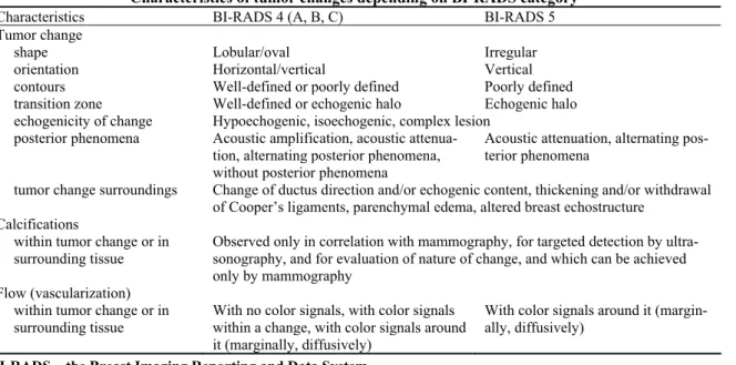
Table 1 Characteristics of tumor changes depending on BI-RADS category
Characteristics BI-RADS 4 (A, B, C) BI-RADS 5
Tumor change
shape Lobular/oval Irregular
orientation Horizontal/vertical Vertical
contours Well-defined or poorly defined Poorly defined transition zone Well-defined or echogenic halo Echogenic halo echogenicity of change Hypoechogenic, isoechogenic, complex lesion
posterior phenomena Acoustic amplification, acoustic attenua-tion, alternating posterior phenomena, without posterior phenomena
Acoustic attenuation, alternating pos-terior phenomena
tumor change surroundings Change of ductus direction and/or echogenic content, thickening and/or withdrawal of Cooper’s ligaments, parenchymal edema, altered breast echostructure
Calcifications
within tumor change or in surrounding tissue
Observed only in correlation with mammography, for targeted detection by ultra-sonography, and for evaluation of nature of change, and which can be achieved only by mammography
Flow (vascularization) within tumor change or in surrounding tissue
With no color signals, with color signals within a change, with color signals around it (marginally, diffusively)
With color signals around it (margin-ally, diffusively)
BI-RADS – the Breast Imaging Reporting and Data System.
Introduction
Breast cancer is a leading disease by mortality in Ser-bian women regardless age and it is a leading cause of early death in women between 25 and 44 years of age in Serbia 1. Mammography is still the “gold standard” for breast exami-nation. Screening mammography has certain limitations as well. First of all, about 20% of cancers present during mam-mography examination may be overlooked. This happens more often in young women due to density of their breast pa-renchyma 2. Then, if we take into account the amount of ra-diation in a ten-year time period starting at the age of 40, cancer induced by radiation will be a cause of death at most in 8 women in 100,000 performed mammography examina-tions 3. Ultrasonography is an additional diagnostic method. Not only that ultrasound can make the difference between cystic and solid changes, as it was a case at the beginning of its use, but it can also make the differential diagnosis in terms of benign-malignant. The main objection of ultrasono-graphic examination in early detection of cancer is inability to recognize microcalcifications. According to the latest pa-pers, microcalcifications may be viewed in 70%, and in case of cancer itself in 90% of cases by new ultrasonographic de-vices. The Breast Imaging Reporting and Data System (BI-RADS) was developed by American College of Radiology (ACR) in 1993, in order to standardize mammography re-ports and to enable easier communication between clinical practitioners dealing with this issue. BI-RADS classification has been applied in mammography only, and it appeared for two breast imaging modalities in the fourth revision of BI-RADS atlas: breast ultrasonography and magnetic resonance imaging (MRI). BI-RADS system is aimed to assess the risk, whether a viewed change is malignant, ie the following could be advised: biopsy, frequent radiology follow-ups or regular preventive examinations. The essence of BI-RADS nomen-clature is a final radiology report with a clearly numerically
indicated conclusion. A motive for this paper was the fact that ultrasonography BI-RADS lexicon has had short history and that there have been fewer data on its use as compared to mammography 4.
Criteria for assessment of pathological changes in the breast, diagnosed by ultrasonography examination are as fol-lows: shape (round, oval or irregular), orientation (horizontal or vertical), contours (very well-defined, poorly defined, an-gular, micro-lobular and spiculated), transition zone (well-defined or echogenic halo), echogenicity (non-echogenic, hyperechogenic, complex lesion, hypoechogenic and isoechogenic), posterior phenomena (without posterior phe-nomena, acoustic amplification, acoustic attenuation, alter-nating posterior phenomena), tissue around lesion (ductis, di-rection of Cooper’s ligaments, parenchymal edema, skin and impaired architectonic of tissue), calcifications, special ex-amples (grouped cysts, complicated cysts), flow (without color signals, with color signals and with color signals around the change) (Table 1).
BI-RADS classification in breast diseases defines whether a change detected in a breast carries a risk of malig-nancy and whether biopsy of that change is indicated. For BI-RADS 4 (A, B, C) and BI-RADS 5 categories of ultra-sonographic finding, pathohistological (PH) verification is necessary, which yields an appropriate indicator of BI-RADS classification accuracy. Therefore, the aim of this study was to categorize breast ultrasonographic finding into BI-RADS 4 (A, B, C) and BI-RADS 5 categories and correlation be-tween BI-RADS 4 (A, B, C) and BI-RADS 5 categories with PH finding of a breast change.
Methods
From the Registry of Cancer from the Institute for On-cology and Radiology of Serbia, and from radiologycal re-ports of ultrasonographic breast examinations performed in the Clinic for Radiotherapy and Radiology Diagnostics in the same institution, a group of 30 women was created, having clinical, mammographic and ultrasound breast examination followed by surgical biopsy with PH verification of a breast change in a period between November 1 2008 and March 31 2009. Criteria for patient selection were clinical, mammo-graphic and ultrasonomammo-graphic. Clinical criteria implied: premenopausal and postmenopausal patients; breast cancer diameter up to 3 cm or resistance with the third dimension regardless dimensions, but without engagement of the skin [T1 and T2 category as per tumor-nodus-metastasis (TNM) classification], status of regional lymph nodes N0, without distant metastasis (M0), mammographic criteria: a change scored as BI-RADS 0 based on standard mammography in two directions, ie a change that is not completely defined and which requires additional diagnostics; and ultrasonographic criteria: additional diagnostic procedures, additional ultra-sonography examination, clinically and mammographically, detected a change scored as BI-RADS 4A (slightly suspi-cious of malignancy), BI-RADS 4B (moderately suspisuspi-cious of malignancy), BI-RADS 4C (medium suspicious of malig-nancy) and BI-RADS 5 (highly suspicious of maligmalig-nancy) according to BI-RADS classification. Ultrasonographic ex-amination of breasts was performed with a 7.5 MHz probe (Sonoview, Acuson device). Before ultrasonographic exami-nation of breasts, the oncologists clinically examined breasts of every patient, and mammography in two directions was performed and analyzed by the radiologist (analogue mam-mography Lorad M-4, Hologic and digital mammam-mography Selenia, Hologic). Ultrasonographic examination of breasts was performed with the standard approach, with a patient be-ing in supination and lateral decubitus position, with exami-nation of regional lymph nodes and by using power Doppler color signalization. PH analysis implied ex tempore evalua-tion of preparaevalua-tions obtained by surgical biopsy, and than a standard analysis of preparations colored with hematoxylin eosin (HE). The results are presented in Tables and as graphs. Evaluation of normal distribution as per age of pa-tients revealed that data are homogenous and that parameter methods (χ2-test, level of significance p < 0.05) can be used for further comparisons.
Results
Sonographic morphology of cancer in BI-RADS 5 cate-gory is of the stellate type: irregular tumor change, with hypoechogenic, heteroechogenic centre, hyperechogenic border, acoustic posterior attenuation, interruption of liga-ments and fascia, with removing hypoechogenicity of subcu-taneous fat tissue and thick skin (Figure 1).
Figures 1 and 2 depict ultrasound features of breast masses categorized as BI-RADS 5 and BI-RADS 4A, respectively.
Fig. 1 – Ultrasound image of breast masses categorized as Breast Imaging Reporting and Data System (BI-RADS) 5.
Fig. 2 – Ultrasound image of breast masses categorized as Breast Imaging Reporting and Data System (BI-RADS) 4A.
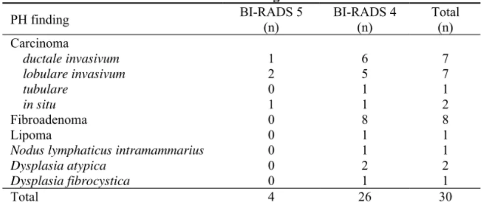
Table 2 Pathohistological (PH) finding of tumors in relation to BI-RADS 4 and
BI-RADS 5 categories
PH finding BI-RADS 5
(n)
BI-RADS 4 (n)
Total (n) Carcinoma
ductale invasivum 1 6 7
lobulare invasivum 2 5 7
tubulare 0 1 1
in situ 1 1 2
Fibroadenoma 0 8 8
Lipoma 0 1 1
Nodus lymphaticus intramammarius 0 1 1
Dysplasia atypica 0 2 2
Dysplasia fibrocystica 0 1 1
Total 4 26 30
BI-RADS – Breast Imaging Reporting and Data System.
Table 3 Pathohistological (PH) finding of tumors in relation to BI-RADS 4A, B, C and BI-RADS 5 categories
PH finding BI-RADS 5
(n)
BI-RADS 4C (n)
BI-RADS 4B, (n)
BI-RADS 4A, (n)
Total n (%)
Carcinoma mammae 4 5 6 2 17 (56.8)
Fibroadenoma 0 0 1 7 8 (26.6)
Lipoma 0 0 0 1 1 (3.3)
Nodus lymphaticus intramammaris 0 1 0 0 1 (3.3)
Dysplasia atypical 0 0 1 1 2 (6.7)
Dysplasia fibrocystica 0 0 1 0 1 (3.3)
Total 4 6 9 11 30 (100)
BI-RADS – Breast Imaging Reporting and Data System.
Table 4 Benign and malignant changes in relation to BI-RADS 4A, B, C and BI-RADS 5
Pathological finding (n) BI-RADS types
benign malignant Total
BI-RADS 4 (n = 26)
A 9 2 11
B 3 6 9
C 1 5 6
BI-RADS 5 (n = 4) 0 4 4
Total 13 17 30
BI-RADS – Breast Imaging Reporting and Data System.
of fibroadenoma in BI-RADS 4 while each finding in pa-tients with BI-RADS 5 was cancer.
Analysis of a total number of PH findings in the group of 30 patients (Table 3) also revealed a highly statistically significant difference (F = 11.278; df = 5; p < 0.01), resulting from the greater presence of cancer (56.7%) and fibroade-noma (26.7%) as compared to other types of findings, and particularly relatively rare lipomas, fibrocystic dysplasia and lymph nodes in breasts (by 3.3%).
In 6 patients with BI-RADS 4C category, PH findings were as follows: invasive ductal cancer in three patients, in-vasive lobular cancer in two, and intramammary lymph node hyperplasia in one patient.
In 9 patients with BI-RADS 4B category, PH findings were as follows: invasive lobular cancer in three patients, ductal cancer in two, tubular cancer (one patient), atypical lobular hyperplasia (one patient), fibroadenoma (one patient)
and fibrocystic dysplasia (one patient).
In 11 patients with BI-RADS 4A category, PH findings were as follows: fibroadenoma in seven patients, lipoma i one patient, invasive ductal cancer one patient, ductal cancer
in situ in one patient, and ductal hyperplasia with atypia in one patient.
If PH findings assigned into two categories (benign and malignant) are compared with BI-RADS categories (Table 4) we get a statistically significant difference (F = 6.188; df = 6;
If PH findings are compared with BI-RADS findings assigned into only two gradations (Table 4) we get a highly statistically significant difference (p = 0.002; p <0.01), re-sulting from fact that BI-RADS 5 (100.0%) prevails in pa-tients with malignant tumors, while BI-RADS 4 is present in an equal number of benign and malignant tumors.
When we consider age of the patients in relation to BI-RADS findings assigned into only two gradations (Table 5) we get a highly statistically significant difference (p = 0.002;
p < 0.01), resulting from the fact that patients from BI-RADS 5 group were older than 50 years of age, while only 50% of patients from BI-RADS 4 group were older than 50.
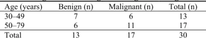
If age of the patients is compared with PH findings as-signed into two gradations (benign/malignant) (Table 6) we get a highly statistically significant difference (p = 0.032; p < 0.05), resulting from the fact that 53.8% of patients from the group with benign tumors were younger than 50 years of age, while this part was only 35.3% of patients with malig-nant findings; accordingly they were much older.
Table 5 Age of the patients in relation to
BI-RADS 4 and BI-RADS 5 categories Age (years) BI-RADS 5 (n) BI-RADS 4 (n) Total (n)
30–49 0 13 13
50–79 4 13 17
Total 4 26 30
BI-RADS – Breast Imaging Reporting and Data System.
Table 6 Age of the patients in relation to pathohistological (PH)
finding of either benign or malignant changes Age (years) Benign (n) Malignant (n) Total (n)
30–49 7 6 13
50–79 6 11 17
Total 13 17 30
Discussion
Ultrasonographic evaluation of breast changes, as a method complemented to clinical examination and to either diagnostic (“symptomatic” breast) or screening mammogra-phy (“asymptomatic” breast) can identify malignancy in some cases, which would otherwise be unidentified, which particularly relates to the glandular structure of breasts be-cause sensitivity of mammography reduces as density of glandular breast tissue becomes higher 5, 6. When it is about ultrasound descriptors according to data in the literature, the highest objectivity and concordance in the assessment among various physicians performing examination, is for a criterion of tumor change orientation (horizontal, ie in parallel with skin, characteristic for benign changes, or vertical, typical for malignant changes). The least accordance is found in the as-sessment of change contour (very well-defined contours usu-ally in benign changes, poorly defined usuusu-ally in malignant changes) and its echogenicity (nonechogenic and hyperecho-genic are benign changes, while hypoechohyperecho-genic, isoecho-genic changes and complex lesions can be seen in both types
of changes, but they are more suspicious of malignant na-ture) while shape, surrounding tissue and posterior phenom-ena are between these extremes 7. According to pathohis-tological findings in our study, breast cancer was identified in 17 out of 30 women (56.6%), invasive ductal carcinoma in 7 women, invasive lobular carcinoma also in 7 women, tubu-lar carcinoma in one woman and ductal carcinoma in situ in 2 women. Fibroadenoma was identified in 8 women (26.6%), dysplasia with atypia in two women (6.7%), while lipoma, intramammary lymph node or fibrocystic dysplasia was pathohistological finding in other patients (by 3.3%). If pa-tients are assigned into two groups: the first one of 49-year-old patients and younger, and the second one of 50-year-49-year-old patients and older in relation to PH finding of be-nign/malignant change, we will get a statistically significant difference because 53.8% of patients from the group with benign changes are younger than 49 years, while only 35.3% of patients have malignant finding. This information is con-sistent with data in the literature stating that cancer rate sud-denly increases after 40 years of age and with data for Cen-tral Serbia stating that in less than one fourth of the total number of women breast cancer was diagnosed before their 50 years of age 8. BI-RADS 4 changes were six times more common reason for biopsy than BI-RADS 5 (p < 0.01) which is consistent with data from the literature. BI-RADS 4 cate-gory is a change with ultrasonographic characteristics having small (A), moderate (B) or medium (C) risk of malignancy. A supposed risk of cancer is 3–94% in this category. BI-RADS 5 is a change of ultrasonographic finding with high malignancy risk (risk of cancer is higher than 95%) and bi-opsy is required 9.
Therefore, this paper proves the correlation between BI-RADS 4 and 5 categories and PH finding (p < 0.05). Every tumor in BI-RADS 5 category was cancer (4/4), while none of benign tumors belonged to RADS 5 category. In RADS 4 category, in malignant tumors, it was found that BI-RADS 4 B and C prevail (64.7%), while BI-BI-RADS 4A pre-vail in benign tumors (69.2%), being a very rare finding in malignant tumors (11.8%). Therefore, it could be said that a BI-RADS 4A finding is almost always related to benign changes, primarily to fibroadenoma, and all other with ma-lignant (specificity for BI-RADS 5 is 100.0%, for BI-RADS 4C 83.3%, and for BI-RADS 4B 67%).
A PH finding was invasive ductal cancer in one patient, then ductal cancer in situ in one, and invasive lobular cancer in two out of four patients with BI-RADS 5 category.
According to the literature, this is a typical ultrasono-graphic view of malignant tumors, with specificity for cancer up to 98%. Pathohistologically, it corresponds to cancers with pronounced desmoplastic reaction and it most often confirms invasive ductal, tubular or lobular cancer.
ultra-sonographic finding being nodular cancer type corresponds to dominant cellularity tumors and it is mostly found in inva-sive lobular cancer and invainva-sive ductal cancer 3.
Moreover, according to a new, molecular classification of breast cancers, triple-negative breast cancers [estrogen-receptor negative, progesterone-[estrogen-receptor negative and human epidermal receptor (HER)2 negative cancer], have mostly nodular type of expression in ultrasonographic finding with
no necessary elements for a benign change 10.
Triple-negative breast cancer are aggressive tumors with poor prog-nosis, and their frequency is higher in women younger than 50 years of age 11.
Intramammary lymph node certainly belongs to a be-nign finding (BI-RADS 2). The basis of certainly bebe-nign change is visible fat tissue within lymph node hilus 12. How-ever, in case of lymph node hyperplasia, when it crosses lon-gitudinal diameter of 1 cm, then in atypical localization (in-ner quadrants) or unfavorable contrast in relation to basic structure of breasts, and accordingly its poor visualization, intramammary lymph node may simulate a malignant change 13, 14.
The BI-RADS 4B group presents heterogeneity of pathohistological and sonomorphological characteristics by four types: nodular form (previously mentioned), well-bordered tumor with pseudocapsule, cystic tumor type and diffuse infiltrative growth tumors.
The pseudobenign aspect of well-defined tumor with pseudocapsule implies: circular or oval shape, well-defined contours, non-homogeneous echotexture and pseudocapsule. According to the literature, this type is particularly character-istic for medullary carcinoma, when they look like cysts with thick content and acoustic amplification. The probability of malignancy in this sonomorphological type is 1–4% at more than 40 years of age 3.
Structurally changed zone, without defined border, an-gular and dilated ductus, hypoechogenic zones with acoustic shadow are viewed in ultrasonographic finding of diffuse in-filtrative growth tumor. This way of sonographic expression is most common in lobular cancer, confirmed in three pa-tients in our group with BI-RADS 4B category. During tu-mor growth, three-dimensional tutu-mor change does not de-velop initially, but most often focal nodularity or parenchy-mal asymmetry, like dysplastic changes, and therefore tumor is diagnosed in its late stage 14.
Cystic cancer is cystic with intracystic proliferation (complex – semisolid, semicystic lesion) by type. According to the literature, it is a lesion with low frequency of occur-rence (less than 1%) 3. In our group, one patient had
patho-histological finding Dysplasia polycystica proliferativa
mammae. Four types of cystic lesions are listed in BI-RADS atlas: simple cyst, grouped microcysts, complicated cyst and complex lesion. Simple cyst belongs to BI-RADS 2 category. Grouped microcysts belong to BI-RADS 3 if they are non-palpable, while palpable cysts are subjected to fine needle aspiration. Complicated cysts (not necessarily indicating in-flamed cysts) refer to hypoechogenic lesion with other char-acteristics of a benign change. Nonpalpable cysts belong to BI-RADS 3 category, and palpable are subjected to fine
nee-dle aspiration. Complex lesion refers to the presence of solid and cystic component and it can belong to: intracystic papil-loma, cancer, cystic degeneration of either malignant or be-nign tumors and it is classified into BI-RADS 4 category.
The aforementioned overlaps of sonomorphological types seen in this type of cancer with benign changes are the reason for biopsy and pathohistological evaluation, but false negative findings are possible, as well.
Sonomorphological pseudobenign tumor type with cap-sule prevailed in the group with BI-RADS 4A category, while the most common PH finding was fibroadenoma.
Overlapping of clinical, mammographic and sono-graphic findings of fibroadenoma with malignant changes is possible. Therefore, a question remains on the algorithm of diagnostics in palpable change of clinically benign aspect, which is mammographically a type of circular, oval or lobu-lar and well-defined shadow. In this case, ultrasonographic diagnostics is a necessary additional modality of examina-tion, after clinical examination and mammography. If a change is of the solid type, not cystic, a finding is defined as solid tumor, with sonomorphologically benign characteris-tics, with low risk of malignancy (BI-RADS 4A category) and core needlebiopsy of change is indicated (Figure 2). The probability of malignancy is excluded only if fibroadenoma is pathohistologically proved. Therefore, fibroadenoma is significantly present in BI-RADS 4A in this study. A com-mon incidental sonographic finding of non-palpable solid tumors with benign aspect, with size less than 1 cm, has de-fined another algorithm over time, and it differs from the abovementioned algorithm so far as it is related to nonpalpa-ble change. Namely, a retrospective analysis revealed that risk of malignancy for nonpalpable changes being sono-graphically a solid type and benign morphology is less than 1–2%. Therefore, the approach is sonographic monitoring in 2 years' time every 6 months, as BI-RADS 3 category, and in case of a stationary finding, it should be translated into BI-RADS 2 category (certainly benign change) after two years of monitoring, without indications for biopsy 15.
Ductal carcinoma in situ is normally nonpalpable, sub-clinical change, detected by mammography during preventive examinations, primarily through microcalcifications typical for malignant changes. However, it was found in two patients with palpable change in our group of patients, as RADS 5 and BI-RADS 4A ultrasound category. Ductal carcinoma in situ may sometimes clinically manifest as palpable resistance or nodular-ity. Non-specific cystic or solid lesion, not well bordered hypoechogenic change, microlobular tumor, ductus dilatation or calcifications, are present in the ultrasonographic finding. It is hard to differentiate ductal carcinoma in situ without typical ra-diologic suspicious calcifications on mammography from be-nign lesions only by ultrasonography, so further cytological or pathohistological evaluation is necessary 16.
Conclusion
six times more common cause for biopsy than BI-RADS 5 category. BI-RADS 5 and BI-RADS 4 categories are the fol-lowing pathohistological types of lesions: breast cancer (56.7%), fibroadenoma (26.7%), dysplasia with atypia (6.7%) and lipoma, intramammary lymph node or fibrocystic dyspla-sia by 3.3%. There is a correlation between BI-RADS 4 and 5 categories and pathohistological findings: BI-RADS 4A find-ing is almost always related to benign changes, primarily to fi-broadenoma, and all other with malignant (specificity for RADS 5 is 100.0%, for RADS 4C 83.3%, and for
BI-RADS 4B 67%). BI-BI-RADS 4 and 5 categories and age of women are related to pathohistological finding: benign changes are more present (53.8% of all benign changes) than malignant (35.3% of all malignant changes) in 49 year-old women and younger. Heterogeneity of pathohistological find-ings in BI-RADS 4 category with the domination of malignant changes in BI-RADS 4B and BI-RADS 4C groups, as well as only malignant changes in BI-RADS 5 category confirm ne-cessity of pathohistological verification of lesions from these categories, particularly in women older than 50 years of age.
R E F E R E N C E S
1. Milošević Z. Newspapers mammography in the diagnosis of breast cancer. In: Nešković-Konstantinović Z, Borojević N, Vučković-Dekić Lj, editors. Newspapers in the diagnosis and treatment of breast cancer. Belgrade: Akademija medicinskih nauka Srpskog lekarskog društva, Institut za onkologiju i radiologiju Srbije; 2008. p. 41−52. (Serbian)
2. Crystal P, Strano SD, Shcharynski S, Koretz MJ. Using sonography to screen women with mammographically dense breasts. AJR Am J Roentgenol 2003; 181(1): 177−82.
3. Pichler E. Ultrasound breast atlas: differential diagnosis and intervention techniques. Zagreb: Školska knjiga; 2005. (Croatian) 4. Levy L, Suissa M, Chiche JF, Teman G, Martin B. BIRADS
ultra-sonography. Eur J Radiol 2007; 61(2): 202−11.
5. Dobrosavljević A, Brković V, Vujković B, Milošević Z. Basic constitutional and reproductive parameters in optimalise application of mammography in the diagnosis of breast diseases. Medicinski podmladak 2004; 55: 61−3. (Serbian)
6. Milošević Z. Malignant tumors of the breast. In: Goldner B, Milošević Z, Jovanović T, editors. Mammography in the diagnosis of breast diseases. Belgrade: Velarta; 2001. p. 217−82. (Serbian)
7. Park CS, Lee JH, Yim HW, Kang BJ, Kim HS, Jung JI, et al. Observer Agreement Using the ACR Breast Imaging Reporting and Data System (BI-RADS)-Ultrasound, First Edition (2003). Korean J Radiol 2007; 8(5): 397−402.
8. Jovićević BA. Epidemiology and prevention of breast cancer. In: Nešković-Konstantinović Z, Borojević N, Vučković-Dekić Lj, editors. Newspapers in the diagnosis and treatment of breast cancer. 2nd
ed. Belgrade: Akademija medicinskih nauka Srpskog lekarskog društva i Institut za onkologiju i radiologiju Srbije. 2008. p. 11−24. (Serbian)
9. American College of Radiology. ACR Breast Imaging Reporting and Data System, Breast Imaging Atlas. 4th ed. Reston, VA: American College of Radiology; 2003.
10. Stavros AT, Thickman D, Rapp CL, Dennis MA, Parker SH, Sisney GA. Solid breast nodules: use of sonography to distinguish be-tween benign and malignant lesions. Radiology 1995; 196(1): 123−34.
11. Ko ES, Lee BH, Kim H, Noh W, Kim MS, Lee S. Triple-negative breast cancer: correlation between imaging and pathological find-ings. Eur Radiol 2009; 20(5): 1111−7.
12. Radisky ES, Radisky DC. Matrix metalloproteinase-induced epithe-lial-mesenchymal transition in breast cancer. J Mammary Gland Biol Neoplasia 2010; 15(2): 201−12.
13. Kinoshita T, Yashiro N, Yoshigi J, Ihara N, Fukuma E, Narita M. In-flammatory intramammary lymph node mimicking the malignant lesion in dynamic MRI: a case report. Clin Imaging 2002; 26(4): 258−62.
14. Tabár L, Duffy SW, Vitak B, Chen HH, Prevost TC. The natural his-tory of breast carcinoma: what have we learned from screening. Cancer 1999; 86(3): 449−62.
15. Graf O, Helbich TH, Hopf G, Graf C, Sickles EA. Probably benign breast masses at US: is follow-up an acceptable alternative to bi-opsy. Radiology 2007; 244(1): 87−93.
16. Izumori A, Takebe K, Sato A. Ultrasound findings and histological features of ductal carcinoma in situ detected by ultrasound exami-nation alone. Breast Cancer 2010; 17(2): 136−41.