Positive predictive values of Breast Imaging Reporting
and Data System (BI-RADS
®) categories 3, 4 and 5 in breast
lesions submitted to percutaneous biopsy
*
Valores preditivos positivos das categorias 3, 4 e 5 do Breast Imaging Reporting and Data System (BI-RADS®) em lesões mamárias submetidas a biópsia percutânea
Gustavo Machado Badan1, Décio Roveda Júnior2, Carlos Alberto Pecci Ferreira3, Felipe Augusto
Trocoli Ferreira4, Eduardo de Faria Castro Fleury5, Mário Sérgio Dantas do Amaral Campos4,
Rodrigo de Oliveira Seleti6, Hélio da Cruz Júnior7
Objective: To evaluate the BI-RADS as a predictive factor of suspicion for malignancy in breast lesions by correlating radiological with histological results and calculating the positive predictive value for categories 3, 4 and 5 in a breast cancer reference center in the city of São Paulo. Materials and Methods: Retrospective, analytical and cross-sectional study including 725 patients with mammographic and/or sonographic findings classified as BI-RADS categories 3, 4 and 5 who were referred to the authors’ institution to undergo percutaneous biopsy. The tests results were reviewed and the positive predictive value was calculated by means of a specific mathematical equation. Results: Positive predictive values found for categories 3, 4 and 5 were respectively the following: 0.74%, 33.08% and 92.95%, for cases submitted to ultrasound-guided biopsy, and 0.00%, 14.90% and 100% for cases submitted to stereotactic biopsy. Conclusion: The present study demonstrated high suspicion for malignancy in lesions classified as category 5 and low risk for category 3. As regards category 4, the need for systematic biopsies was observed.
Keywords: Breast cancer; BI-RADS; Percutaneous biopsy; Positive predictive value; Histological diagnosis.
Objetivo: Avaliar o sistema BI-RADS como fator preditivo de suspeição para malignidade em lesões mamárias, corre-lacionando os achados radiológicos e os resultados histológicos por meio do cálculo do valor preditivo positivo das ca-tegorias 3, 4 e 5 em serviço de referência em diagnóstico e tratamento de câncer de mama da cidade de São Paulo.
Materiais e Métodos: Estudo retrospectivo, analítico e transversal contendo casuística de 725 pacientes com acha-dos mamográficos e/ou ultrassonográficos classificaacha-dos nas classes 3, 4 e 5 do BI-RADS e que foram encaminhadas para realização de biópsia percutânea. Os exames foram revisados e o cálculo do valor preditivo positivo foi feito utili-zando-se equação matemática específica. Resultados: Os valores preditivos positivos encontrados das categorias 3, 4 e 5 foram 0,74%, 33,08% e 92,95%, respectivamente, para os casos de biópsias orientadas pelo ultrassom, e 0,00%, 14,90% e 100% para os casos orientados por estereotaxia. Conclusão: Este estudo demonstrou alta suspeição para malignidade em lesões classificadas na categoria 5 e diminuto risco para a categoria 3. Quanto à categoria 4, ficou constatada a necessidade de biópsias sistemáticas.
Unitermos: Câncer de mama; BI-RADS; Biópsia percutânea; Valor preditivo positivo; Diagnóstico histológico.
Abstract
Resumo
* Study developed at Faculdade de Ciências Médicas da Santa Casa de São Paulo, São Paulo, SP, Brazil.
1. Specialist in Breast Imaging, Second Radiologist Assis-tant, Breast Radiology Group of Santa Casa de Misericórdia de São Paulo, São Paulo, SP, Brazil.
2. PhD, Professor of Radiology, Director of the Department of Imaging Diagnosis, Santa Casa de Misericórdia de São Paulo, São Paulo, SP, Brazil.
3. Physician Assistant, Coordinator, Breast Imaging Service, Department of Medical Practice, Faculdade de Medicina da Santa Casa de São Paulo, São Paulo, SP, Brazil.
4. MDs, Specialists in Radiology, Second Radiologist Assis-tants, Santa Casa de Misericórdia de São Paulo, São Paulo, SP, Brazil.
5. PhD, Professor of Radiology, Second Radiologist Assis-tant, Santa Casa de Misericórdia de São Paulo, São Paulo, SP, Brazil.
Badan GM, Roveda Júnior D, Ferreira CAP, Ferreira FAT, Fleury EFC, Campos MSDA, Seleti RO, Cruz Júnior H. Positive predictive values of Breast Imaging Reporting and Data System (BI-RADS®) categories 3, 4 and 5 in breast lesions submitted to percutaneous biopsy.
Radiol Bras. 2013 Jul/Ago;46(4):209–213.
veloped and developing countries. About 226,870 new cases are expected, and 39,510 women will die because of breast cancer in the United States of America in 2012(1). According to Instituto Nacional de
Câncer (INCA) estimates of cancer inci-dence in Brazil this year, 52,680 new cases are expected, with an estimated risk of 52 cases in 100 thousand women(2).
According to extensive observational series, the rate of breast cancer mortality has decreased to 31%, primarily due to the
INTRODUCTION
Breast cancer is the most frequent type of cancer in women worldwide, both in
de-6. MD, Specialist in Radiology, Fellow Master degree in Bre-ast Imaging, Santa Casa de Misericórdia de São Paulo, São Paulo, SP, Brazil.
7. Fellow Master degree in Breast Imaging, Santa Casa de Misericórdia de São Paulo, São Paulo, SP, Brazil.
Mailing Address: Dr. Gustavo Machado Badan. Rua Loureiro da Cruz, 121, ap. 121, Aclimação. São Paulo, SP, Brazil, 01529-020. E-mail: [email protected].
contribution of yearly mammographic screening programs which have lead to early detection of the disease in a consid-erable number of cases(3–5).
The Colégio Brasileiro de Radiologia e Diagnóstico por Imagem (CBR) (Brazilian College of Radiology and Imaging Diagno-sis) and the National Commission on Mam-mography recommend yearly mammo-graphic screening for every women be-tween 40 and 69 years, and on an individual basis after that age range(6).
However, the mammographic screening has been followed by a high number of biopsies, although mammographic findings considered as suspicious for malignancy may correspond to benign alterations. Thus, despite the high mammographic sensitivity and specificity, the positive predictive value (PPV) of biopsies reveals malignancy in only 15% to 40% of cases(7,8).
In order to reduce the disagreement in breast images interpretation, and to stan-dardize reports and respective recommen-dations, in 1993, the American College of Radiology published the Breast Imaging Reporting and Data System (BI-RADS®)(9),
which is currently in its fourth edition(10,11).
The present study was aimed at evalu-ating the BI-RADS as a predictive factor for suspicion of malignancy in breast le-sions by correlating radiological findings with histological results in a breast cancer reference hospital in São Paulo, SP. Brazil. Such a comparison was undertaken by means of PPV calculation for BI-RADS categories 3, 4 and 5 in order to improve the management of abnormal imaging find-ings. As a secondary objective, the authors have determined the PPV of morphologi-cal characteristics of the breast lesions most frequently associated with malignancy.
MATERIALS AND METHODS
By means of a retrospective, analytical and cross sectional study approved by the Committee for Ethics in Research of the In-stitution, and developed in the Breast Im-aging Service at Santa Casa de Misericór-dia de São Paulo, the authors evaluated his-tological results of 725 patients with mean age of 49 years (age range = 16 to 84 years). Such patients presented palpable or non palpable breast lesions at mammography
and/or ultrasonography, classified as BI-RADS categories 3, 4 and 5, and were re-ferred for fine needle aspiration biopsy (FNAB), core biopsy or vacuum-assisted core biopsy in the period from February 1st, 2011 to July 31, 2012.
The patients had their results reviewed on the day of the procedure, and the find-ings were reclassified according the BI-RADS categories by a clinical team with more than ten-year experience in breast imaging.
The cases reclassification was based on a review of the mammographic images and in cases of ultrasonography-guided biopsy a new sonographic study was performed. Finally, the BI-RADS lexicon for breast lesions was utilized, and then the final cat-egorization was done. Microcalcifications were assessed according their morphology (punctiform, pleomorphic or fine pleomor-phic and liner-branching), and according their distribution (clustered or segmental). Nodules were classified according their shape (ovoid, round or irregular) and mar-gins (circumscribed, non circumscribed and spiculated). Among lesions with non circumscribed margins, the lesions with spiculated margins were separately evalu-ated considering their high association with malignancy. Also, focal asymmetry, with or without associated findings (microcalci-fications or distortion), and architectural distortion were evaluated.
In the present study, the exclusion cri-teria were the following: a) radiological findings classified as BI-RADS 2; b) cases considered unsatisfactory for purposes of cytological analysis.
Core biopsies were performed with 12-gauge needles, collecting eight samples per procedure, and the vacuum-assisted biop-sies (Surus system), with 9-gauge needles, both under digital stereotactic guidance (Lorad Multicare Platinum; Hologic). As regards ultrasonography-guided core bi-opsy (EnVisor Ultrasound System; Philips Healthcare), 14-gauge needles were uti-lized, collecting four samples per proce-dure. Ultrasonography was the method of choice for guidance in FNAB. In all the cases of biopsied microcalcifications, the specimens were submitted to radiography and considered satisfactory as calcifica-tions were detected.
The PPVs were calculated and final re-sults were compared with those in the lit-erature.
RESULTS
Among the 725 lesions submitted to percutaneous biopsy, 133 were excluded for being reclassified as BI-RADS 2, and other 12 cases with unsatisfactory material were eliminated. So, 580 procedures met all the inclusion criteria.
In this total of cases, the percentage dis-tribution of imaging studies according their BI-RADS classification showed 47.58% (276) for category 3, 39.65% (230) for cat-egory 4, and 12.75% (74) for catcat-egory 5.
Most of the interventional procedures, represented by 64.82% (376) of cases, was performed by means of core biopsy, and 35.18% (204) by FNAB.
The histological results demonstrated 77.59% negative for malignancy, and 22.41% positive for malignancy (Table 1). Among the results negative for malignancy (450 cases), the most prevalent were fibro-adenomas (36.66%) and fibrocystic alter-ations (30.44%), while results positive for malignancy (130 cases) were represented by invasive ductal carcinoma (67%), followed by invasive lobular carcinoma (10.7%) and by in situ ductal carcinoma (7.7%).
The data indicate a predominance of be-nign results in patients classified as BI-RADS 3, both at mammography (100.00%) and at ultrasonography (99.26%). The PPV of BI-RADS category 4 was 14.90% for the cases of stereotactic biopsy, and 33.08% for cases of ultrasonography-guided biopsy. For BI-RADS category 5, the authors ob-tained PPV of 100.00% at mammography and 92.25% at ultrasonography (Tables 2 and 3).
One of the two cases classified as BI-RADS 3 and positive for neoplastic cells is represented on Figure 1. And among the five cases classified as BI-RADS 5 with histological results negative for malig-nancy, one is shown on Figure 2.
Table 2 Distribution of cases of percutaneous stereotactic biopsies, in agreement with the radiological BI-RADS classification and histopathological diagnosis of benignity or malignancy.
BI-RADS
3 4 5 Total
Histopathological diagnosis
Benign Malignant Total
N
5 80 0 85
%
100.00 85.10
0.00 77.97
N
0 14
3 17
%
00.00 14.90 100.00
22.03
N
5 94
3 102
%
4.90 92.16
2.94 100.00
BI-RADS 3: VPP = 0.00%; BI-RADS 4: VPP = 14.90%; BI-RADS 5: VPP = 100.00%. N, number of cases.
Table 3 Distribution of cases of percutaneous ultrasonography-guided biopsies, in agreement with the radiological BI-RADS classification and histopathological diagnosis of benignity or malignancy.
BI-RADS
3 4 5 Total
Histopathological diagnosis
Benign Malignant Total
N
269 91
5 365
%
99.26 66.92 7.05 76.36
N
2 45 66 113
%
0.74 33.08 92.95 23.64
N
271 136 71 478
%
56.70 28.45 14.85 100.00
BI-RADS 3: VPP = 0.74%; BI-RADS 4: VPP = 33.08%; BI-RADS 5: VPP = 92.95%. N, number of cases.
DISCUSSION
Since 1993, when the BI-RADS was published, innumerable studies have been developed to correlate imaging findings with histopathological results(9).
Although the BI-RADS itself recom-mends that biopsy is not performed in pa-tients with lesions classified as BI-RADS category 3, such a procedure is performed in a high number of cases. The main rea-sons for the practice of biopsy include pa-tients’ anxiety, physicians’ insecurity and presence of risk factors for breast cancer. With the objective of providing mastolo-gists with further information to select pa-tients eligible for biopsy, studies in the lit-erature have tried to establish the PPVs of BI-RADS categories 3, 4 and 5(12).
Studies in the literature have reported PPV for category 3 between 0% e 3%, while for category 4it has ranged between 15% and 40%, and for category 5, between 81% and 97%(7,8,12–17).
Table 1 Histopathological result of percutaneous biopsies of lesions classified as BI-RADS categories 3, 4 and 5. Histopathological result
Findings negative for malignancy
Precursor lesions/lesions with increased breast cancer risk
Positive for malignancy
Histological type
Fibroadenoma Fibrocystic alteration
Ductal hyperplasia without atypias Steatonecrosis
Mastitis Adenosis Apocrine cyst Fibrosclerosis Papilloma Sclerosing adenosis Intramammary lymph node Tubular adenoma
Ductal hyperplasia with atypias Complex sclerosing lesion Complex fibroadenoma
Columnar cell lesions with atypias Lobular hyperplasia with atypias
Invasive ductal carcinoma Invasive lobular carcinoma
In situ ductal carcinoma
Mucinous carcinoma Invasive apocrine carcinoma Lesion of malignant nature Invasive papilliferous carcinoma Invasive cribriform carcinoma
Number of cases
165 137 60 17 16 9 7 6 6 2 2 1
13 4 3 1 1
87 14 10 5 5 5 3 1
Percentage
28.45% 23.62% 10.34% 2.93% 2.76% 1.55% 1.20% 1.04% 1.04% 0.35% 0.35% 0.17%
2.24% 0.69% 0.52% 0.17% 0.17%
As the present study results are corre-lated with those in the literature, one ob-serves that in spite being within the ex-pected ranges for their respective catego-ries, particularly in category 3, the PPVs ap-proximated the inferior threshold (PPV of 0.0% for mammography and 0.74% for ul-trasonography), while in category 5 PPV of
92.95% was observed in cases of ultra-sonography-guided biopsies, approximat-ing the superior threshold reported by the studies and 100% for stereotactic biopsies, which demonstrates a good accuracy of the team in the BI-RADS categorization.
The categorization according the BI-RADS may present limitations due to
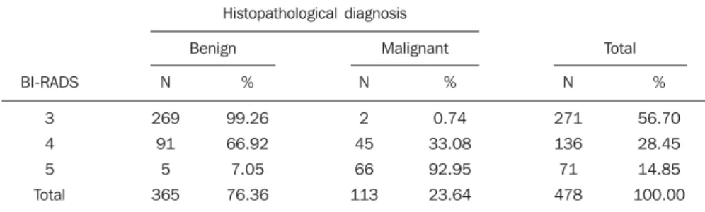
de-Figure 1. Non palpable nodules located in the axillary extension of the left breast in a 63-year-old woman. Ultrasonography demonstrates an ovoid, hypoechoic, circumscribed nodule measuring 9 × 6 × 8 mm, with homogeneous echotexture, parallel orientation, without posterior acoustic shadowing. Category BI-RADS 3. FNAB demonstrated the presence of cells positive for malignancy. Surgery revealed a mucinous carcinoma measuring 7 mm with negative sentinel lymph node.
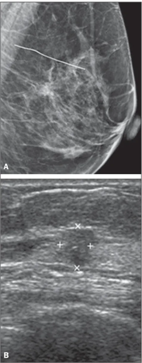
Figure 2. Non palpable nodule in the retroareolar region of the left breast in a 66-year-old woman previously submitted to surgery for a benign nod-ule in the same breast. A: Mammography demon-strates the presence of an irregular, isodense nod-ule with spiculated margins. Category BI-RADS 5. B: Ultrasonography demonstrates irregular, hypo-echoic nodule with angulated margins, measuring 6 mm, with heterogeneous echotexture, non par-allel orientation, and posterior acoustic shadowing. Category BI-RADS 5. Core biopsy revealed adenosis and fibrocystic alterations. Considering the absence of anatomo-radiological correlation, the patient underwent surgery that revealed the same anato-mopathological finding.
B A
Table 4 BI-RADS categorization of sonographic findings and attribution of PPVs of the morphological characteristics of lesions.
BI-RADS category
3
4
5
Ultrasonographic findings
Ovoid nodule with circumscribed margins Thick-walled cyst or clustered cysts
Solid nodule with noncircumscribed margins Complex cyst
Irregular nodule with spiculated margins
Total
248 23
118 18
71
Positive for malignancy
2 0
43 2
66
PPV
0.80% 0%
36.44% 11.11%
92.95%
Table 5 BI-RADS categorization of mammographic findings and attribution of PPVs of the morphological characteristics of lesions.
BI-RADS category
3
4
5
Mammographic findings
Clustered punctiform microcalcifications Focal asymmetry without associated findings
Clustered pleomorphic microcalcifications Architectural distortion
Focal asymmetry with punctiform calcifications
Fine pleomorphic and segmental branching microcalcifications
Total
4 1
79 13 2
3
Positive for malignancy
0 0
10 4 0
3
PPV
0% 0%
12.65% 30.77% 0%
100%
ficient training of radiologists, influencing PPV results(18,19). For this reason, all the
In the present study, the PPV for BI-RADS category 4 was 33.08% in the cases with ultrasonography-guided biopsy, within the expected average reported in the literature; and 14.9% in the cases with ste-reotactic biopsy, an index similar to the values found by Melhado et al.(17).
Histo-pathological analyses of breast biopsies of women whose mammograms revealed sus-picious microcalcifications diagnosed breast cancer in 10% to 40% of cases(20). In
the present study, among 82 biopsied cases (BI-RADS 4 and 5), 13 were malignant le-sions, revealing PPV of 15.85%. Pleomor-phic and linear branching lesions, particu-larly those with segmental distribution, are considered as highly suspicious(21,22) and,
according to Liberman et al., reveal malig-nancy in 81% to 92% of cases(14). In the
present study, three cases presented such characteristics, and all of them were posi-tive for malignancy.
In several studies in the literature, mor-phological criteria with higher PPV in re-lation to nodules were spiculated margins, followed by irregular shape ranging from 70% to 92%(14,23–26). For irregular and
spicu-lated nodules, the authors have found PPV of 94.28%, a value similar to those reported in the literature. On the other hand, reports in the literature indicate that a nodule with circumscribed margin is the criterion that is most frequently associated with benig-nity, in agreement with the present study results (99.1% of benign cases)(23,27–29).
CONCLUSION
The present study has demonstrated agreement with data in the literature regard-ing the prediction of a benign or malignant nature of breast lesions, with the objective of improving the appropriate management of lesions while optimizing the practice of biopsies. Among the morphological crite-ria, spiculated margins of nodules and seg-mental, fine pleomorphic and branching microcalcifications are most frequently associated with malignancy.
Considering the present study results, one can affirm with certainty that lesions classified as BI-RADS category 5 are highly suspicious of malignancy, and that
the major contribution is related to a con-servative management of breast lesions classified as mammographic and/or sono-graphic BI-RADS category 3, in order to avoid unnecessary biopsies. As regards BI-RADS category 4, the authors have found out the necessity of systematic biopsies.
REFERENCES
1. National Cancer Institute. Surveillance, Epidemi-ology and End Results (SEER). [acessado em 13 de agosto de 2012]. Disponível em: http://seer. cancer.gov.
2. Ministério da Saúde. Instituto Nacional de Câncer. Estimativa 2012: incidência de câncer no Brasil. [acessado em 16 de agosto de 2012]. Disponível em: http://www.inca.gov.br/estimativa/2012. 3. Tabár L, Vitak B, Chen TH, et al. Swedish
two-county trial: impact of mammographic screening on breast cancer mortality during 3 decades. Ra-diology. 2011;260:658–63.
4. Kopans DB, Smith RA, Duffy SW. Mammo-graphic screening and “overdiagnosis”. Radiol-ogy. 2011;260:616–20.
5. Miranda CMNR, Santos CJJ, Maranhão CPM, et al. A tomografia computadorizada multislice é
ferramenta importante para o estadiamento e se-guimento do câncer de mama? Radiol Bras. 2012; 45:105–12.
6. Urban LABD, Schaefer MB, Duarte DL, et al. Recomendações do Colégio Brasileiro de Radio-logia e Diagnóstico por Imagem, da Sociedade Brasileira de Mastologia e da Federação Brasileira das Associações de Ginecologia e Obstetrícia para rastreamento do câncer de mama por métodos de imagem. Radiol Bras. 2012;45:334–9.
7. Kestelman FP, Souza GA, Thuler LC, et al. Breast Imaging Reporting and Data System – BI-RADS®:
valor preditivo positivo das categorias 3, 4 e 5. Revisão sistemática da literatura. Radiol Bras. 2007;40:173–7.
8. Hall FM, Storella JM, Siverstone DZ, et al. Non-palpable breast lesions: recommendations for bi-opsy based on suspicion of carcinoma at mam-mography. Radiology. 1988;167:353–8. 9. American College of Radiology. Breast Imaging
Reporting and Data System (BI-RADS). Reston, VA: American College of Radiology; 1993.
10. American College of Radiology. The ACR Breast Imaging Reporting and Data System (BI-RADS®).
4th ed. Reston, VA: American College of Radiol-ogy; 2003.
11. Chala LF, Barros N. ACR BI-RADS na ultra-so-nografia. Radiol Bras. 2004;37(2):iii–iv.
12. Roveda Jr D, Piato S, Oliveira VM, et al. Valores preditivos das categorias 3, 4 e 5 do sistema BI-RADS em lesões mamárias nodulares não palpá-veis avaliadas por mamografia, ultra-sonografia e ressonância magnética. Radiol Bras. 2007;40: 93–8.
13. Raza S, Chikarmane SA, Neilsen SS, et al. BI-RADS 3, 4, and 5 lesions: value of US in man-agement – follow-up and outcome. Radiology. 2008;248:773–81.
14. Liberman L, Abramson AF, Squires FB, et al. The Breast Imaging Reporting and Data System: posi-tive predicposi-tive value of mammographic features and final assessment categories. AJR Am J Roentgenol. 1998;171:35–40.
15. Lacquement MA, Mitchell D, Hollingsworth AB. Positive predictive value of the Breast Imaging Reporting and Data System. J Am Coll Surg. 1999;189:34–40.
16. Orel SG, Kay N, Reynolds C, et al. BI-RADS cat-egorization as a predictor of malignancy. Radiol-ogy. 1999;211:845–50.
17. Melhado CV, Alvares RB, Almeida JO. Correla-ção radiológica e histológica de lesões mamárias não palpáveis em pacientes submetidas a marca-ção pré-cirúrgica, utilizando-se o sistema BI-RADS. Radiol Bras. 2007;40:9–11.
18. Liberman L, Menell JH. Breast Imaging Report-ing and Data System (BI-RADS). Radiol Clin North Am. 2002;40:409–30.
19. Godinho ER, Koch HA. Breast Imaging Report-ing and Data System (BI-RADS): como tem sido utilizado? Radiol Bras. 2004;37:413–7.
20. CoÕar ZS, Çetin M, Tepe TK, et al. Concordance of mammographic classifications of microcalci-fications in breast cancer diagnosis: utility of the Breast Imaging Reporting and Data System (fourth edition). Clin Imaging. 2005;29:389–95. 21. Burnside ES, Ochsner JE, Fowler KJ, et al. Use of microcalcification descriptors in BI-RADS 4th edition to stratify risk of malignancy. Radiology. 2007;242:388–95.
22. Nascimento JHR, Silva VD, Maciel AC. Acurácia dos achados mamográficos do câncer de mama: correlação da classificação BI-RADS e achados histológicos. Radiol Bras. 2010;43:91–6. 23. Calas MJG, Koch HA, Dutra MVP.
Ultrassono-grafia mamária: avaliação dos critérios ecográfi-cos na diferenciação das lesões mamárias. Radiol Bras. 2007;40:1–7.
24. Bérubé M, Curpen B, Ugolini P, et al. Level of suspicion of a mammographic lesion: use of fea-tures defined by BI-RADS lexicon and correla-tion with large-core breast biopsy. Can Assoc Radiol J. 1998;49:223–8.
25. Mendez A, Cabanillas F, Echenique M, et al. Mammographic features and correlation with bi-opsy findings using 11-gauge stereotactic vacuum-assisted breast biopsy (SVABB). Ann Oncol. 2004;15:450–4.
26. Stavros AT, Thickman D, Rapp CL, et al. Solid breast nodules: use of sonography to distinguish between benign and malignant lesions. Radiol-ogy. 1995;196:123–34.
27. Zonderland HM, Hermans J, Coerkamp EG. Ul-trasound variables and their prognostic value in a population of 1103 patients with 272 breast cancers. Eur Radiol. 2000;10:1562–8. 28. Murad M, Bari V. Ultrasound differentiation of
benign versus malignant solid breast masses. J Coll Physicians Surg Pak. 2004;14:166–9.