171
Positive predictive value of BI-RADS categories 3, 4 and 5
Radiol Bras. 2010 Mai/Jun;43(3):171–174 Original Article • Artigo Original
Positive predictive value of Breast Imaging Reporting
and Data System (BI-RADS
®) categories 3, 4 and 5*
Valor preditivo positivo das categorias 3, 4 e 5 do Breast Imaging Reporting and Data System (BI-RADS®)
Gérson Luís Medina Prado1, Maria Tereza Paraguassú Martins Guerra2
OBJECTIVE: The present study was aimed at evaluating BI-RADS® 3, 4 and 5 categories as positive predictive value for malignancy of non-palpable breast lesions, correlating mammographic and histopathological findings. MATERIALS AND METHODS: In the period from July/2005 to March/2008, 371 patients with mammograms classified as BI-RADS categories 3, 4 and 5 were referred to a center of reference in cancer treatment in Terezina, PI, Brazil, for histopathological investigation and had their mammograms reviewed. Among these 371 patients, 265 were submitted to core-biopsy and 106 to preoperative needle localization. RESULTS: Mammograms were classified as follows: 11.32% category 3, 76.28% category 4 and 12.4% category 5. The histopathological studies demonstrated 24% of results positive for malignancy. Positive predictive values for categories 3, 4 and 5 were, respectively, 7.14%, 16.96% and 82.61%. Positive predictive values were separately calculated for core-biopsies (7.14%, 15.76%, and 76.47%) and pre-surgical needle localization (7.14%, 20%, 100%). CONCLUSION: Malignant findings were underestimated, and benign findings were overestimated by mammographic reports, thus resulting in some unnecessary invasive procedures.
Keywords: BI-RADS; Mammography; Cancer; Breast.
OBJETIVO: O objetivo deste trabalho foi avaliar o BI-RADS® como fator preditivo de suspeição de maligni-dade em lesões mamárias não palpáveis nas categorias 3, 4 e 5, correlacionando as mamografias com os re-sultados histopatológicos através do cálculo do valor preditivo positivo do exame mamográfico. MATERIAIS E MÉTODOS: Trezentas e setenta e uma pacientes encaminhadas a um serviço de referência em tratamento de câncer em Teresina, PI, para realização de exames histopatológicos em mama no período de julho de 2005 a março de 2008, por terem mamografia de categorias 3, 4 ou 5, tiveram seus exames revisados. Das 371 pacientes, 265 foram submetidas a biópsia por agulha grossa e 106, a marcação pré-cirúrgica. RESUL-TADOS: Em relação às mamografias, 11,32% foram classificadas como categoria 3, 76,28% como catego-ria 4 e 12,4% como categocatego-ria 5. Os resultados histológicos demonstraram 24% de exames positivos para malignidade. Os valores preditivos positivos das categorias 3, 4 e 5 foram, respectivamente, de 7,14%, 16,96% e 82,61%. Foram calculados os valores preditivos positivos, separadamente, para as biópsias per-cutâneas (7,14%, 15,76%, 76,47%) e para as marcações pré-cirúrgicas (7,14%, 20%, 100%). CONCLU-SÃO: Achados malignos foram subestimados pelo laudo radiológico e houve superestimação de achados benignos, o que resultou na realização desnecessária de alguns procedimentos invasivos.
Unitermos: BI-RADS; Mamografia; Câncer; Mama.
Abstract
Resumo
* Study developed at the Unit of Radiology of São Marcos Hos-pital – Universidade Estadual do Piauí (UESPI), Teresina, PI, Brazil.
1. PhD, MD, Radiologist, Substitute Professor at Faculdade de Ciências Médicas da Universidade Estadual do Piauí (UES-PI), Teresina, PI, Brazil.
2. Graduate Student of Medicine, Universidade Estadual do Piauí (UESPI), Teresina, PI, Brazil.
Mailing address: Maria Tereza Paraguassú Martins Guerra. Rua 24 de Janeiro, 2139. Teresina, PI, Brazil, 64018-650. E-mail: [email protected]
The reporting standardization devel-oped in 1993 by the American College of Radiology (ACR) – Breast Imaging Re-porting and Data System (BI-RADS®),
cur-rently in its fourth edition –, is an attempt to standardize the reading and reporting of mammographic images, improving the communication among the different health professionals involved in the diagnosis and treatment of breast cancer, aiding in the investigation and follow-up of patients(4).
According to the fourth edition of BI-RADS(5), the classification of
mammo-grams is based on the level of suspicion of Prado GLM, Guerra MTPM. Positive predictive value of Breast Imaging Reporting and Data System (BI-RADS®) categories 3,
4 and 5. Radiol Bras. 2010;43(3):171–174.
deaths per year(1). In Brazil, breast cancer is
prevalent in women aged between 40 and 69 years, and is the leading cause of female deaths(2).
According to the Estimates of Cancer Incidence in Brazil for 2010, published by Instituto Nacional de Câncer (INCA), the number of new breast cancer cases ex-pected for the country in 2010 corresponds to 49,240, with an estimated risk of 49 cases for every 100,000 women(3).
0100-3984 © Colégio Brasileiro de Radiologia e Diagnóstico por Imagem
INTRODUCTION
Breast cancer is the second most fre-quent type of tumor worldwide and the first one among women, with more than 10 mil-lion new cases and more than 6 milmil-lion
172
Prado GLM, Guerra MTPM
Radiol Bras. 2010 Mai/Jun;43(3):171–174 the lesion as follows: category 1 (negative);
category 2 (benign findings); category 3 (probably benign findings); category 4 (findings suspicious for malignancy); cat-egory 5 (findings highly suggestive of malignancy). Lesions requiring further evaluation with, for example, ultrasonog-raphy, are classified as category 0, and those with a previously confirmed malig-nant histopathological diagnosis, as cat-egory 6.
Category 4 has been subdivided into 4A, 4B and 4C. All the categories must reflect the radiologist’s level of suspicion for malignancy and correspond exactly to the possibility of malignancy confirmed by subsequent studies such as plain mammog-raphy, mammography with supplementary views, ultrasonography with or without Doppler, and magnetic resonance imag-ing(4,6,7).
The present study was aimed at evalu-ating BI-RADS categories 3, 4 and 5 as positive predictive value for malignancy of non-palpable breast lesions, correlating mammographic and histopathological find-ings, at the division of radiology of a cen-ter of reference in cancer treatment in Teresina, PI, Brazil. Such comparison was based on the calculation of the positive predictive value (PPV) of mammographic images.
MATERIALS AND METHODS
All the patients referred to the division of radiology of a center of reference in can-cer treatment to be submitted to invasive procedures for histopathological investiga-tion of breast lesions classified as BI-RADS categories 3, 4 and 5, in the period from July 2005 to March 2008, had their mammograms reviewed independently of the origin of such studies. Patients with mammograms classified as BI-RADS 0, 1, 2, 6, or with incomplete mammographic reports (without type and size of findings), were excluded.
The following data were collected: pa-tients’ origin and age, site of the finding (right/left breast and quadrant), type of finding and respective BI-RADS category. Among the patients, 265 underwent core biopsy with 12-14 gauge needle guided either by digital stereotaxy (Mammomat
3000 Nova/Opdima – Siemens; Erlangen, Germany) or ultrasonography (Logiq 7 – General Electric Medical Systems; Mil-waukee, WI, USA), and 106 were submit-ted to preoperative needle localization ei-ther by digital stereotaxy (Mammomat 3000 Nova/Opdima) or ultrasonography (Logiq 7).
All the above mentioned procedures were performed by a single radiologist, Board Certified by the Colégio Brasileiro de Radiologia e Diagnóstico por Imagem (CBR). Data regarding the histological analyses were also collected. The PPVs were calculated and the final results were compared with available data in the litera-ture.
RESULTS
Data regarding 426 invasive procedures were collected. Among them 371 matched the inclusion criteria. Most of the patients (73.85%) lived in the state of Piauí. The patients’ age range was from 23 to 89 years – mean age, 52.49 years (51.31 years among those with benign histopathological results, and 56.21 years among those with malignant results). Table 1 demonstrates the correlation between age and risk for breast cancer.
The majority of the procedures (208 of 371) were performed in the right breast and
in three patients both breasts were in-volved. The distribution according to af-fected quadrant demonstrated a highest incidence on the lateral upper quadrant (171 mammograms). Ten mammograms presented findings on two quadrants, and one, in three quadrants. Thus, on 371 mammograms, 383 quadrants were af-fected.
The distribution according to BI-RADS, demonstrated a predominance of category 4 findings (Table 2).
Masses, microcalcifications, cystic le-sions and asymmetric densities were men-tioned as indications for submission to in-vasive procedures (Table 3).
The majority of the invasive procedures (71.43%) were performed by means of core biopsy, and preoperative needle localiza-tion was performed in 28.57% of the pro-cedures.
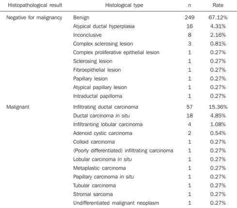
Histological studies demonstrated the following results: 76% negative for malig-nancy and 24% positive for maligmalig-nancy (Table 4).
Positive predictive values for categories 3, 4 and 5 were, respectively, 7.14%, 16.96% and 82.61% (Table 5).
In the calculation of the PPV for the category 4 subcategories, the 69 studies whose reports failed to indicate the subcat-egory were not taken into consideration (Table 6).
Table 1 Age versus risk for breast cancer.
Age
≤ 40 41–50
51–60
> 60
Malignant
5
25
30
29
Benign
42
112
69
59
Relative risk (confidence interval)
1
1.71 (0.69–4.22)
2.84 (1.18–6.87)
3.09 (1.28–7.47)
Odds ratio (confidence interval)
1
1.87 (0.67–5.21)
3.65 (1.31–10.14)
4.12 (1.47–11.54) p
0.222
0.009
0.004
Table 2 BI-RADS categories.
BI-RADS
3
4*
4A
4B
4C
5
Total
n
42
69
112
66
36
46
371
Rate
11.32%
18.60%
30.19%
17.79%
9.70%
12.40%
100%
* Subcategory (4A, 4B or 4C) was not specified.
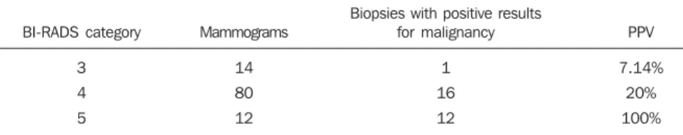
The PPVs were separately calculated for core biopsies and preoperative needle localization (Tables 7 and 8).
DISCUSSION
173
Positive predictive value of BI-RADS categories 3, 4 and 5
Radiol Bras. 2010 Mai/Jun;43(3):171–174
In the present study, the right breast was most affected in 56.06% of cases, and so did the lateral upper quadrant (171 in 383 affected quadrants). The presence of a mass was most frequent for invasive procedures (59.57% of cases). In the present study, the first most frequent type of cancer was in-filtrating ductal carcinoma (15.36% of cases), the second, ductal carcinoma in situ (4.85% of cases). The mean age of patients with cancer was higher than the mean age of patients with benign lesions, and Table 1 demonstrates, through the relative risk and odds ratio, that advanced age is a risk factor for the disease.
The BI-RADS suggests values < 2% chance of malignancy for category 3, and > 95% for category 5. Category 4 must in-dicate 23%–30% chance of malignancy(5).
In the present study, 23.99% of patients submitted to histopathological study pre-sented malignant lesions, i.e., the global PPV was of 23,99%. In the USA, this value ranges between 15% and 40%(8–10).
In the present study, 42 patients pre-sented probably benign findings (BI-RADS 3), three presented positive results for malignancy; thus the PPV remained above the recommended rate (7.14% > 2%), which is within the mean rate reported by studies in the literature where this rate ranges between 0% and 8%. For category 4, the authors found a PPV of 16.96%, while in the literature this rate ranges be-tween 4% and 63%. The PPV was sepa-rately calculated for subcategories 4A (8.04%), 4B (15.15%) and 4C (41.67%). The PPV of 82.61% for category 5 is within the expected range as compared with the several studies in the literature which report this parameter ranging between 54% and 100%(11–22).
The above mentioned values reflect the correlation between mammographic find-ings and results of two types of invasive procedures, namely, core biopsy and pre-operative needle localization.
Once values are separately calculated for each type of invasive procedure, even lower PPVs are found for the cases submit-ted to core biopsy, as follows: for category 3, 7.14% (0% to 4% in the literature); for category 4, 15.76% (4% to 20% in the lit-erature); and for category 5, 76.47% (54% to 92% in the literature)(13,14,18–21). Table 3 Mammographic findings.
Finding
Mass
Microcalcifications
Asymmetrical density
Mass + microcalcifications
Cyst
Asymmetrical density + microcalcifications
Complex cystic lesion
Total
221
104
31
8
3
2
2
Malignant
57
24
6
2
0
0
0
PPV
25.79%
23.08%
19.35%
25%
0%
0%
0%
PPV, Positive predictive value.
Table 4 Histopathological result.
Histopathological result
Negative for malignancy
Malignant
Histological type
Benign
Atypical ductal hyperplasia
Inconclusive
Complex sclerosing lesion
Complex proliferative epithelial lesion
Sclerosing lesion
Fibroepithelial lesion
Papillary lesion
Atypical papillary lesion
Intraductal papilloma
Infiltrating ductal carcinoma
Ductal carcinoma in situ
Infiltranting lobular carcinoma
Adenoid cystic carcinoma
Colloid carcinoma
(Poorly differentiated) infiltrating carcinoma
Lobular carcinoma in situ
Metaplastic carcinoma
Papillary carcinoma in situ
Tubular carcinoma
Stromal sarcoma
Undifferentiated malignant neoplasm
n
249
16
8
3
1
1
1
1
1
1
57
18
4
2
1
1
1
1
1
1
1
1
Rate
67.12%
4.31%
2.16%
0.81%
0.27%
0.27%
0.27%
0.27%
0.27%
0.27%
15.36%
4.85%
1.08%
0.54%
0.27%
0.27%
0.27%
0.27%
0.27%
0.27%
0.27%
0.27%
Table 6 Mammogram PPV – BI-RADS 4.
BI-RADS 4 subcategory
4A
4B
4C
Mammograms
112
66
36
Biopsies with positive results for malignancy
9
10
15
VPP
8.04%
15.15%
41.67%
PPV, Positive predictive value. Table 5 Mammograms PPV.
BI-RADS category
3
4
5
Mammograms
42
283
46
Biopsies with positive results for malignancy
3
48
38
PPV
7.14%
16.96%
82.61%
174
Prado GLM, Guerra MTPM
Radiol Bras. 2010 Mai/Jun;43(3):171–174 As only the cases submitted to
preopera-tive needle localization were taken into consideration, the following values were found: 7.14% for category 3 (0% to 5% in the literature); 20% for category 4 (26% to 34% in the literature); and 100% for cat-egory 5 (81% to 97% in the literature)(12, 13,16,17).
The difference between PPVs for core biopsies and for preoperative needle local-ization corroborate the values reported in the literature which demonstrate that the PPV for mammography is higher as the finding is submitted to preoperative needle localization. Among the lesions diagnosed as atypical ductal hyperplasias at 14 gauge needle core biopsy, 20% to 56% corre-spond to carcinomas at surgical biopsy(23).
It should be taken into consideration that the BI-RADS may present limitations related to the classification itself and the training of radiologists involved in the uti-lization of this system(24,25).
CONCLUSION
The present study demonstrated dis-crepancies between the BI-RADS classifi-cation and histopathological results for findings in patients submitted to invasive diagnostic procedures at a center of refer-ence for cancer treatment in Teresina, PI, which, in some cases demonstrated to be unnecessary, particularly for findings
clas-sified as BI-RADS 4 and 5. Additionally, underestimation of findings classified as BI-RADS 3 was observed.
REFERENCES
1. National Cancer Institute. Surveillance, Epidemi-ology and End Results (SEER). [acessado em 16 de maio de 2008]. Disponível em: http://seer. cancer.gov
2. Ministério da Saúde. Instituto Nacional de Cân-cer. Estimativa 2006: incidência de câncer no Brasil. [acessado em 16 de maio de 2008]. Dis-ponível em: http://www.inca.gov.br/estimativa/ 2006
3. Ministério da Saúde. Instituto Nacional de Câncer. Estimativa 2009: incidência de câncer no Brasil. [acessado em 7 de dezembro de 2009]. Disponí-vel em: http://www.inca.gov.br/estimativa/2010 4. American College of Radiology. The ACR breast imaging reporting and data system (BI-RADS) [web source]. November 11, 2003. [cited 2004 Feb 27]. Available from: http://www.acr.org/de-partments/stand_accred/birads/contents.html 5. American College of Radiology. Breast Imaging
Reporting and Data System (BI-RADS®). 4th ed.
Reston, VA: American College of Radiology; 2003.
6. Nascimento JHR, Silva VD, Maciel AC. Acurá-cia dos achados ultrassonográficos do câncer de mama: correlação da classificação BI-RADS® e
achados histológicos. Radiol Bras. 2009;42:235– 40.
7. Schmillevitch J, Guimarães Filho HA, De Nicola H, et al. Utilização do índice de resistência vascu-lar na diferenciação entre nódulos mamários be-nignos e malignos. Radiol Bras. 2009;42:241–4.
8. Ciatto S, Cataliotti L, Distante V. Nonpalpable le-sions detected with mammography: review of 512 consecutive cases. Radiology. 1987;165:99–102.
9. Cyrlak D. Induced costs of low-cost screening mammography. Radiology. 1988;168:661–3. 10. Hall FM, Storella JM, Silverstone DZ, et al.
Nonpalpable breast lesions: recommendations for biopsy based on suspicion of carcinoma at mam-mography. Radiology. 1988;167:353–8.
11. Lacquement MA, Mitchell D, Hollingsworth AB. Positive predictive value of the Breast Imaging Reporting and Data System. J Am Coll Surg. 1999;189:34–40.
12. Orel SG, Kay N, Reynolds C, et al. BI-RADS cat-egorization as a predictor of malignancy. Radiol-ogy. 1999;211:845–50.
13. Liberman L, Abramson AF, Squires FB, et al. The breast imaging reporting and data system: posi-tive predicposi-tive value of mammographic features and final assessment categories. AJR Am J Roentgenol. 1998;171:35–40.
14. Bérubé M, Curpen B, Ugolini P, et al. Level of suspicion of a mammographic lesion: use of fea-tures defined by BI-RADS lexicon and correla-tion with large-core breast biopsy. Can Assoc Radiol J. 1998;49:223–8.
15. Kestelman FP, Canella EO, Arvellos NA, et al. Classificação radiológica nas lesões não-palpá-veis da mama. Análise de resultados do Hospital do Câncer III – INCA-MS. Radiol Bras. 2001;34 (Supl 1):20.
16. Ball CG, Butchart M, MacFarlane JK. Effect on biopsy technique of the breast imaging reporting and data system (BI-RADS) for nonpalpable mammographic abnormalities. Can J Surg. 2002; 45:259–63.
17. Tan YY, Wee SB, Tan MP, et al. Positive predic-tive value of BI-RADS categorization in an Asian population. Asian J Surg. 2004;27:186–91.
18. Tate PS, Rogers EL, McGee EM, et al. Stereotac-tic breast biopsy: a six-year surgical experience. J Ky Med Assoc. 2001;99:98–103.
19. Margolin FR, Leung JW, Jacobs RP, et al. Percu-taneous imaging-guided core breast biopsy: 5 years’ experience in a community hospital. AJR Am J Roentgenol. 2001;177:559–64. 20. Travade A, Isnard A, Bagard C, et al.
Macro-biopsies stéréotaxiques par système à aspiration 11-G: à propos de 249 patientes. J Radiol. 2002; 83(9 Pt 1):1063–71.
21. Mendez A, Cabanillas F, Echenique M, et al. Mammographic features and correlation with biopsy findings using 11-gauge stereotactic vacuum-assisted breast biopsy (SVABB). Ann Oncol. 2004;15:450–4.
22. Zonderland HM, Pope TL Jr, Nieborg AJ. The positive predictive value of the breast imaging reporting and data system (BI-RADS) as a method of quality assessment in breast imaging in a hospital population. Eur Radiol. 2004;14: 1743–50.
23. Liberman L. Percutaneous image-guided core breast biopsy. Radiol Clin North Am. 2002;40: 483–500.
24. Liberman L, Menell JH. Breast imaging report-ing and data system (BI-RADS). Radiol Clin North Am. 2002;40:409–30.
25. Godinho ER, Koch HA. Breast Imaging Report-ing and Data System (BI-RADS™): como tem sido utilizado? Radiol Bras. 2004;37:413–7. Table 7 PPV of mammograms that led to core biopsy.
BI-RADS category
3
4
5
Mammograms
28
203
34
Biopsies with positive results for malignancy
2
32
26
PPV
7.14%
15.76%
76.47%
PPV, Positive predictive value.
Table 8 PPV of mammograms that led to preoperative needle localization.
BI-RADS category
3
4
5
Mammograms
14
80
12
Biopsies with positive results for malignancy
1
16
12
PPV
7.14%
20%
100%