Refinement of the Central Steps of Substrate
Transport by the Aspartate Transporter
GltPh: Elucidating the Role of the Na2
Sodium Binding Site
SanthoshKannan Venkatesan1, Kusumika Saha1, Azmat Sohail1, Walter Sandtner1, Michael Freissmuth1, Gerhard F. Ecker2, Harald H. Sitte1, Thomas Stockner1*
1Institute of Pharmacology, Center for Physiology and Pharmacology, Medical University of Vienna, Vienna, Austria,2Division of Drug Design & Medicinal Chemistry, Department of Pharmaceutical Chemistry, University of Vienna, Vienna, Austria
*thomas.stockner@meduniwien.ac.at
Abstract
Glutamate homeostasis in the brain is maintained by glutamate transporter mediated accu-mulation. Impaired transport is associated with several neurological disorders, including stroke and amyotrophic lateral sclerosis. Crystal structures of the homolog transporter GltPh fromPyrococcus horikoshiirevealed large structural changes. Substrate uptake at the atomic level and the mechanism of ion gradient conversion into directional transport remained enigmatic. We observed in repeated simulations that two local structural changes regulated transport. The first change led to formation of the transient Na2 sodium binding site, triggered by side chain rotation of T308. The second change destabilized cytoplasmic ionic interactions. We found that sodium binding to the transiently formed Na2 site ener-gized substrate uptake through reshaping of the energy hypersurface. Uptake experiments in reconstituted proteoliposomes confirmed the proposed mechanism. We reproduced the results in the human glutamate transporter EAAT3 indicating a conserved mechanics from archaea to humans.
Author Summary
We used the archaeal homolog GltPh of the human glutamate transporters to refine our understanding how large scale conformational changes are translated into substrate trans-location. We identified the structural changes that accompany substrate transport and convert the energy stored in the ion gradient into a directional transport. Insights into the mechanics of these transporters are likely to increase our understanding of how they con-tribute to excitotoxicity and to develop drugs, which preclude the underlying accumula-tion of glutamate in the synaptic cleft.
OPEN ACCESS
Citation:Venkatesan S, Saha K, Sohail A, Sandtner W, Freissmuth M, Ecker GF, et al. (2015) Refinement of the Central Steps of Substrate Transport by the Aspartate Transporter GltPh: Elucidating the Role of the Na2 Sodium Binding Site. PLoS Comput Biol 11 (10): e1004551. doi:10.1371/journal.pcbi.1004551
Editor:Marco Punta, Pierre and Marie Curie University (UPMC), FRANCE
Received:March 30, 2015
Accepted:August 12, 2015
Published:October 20, 2015
Copyright:© 2015 Venkatesan et al. This is an open access article distributed under the terms of the
Creative Commons Attribution License, which permits unrestricted use, distribution, and reproduction in any medium, provided the original author and source are credited.
Data Availability Statement:All relevant data are within the paper and its Supporting Information files.
for patients.
The aspartate transporter fromPyrococcus horikoshii(GltPh) [10–13] is a homolog of the mammalian EAATs. GltPh and human EAATs share ~36% sequence identity. Sequence con-servation is higher for residues implicated in substrate and ion binding [14–18]. The first crys-tal structure of the trimeric GltPh was solved in the outward-occluded state [10], followed by structures determined in three additional conformations [11–13,19]: outward-open, inward-occluded and intermediate, revealing a large scale translational motion during substrate trans-port [12]. The transport domain contains substrate and ion binding sites, while the trimeriza-tion domain mediates the contact between the protomers. Functrimeriza-tional studies indicated that transport of one substrate molecule is coupled to the co-transport of three sodium ions [20,21]. The respective sodium binding sites were termed Na1, Na2 and Na3.
Simulations of the outward-occluded and the inward-occluded conformation have been used to study binding of substrate and co-transported ions [22–24]. The importance of the HP1 and HP2 gates for restricting the accessibility to the substrate-binding site has been stud-ied using molecular dynamics (MD) simulation [23,25–28]. The Na3 sodium-binding site [23] has been predicted by MD simulations and was experimentally validated in GltPh and EAATs [24,29–31]. The conformational changes of substrate translocation have been studied by motion planning, elastic network model and metadynamics [32–34].
A comparision of the outward and inward facing crystal structures (PDB ID: 2NWX, and 3KBC) suggested a nearly rigid body movement of the transport domain vs. the trimerization domain. We used steered molecular dynamics (SMD) simulations to investigate the substrate translocation step from the outward-facing to the inward-facing conformation. We identified local events that are essential for coupling of the ion gradient to substrate transport and verified our observations by experiments. Repeated SMD simulations of 0.3μs length revealed the exis-tence of an intermediate state, which is characterized by maturation of the Na2 binding site. Maturation required changes in the local environment, which are triggered by rotation of the side chain of T308. We provide biochemical evidence for the localization and the functional role of the Na2 site: (i) mutation of residue T308 increased Kmfor substrate and sodium in
GltPh and in human EAAT3. (ii) The Hill coefficient of sodium dependence was reduced from 2.5 for wild type to 1.7 for T308V and 1.1 for T308A confirming the location of the Na2 site and its role in coupling of the sodium gradient to substrate uptake.
Results
were expected, because the transport rate of GltPh [35] is in the range of 0.3 min-1. Simulations of the outward-occluded state revealed that the HP2 loop opened in the absence of substrate and ions. The observed gate opening is consistent with experimental data, which showed that closing of the HP2 loop requires binding of substrate and sodium ions[23,25,26,36].
Substrate and the sodium ions bound to the Na1 and Na3 sites remained close to their respec-tive initial positions. In contrast, the sodium ion bound to the Na2 site could not be maintained in its position and diffused into the solvent during equilibration. Dissociation of sodium from the Na2 site occurred in all protomers in every simulation of the outward-occluded state (also in the SMD simulations described below). In contrast, it remained bound in the simulations of the inward-occluded state. Several modifications were applied to the release protocol, but we did not obtain a sodium ion stably bound to the crystallographically observed Na2 site. Similar observa-tions were reported by others [22,37]. A Na2 site that was shifted by ~0.3 nm relative to the posi-tion of the crystallographically observed Tl+ion was recently proposed [22].
Global movement of domains during transport
The inward-occluded state was crystallized with the help of a conformation stabilizing dou-ble cysteine mutant (K55C and A364C). We observed a transition to the inward-facing state of at least one protomer in each simulation. Thereby residues K55 and A364 were found to come into close proximity (SeeS4 Fig). A second chain reached the inward-facing state in two instances. The third chain showed only once a transition to the inward facing state. The extra-cellular gate of the A chain is more open in the crystal structure (PDB ID: 2NWX) (See also
S3A–S3C Fig). For simplicity, below we discuss results of only one simulation. Analyses of the other two simulations lead to the same conclusions (S5andS6Figs).
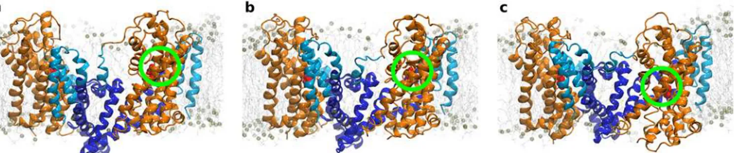
We observed a progression towards the inward-occluded state in two global steps. Repre-sentative snapshots (Fig 2A, 2B and 2C) are depicted at three time-points, representing the out-ward-occluded (at 0 ns), the intermediate (at 152 ns) and the inout-ward-occluded state (at 272 ns). The structural overlap with the crystallized intermediate structure (PDB ID: 3V8G) was
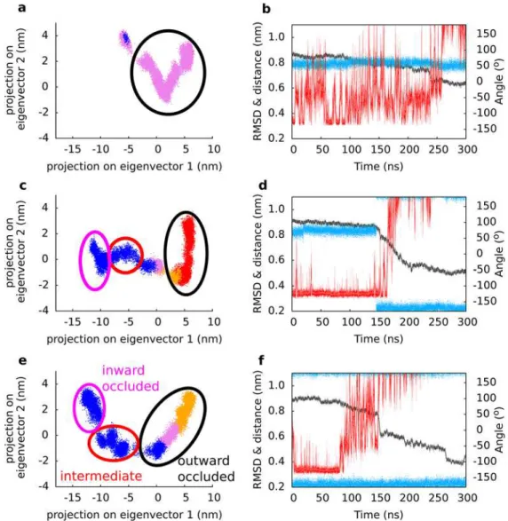
Fig 1. Global structure of GltPh.(a) Membrane embedded trimeric transporter structure, seen from the extracellular site. The transport domain is shown in orange, the trimerization domain in light (membrane exposed) and dark blue (without membrane contacts). The same color code is applied to all figures. The backbone atoms used as pulling groups are highlighted in green and purple. (b-d) Time evolution of transporter geometry is shown of three independent simulations, measuring the structural deviation of each protomer individually (chain A in black, chain B in red, chain C in blue) from the inward-facing crystal structure. (e) Overlay of the inward-facing crystal structure in gray (PDB ID: 3KBC) and the closest conformation extracted from the SMD simulation. The RMSD between the structures was 0.35 nm.
doi:10.1371/journal.pcbi.1004551.g001
Fig 2. Conformation of GltPh during substrate translocation.Snapshots of the transporter in (a) the outward (at 0 ns), (b) the intermediate (at 152 ns) and (c) the inward-facing state (at 272 ns): the transport domain remained in contact with the membrane and translated minimally relative to the membrane normal, while the trimerzation domain moved towards the cell exterior.
largest in the intermediate conformation of our trajectories (S7 Fig). The observed structural rearrangements showed a movement of the transport domain (orange) relative to the trimeri-zation domain (blue). The transport domain is in direct contact with the membrane; it moved as a rigid body (S3 Fig), and translated only minimally relative to the membrane. In contrast, the core of GltPh (TM2, TM4 and TM5), which showed minimal contacts with the membrane, displayed a large movement towards the extracellular side. This was accompanied by a switch in accessibility of the substrate binding site from extracellular to intracellular. We identified two distinct dynamic subdomains within the trimerization domain. A comparison between available crystal structures (PDB ID: 2NWX, 3V8G and 3KBC) suggested that the trimerization domain could bend within helices TM2 and TM5. We observed that the central part of the tri-merization domain moved relative to the transport domain. In contrast, the membrane-exposed helices TM1 and the membrane adjacent parts of TM2 and TM5 did not translocate. Instead, the trimerization domain bent at residues P45-G47, P60 in TM2 and P206-G208 in TM5 to allow the membrane-exposed part to maintain membrane contacts.
The Na2 site matured during substrate transport
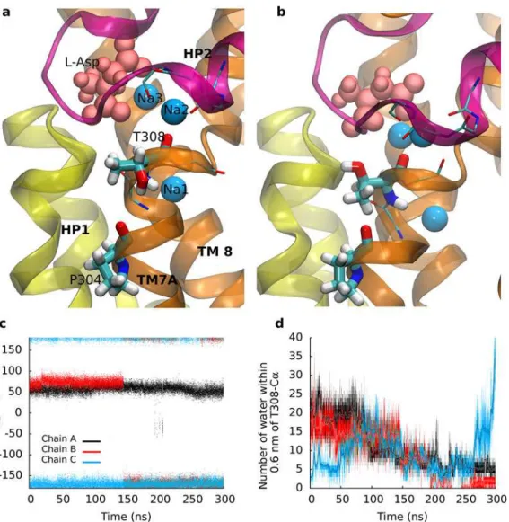
We observed that sodium bound to the Na2 site was unstable in the crystallographically observed position of the outward-occluded state (Fig 3A). The side chain of T308 rotated by 120 degrees in the course of the SMD simulations (Fig 3B). The same re-rotation did not occur in equilibrium simulations. Residue T308 is adjacent to the crystallographically observed Na2 site and located on the last turn of the helix TM7A. The contiguous NMDG motif was impli-cated in substrate and ion binding [15,39]. The hydroxyl group of the T308 side chain formed a hydrogen bond with the backbone carbonyl of P304 on the preceding helical turn. Breaking of the P304-T308 hydrogen bond triggered rotation of the T308 side chain (Fig 3C). The transi-tion correlated with a reduced hydratransi-tion of T308 (Fig 3D).
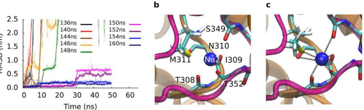
We extracted closely spaced snapshots from before and from after rotation of the T308 side chain to investigate, if partial dehydration and rotation of the T308 side chain improved coor-dination of sodium at the Na2 site. We re-inserted sodium into its original position in the Na2 site and carried out 50 ns long equilibrium MD simulations. The protein was stable in all simu-lations: we observed CαRMSD values between 0.12 and 0.16 nm for the TM helices. The low RMSD showed that the pulling rate of the SMD simulation was slow enough to allow the trans-porter to proceed along the low energy path without significant force induced distortions. Sodium bound to the Na2 site was unstable before rotation of the T308 side chain and dissoci-ated from the Na2 binding site in simulations using starting conformations extracted before rotation of T308 (Fig 4A). In contrast, sodium was stable after rotation. The structural adjust-ments, which were only possible after breaking of the hydrogen bond between T308 and P304, needed a few nanoseconds to propagate through TM7A to form the Na2 sodium binding site, because sodium was found to remain stably bound to the position in the crystal structure (of Tl+) only 6 ns after rotation of T308. We observed that backbone carbonyl oxygen atoms of the last turn of helix TM7A improved coordination of the sodium ion. The fully assembled Na2 binding site was formed by the carbonyl oxygen atoms at the C-terminal ends of helix TM7A, the first helix of the HP2 loop and by the sulfur atom of the M311 side chain. Na2 remained stably coordinated in the inserted position in the simulations started from the last two tested time points (SeeFig 4B and 4C), indicative of a completed Na2 sodium binding site. We also found (using APBS [40]) a considerable increase in the negative potential at the Na2 site.
interacted with the side chain of T308. Thereby, sodium had moved away from the center of the region with negative electrostatic potential created by the helix dipoles of helix TM7A and the first helix of the HP2 loop. We carried out three independent simulations to investigate the possibility that a rotated T308 in the start structure could stabilize the sodium in the Na2 bind-ing site. Sodium shifted to the same position with direct contact to the hydroxyl group of the T308 side chain as proposed earlier [22] in the beginning of each simulation. This position resulted metastable. In two simulations sodium dissociated from this intermediate site after 5 ns and diffused into the water phase. In the third simulation, sodium detached within the first nanosecond, suggesting that this could be a intermediate state on the path of sodium binding to the Na2 site.
Ionic interactions prevent unproductive transport events
The outward-facing transporter is stabilized at the cytosolic side by an interaction network between the trimerization domain and the transport domain. The network includes residues
Fig 3. The Na2 site.(a) Comparison of the Na2 site in the outward-facing conformation before and (b) after rotation of T308. The HP1 re-entrance loop is highlighted in yellow, the HP2 loop in purple. Local changes allowed for an optimal coordination of the bound sodium. (c) Time evolution of the side chain dihedral angle of T308. (d) Level of hydration of T308 measured by the number of water molecules within 0.6 nm from the Cαatom.
E192, Y195 and N199 on helix TM5 (trimerization domain) and residues R287 and K290 on the HP1 loop (transport domain) (Fig 5A and 5B). We observed salt bridges between residues E192 and K290 and between residues N199 and R287. The salt bridge between E192 and K290 was further stabilized by a cation-πinteraction of K290 with the aromatic ring of Y195. The distance of the salt bridge between atoms Cδof E192 and atom Nzof K290 increased from direct residue contact in the outward-facing conformation to a broad distribution around 1.5 nm in the intermediate state (Fig 5C). Opening of the salt bridge (E192-K290) was accompa-nied by rotation of Y195. The distance increased further, when the inward facing state was reached. The second salt bridge between residues N199 and R287 showed similar behavior.
Experimental validation of the Na2 sodium-binding site
Our simulations suggested that (i) the Na2 sodium-binding site forms during substrate translo-cation and that (ii) the side chain of residue T308 is a key player of its maturation. We chal-lenged this hypothesis by experimentally measuring the involvement of T308 in binding and transport. We created three types of mutations: (i) the T308W mutation introduced an amino acid that would be too large to allow for transport; (ii) the smaller T308A and the T308V
Fig 4. Stabilizing sodium to the Na2 site.We extracted closely spaces snapshots before and after the rotation of T308 and carried out 50 ns long simulations. (a) The RMSD of sodium 2 reflects the stability of the reinserted sodium. The time evolution of the RMSD can be classified into two groups: Large RMSD and unbinding before T308 rotation, and sodium bound to the Na2 site after rotation of T308. Panel (b) and (c) show the initial and final structure of the simulation initiated for the snapshot extracted at 160 ns. Sodium 2 was reinserted into the Na2 site and remained stable, coordinated by backbone carbonyl oxygens and the sulfur atom of the M311 side chain.
doi:10.1371/journal.pcbi.1004551.g004
Fig 5. Interactions at the cytosolic site.Interaction network at the cytosolic side (a) in the outward-facing and (b) the intermediate state. (c) Time evolution of the salt bride between E192 and K290. The transporter moves only after opening of the salt bridge.
mutations were designed to remove the hydroxyl group; (iii) the T308S mutation retained the hydroxyl group (Fig 6andTable 1). The T308W mutation abolished transport as predicted (Fig 6A). The T308A and T308V mutations showed increased Michaelis-Menten constant (Km) values for the substrate L-Asp and for the co-transported sodium ions (Fig 6B and 6C).
The transport stoichiometry of GltPh predicts that three sodium ions are co-transported with substrate. A Hill coefficient of ~2.5 for sodium indicates strong cooperativity [20]. This was recapitulated in our experiments: we observed for wild type GltPh a Hill coefficient of 2.5 ± 0.3 for sodium-supported substrate uptake (Fig 6C). The cooperativity was reduced in both mutants, which removed the hydroxyl group, i.e. to 1.1 ± 0.1 and 1.7 ± 0.3 in T308A and T308V, respectively. The drop in cooperativity is consistent with the prediction that the muta-tions affected binding of sodium to the Na2 site. The Kmof substrate uptake was increased
from 119 ± 18 nM in wild type GltPh to 183 ± 26 nM and 220 ± 24 nM for T308A and T308V, respectively. The T308S mutation maintained the hydroxyl group at theγposition. We observed an almost 3 fold decrease in the maximal transport rate (Vmax), but only a very small
change in the Hill coefficient (2.3 ± 0.7). The Kmfor sodium (9.2 ± 2.3 mM) and substrate
(134 ± 27) were unchanged.
GltPh is predicted to share its fold with the human EAATs. Sequence alignments showed that T308 and P304 are conserved between the archeal and the human transporters. We there-fore reasoned that mutations in human EAATs would recapitulate the effects seen in GltPh, if the structure-function relationship was shared. The same mutations (tryptophan, alanine, valine and serine) of corresponding residue T364 were introduced to the human EAAT3. Experiments were carried out in transiently transfected HEK293 cells (Fig 7). Mutations of EAAT3 reproduced the findings seen in GltPh: the Kmfor transport was not changed in
Fig 6. Effects of T308 mutations on substrate uptake.(a) Uptake of [3H] L-aspartate into GltPh containing proteoliposomes. We could not observe
aspartate uptake for the T308W mutation. (b) [3H] L-aspartate uptake into proteoliposomes as a function of [3H] L-aspartate concentration and (c) [3H]
L-aspartate uptake into proteoliposomes as a function of sodium concentration. The data are from three independent experiments; error bars show s.e.m.
doi:10.1371/journal.pcbi.1004551.g006
Table 1. Transport of Na+and L-Asp in GltPh.
Mutant Kmfor Na+(mM) Hill coefficient for Na+uptake Kmfor Asp (nM)
Wild typea 8.4±1.3 2.5±0.3 119±18
T308A 47.6±6.3 1.1±0.1 220±24
T308S 9.2±2.3 2.3±0.7 134±27
T308V 18.4±6.3 1.7±0.3 183±26
T308W N.D. N.D. N.D.
aErrors are represented as s.e.m.
EAAT3-T364S (37 ± 7μM) as compared to wild type EAAT3 (41 ± 4μM), but increased in EAAT3-T364V (79 ± 20μM), in EAAT-T364A (159 ± 48μM) and EAAT3-T364W
(341 ± 82μM). In contrast to GltPh, we found residual transport in EAAT3-T364W, indicating that EAAT3 might be more tolerant.
Discussion
During substrate translocation, accessibility of the substrate binding site changed from the extracellular to the intracellular side. We used SMD simulations to investigate the structural details of this large scale conformational change. Analysis of the conformational landscape revealed the existence of an intermediate state, which is similar to the conformation observed in the crystal structure (PDB ID: 3V8G) [13], showing an RMSD of 0.3–0.4 nm. Binding of substrate and sodium has been shown to induce the closure of the HP2 gate [25,26,36]. We found that the Na2 sodium-binding site is not yet fully formed in the outward-occluded state [12]. The sodium ion escaped in spite of weak restraints, which were imposed to keep it in place. The Na2 site was found to mature during the transition to the intermediate state. The side chain of residue T308 played a pivotal role: breaking of the P304-T308 hydrogen bond weakened the structural stability of the last turn of the TM7A helix (Fig 3). The backbone car-bonyl oxygen atoms could therefore adjust and optimally coordinate the sodium ion in the Na2 site. The structural adaptation increased the negative electrostatic potential at the Na2 site, which became more attractive for the positively charged sodium ion. Sodium binding can neu-tralize the negative potential, thereby removing an energy barrier for the transition to the inter-mediate state. Therefore, we carried out additional MD simulations to confirm that the Na2 site matured during the transition to the intermediate state. Simulations starting from closely spaced snapshots showed that the Na2 site became competent for sodium binding only after breaking of the hydrogen bond between the side chain of T308 and the backbone carbonyl oxy-gen of P304. Our results indicate that breaking of this hydrooxy-gen bond is the key event that trig-gers the structural adjustments that lead to maturation of the Na2 site. This observation is in line with the predicted sequence of binding events in GltPh: sodium 3, followed by sodium 1, substrate and sodium 2 [22,25,26]. Electrophysiological measurements determined that bind-ing to the outward-facbind-ing EAAT3 transporter occurred in at least two steps: (i) fast bindbind-ing of substrate and sodium ion(s) followed by (ii) slow binding of one additional sodium ion [41]. The transporter translocated substrate only after the slow sodium binding event.
Fig 7. Substrate uptake by human EAAT3.Uptake of [3H] L-glutamate into HEK293 cells expressing the
human EAAT3 transporter that carrying the same mutations (A, S, V, and W) at the corresponding residue T364. The data are from three independent experiments; error bars show s.e.m.
Fig 8. Correlation analysis.Panel (a, c, e) show movements of a GltPh protomer along the two largest eigenvectors (translation and rotation of the domains relative to each other) observed in the simulations of protomer A, B and C, respectively. Each dot represents the projection of one frame in the trajectory onto the 2 dimension of eigenvectors 1 (translation) and 2 (rotation). Four distinct periods of the trajectory are color coded: The first part of the trajectory (red) represents the outward-occluded state. A shift to orange marked the rotation of T308. Rotation of side chain of Y195 notifies the transition to the third period (pink). The fourth region shown in blue represents the period after opening of the intracellular interaction network. The intermediate state is reached in the populated region in the middle of the plot, after opening of the intracellular interaction network. Panel (b, d, f) show an overlay of the time evolution of the RMSD (black), the rotational state of the side chain of T308 (blue) and the side chain–side chain distance between E192 and K290 (red) of
protomer A, B and C, respectively.
reached the inward-occluded state. The time points of local changes were color-code mapped onto the 2d projection. The analyses revealed that (i) formation of the Na2 site occurred before the transition to the intermediate state. (ii) Opening of the interaction network was also required prior to formation of the intermediate state. It followed after maturation of the Na2 site. Mutations of residues contributing to the intracellular interaction network (specifically K290A) were shown to reduce substrate transport [42], most likely by destabilizing the out-ward-facing state. We found the conformation of the Y195 side chain to be a sensor for early onset of conformational changes at the inner site of the transporter. The long side chains of E192 and K290 allowed for limited structural adjustment before rupture. Only once the salt bridge opened could substrate translocation proceed. The same results were obtained in the analyses of the other two simulations (S7 Fig). Our correlation analyses therefore suggest that the local changes arethe key switches, which prepare GltPh for the global changes and should therefore control the transition through the transport cycle.
These observations have important implications for the transport mechanism. They predict that the local events represent essential shifts in the energy hypersurface along the transport cycle: (i) binding of substrate and sodium to the Na1 and Na3 site stabilizes the outer gate HP2 loop in the closed state [36]. (ii) Occupation of the Na2 site reduces the energy barrier for the transition to the intermediate state. (iii) Opening of the interaction network at the cytosolic site allows transport and trimerization domain to move relative to each other. Accessibility of the substrate binding site changes from extracellular to intracellular. (iv) Opening of the intracellu-lar gate allows for substrate release and ion dissociation [28]. These events indicate that the role of sodium binding is to modify the energy hypersurface of the transporter and, as a conse-quence, to allow for directional transport. Directionality can be maintained as long as the inter-nal sodium concentration remains low to reset the transporter [43].
We confirmed this mechanism by performing radiotracer flux experiments of wild type and mutant GltPh. Residues P304 and T308 in TM7A are conserved from archaea to humans, pre-serving the hydrogen bonding property. The conservative T308S mutation, which maintains the hydroxyl group, had little effect on Hill coefficient and Km. Removal of the hydroxyl group
(mutated to alanine and valine) was predicted to alter the Kmand the Hill coefficient. We
observed a larger effect for the T308A mutant as compared to the T308V mutant. Valine and threonine are bothβ-branched amino acids and have helix destabilizing properties, while ala-nine does not have the same property, suggesting that an imperfectly stabilized TM7A helix is important for transporter function. These experimental results support the transport mecha-nism model developed from our simulations.
structure of GltPh (PDB-ID: 2NWX) [11] were selected; missing residues (side chains and resi-dues) were added using MODELLER version 9.8 [47]. The third sodium ion was placed into the previously reported ion binding site [23]. GltPh was embedded in a pre-equilibrated POPC bilayer using the g_membed [48] procedure and solvated with water, electro-neutralized and a physiological concentration of NaCl (150 mM). The SPC water model [49] was used. Lennard-Jones interactions were calculated until a cutoff of 1.0 nm. Long range electrostatic interactions were treated using PME [50]. Bond lengths were constrained using LINCS [51]. The tempera-ture was maintained at 310 K applying the v-rescale thermostat [52]; pressure was set to 1 bar and controlled by the Berendsen barostat [53], coupling protein, membrane, and water/ions separately. The assembled system was equilibrated by first relaxing membrane and solvent. The protein was then gradually relaxed by reducing the restraints on the protein atoms in five steps [54] (each 2.5 ns simulation) applying a restraining force on the protein of 1000, 100, 10, 1, 0.1 kJ mol-1nm-2. Production runs were then started. We carried out equilibrium simula-tions starting from two independently prepared inward and the outward-occluded state, and nine simulations starting from snapshots extracted from one SMD simulation.
Steered molecular dynamics
We used SMD simulations [55] to induce a conformational change from the outward- to the inward-facing conformation. We selected the same number of Cαatoms from both the trimerisa-tion and the transport domain. The spring force constant was set to 1000.0 kJ mol-1nm-2, applied only along the membrane normal (Z-direction). Pilot simulations with different force constants indicated that higher force constants would lead to a force profile that showed an increasing level of fluctuations and a larger number of force peaks in the force profile (SeeS1 Fig), indicative for entanglement and forced barrier-crossing events. These force peaks seemed to depend on the applied sprint constant, because they gradually disappeared with decreasing restraining force. Repeated simulations showed that the peaks did not always appear at the same time points. Cor-rect behavior of the protein could therefore only be expected with a force constant of 1000.0 kJ mol-1nm-2. All simulations were repeated three times. Pilot simulations were also performed to identify the optimal pulling velocity. We selected a pulling rate of 0.00001 nm ps-1. This velocity is slow enough to give GltPh time to relax all not coupled degrees of freedom and fast enough to be still accessible to MD simulations. Higher pulling speed resulted in unphysical structural deformations and increasingly larger peaks in the force time contour. We carried out three SMD simulations starting from independently prepared systems.
Plasmids and site-directed mutagenesis
Protein purification and reconstitution
GltPh was purified as described previously [56]. Briefly, membranes were isolated, solubilized with (1%) n-dodecyl-β-D-maltopyranoside (DDM) (Anatrace Detergents), and the protein was purified using Ni-NTA resin. The detergent was then exchanged to (0.15%) n-decyl-β -D-maltopyranoside (DM) (Anatrace Detergents). The protein was reconstituted into liposomes using the method described previously with slight modifications [57]. Escherichia coli total lip-ids (Avanti Polar liplip-ids Inc.) and 1-palmitoyl-2-oleoyl-sn-glycero-3-phosphocholine (Avanti Polar Lipids Inc.) were mixed at a ratio of 3:1, dried under nitrogen and re-suspended in inter-nal buffer (100 mM KCl, 20 mM HEPES, pH 7.5). The lipid mixture was briefly sonicated using a point tip sonicator. The lipid suspension was flash frozen in liquid nitrogen and thawed at room temperature multiple times. Liposomes were formed by extrusion through 400 nm polycarbonate membranes (Avanti Polar Lipids Inc.) and destabilized with Triton X-100 prior to addition of protein at a ratio of 0.25μg protein per mg lipid. The protein-lipid mixture was kept at room temperature for 30 minutes. Detergent was then removed using SM2 biobeads (Biorad). The protein-lipid mixture was incubated and gently agitated with three consecutive batches of biobeads (20 mg ml-1). The biobeads were subsequently removed by filtration. The resulting proteoliposomes were concentrated by centrifugation at 150,000 g for 30 min in a Beckman centrifuge, re-suspended at 100 mg lipid mL-1and either used immediately or flash frozen in liquid nitrogen and stored at−80°C.
Proteoliposome transport assay
Transport of [3H] L-aspartate by wild type and mutant GltPh was assayed by applying the pro-tocol modified from Gaillard et al., 1996 [56]. Briefly, proteoliposomes were loaded with buffer A (100 mM KCl, 20 mM HEPES, pH 7.5) by repeated freeze and thaw cycles followed by extru-sion. The uptake was initiated by diluting the proteoliposomes into buffer B (100 mM NaCl, 20 mM HEPES, pH 7.5, 1μM valinomycin and the indicated concentrations of [3H] L-aspartate) pre-warmed to 30°C. At each time point, a 200μL aliquot was removed and diluted 10 fold into ice cold stop buffer C (100 mM LiCl, 20 mM HEPES, pH 7.5), followed by filtration over nitrocellulose filters (0.22μm pore size, Merck Millipore). The filters were washed three times with 2 mL of ice-cold quench buffer and assayed for radioactivity. Sodium dependence of [3H] L-aspartate transport was measured by varying extraliposomal Na+concentration from 0.1 to 300 mM. Osmolarity was balanced using choline chloride (Sigma Aldrich). [3H] L-aspartate concentrations used for determining the sodium dependence were 100 nM for wild type and around Kmfor other mutants. Background levels of uptake were measured by diluting
proteoli-posomes into buffer A containing 1μM valinomycin and the indicated concentrations of [3H] L-aspartate.
Transport assay of EAAT3 in HEK293 cells
The plasmid PL28 encoding for the human excitatory amino acid transporter 3 (EAAT3) was a kind gift by Peter Larsson (Oregon Health & Science University). The EAAT3 cDNA was cloned into pmGFP-C1. The point mutations T364A, T364V, T364S, and T364W were gener-ated using Quick Change Lightning site-directed mutagenesis kit II (Agilent technologies) and confirmed by DNA sequencing. Human embryonic kidney 293 (HEK293) cells were cul-tured in Dulbecco’s Modified Eagle’s Medium (DMEM), which was supplemented with 10% fetal bovine serum at 37°C and 5% CO2. Cells were transiently transfected with wild type
Supporting Information
S1 Fig. Distance vs. time and force vs. time plot.Panel (a) shows the change in distance over time with respect to the starting conformation of the 2NWX crystal structure, panel (b) shows the time evolution of the force acting on the two domains. The reference point (or reference distance) is changed at a constant velocity in the SMD simulations. The applied force is calcu-lated from the difference between the reference distance and the instantaneous distance using Hook’s law.
(TIFF)
S2 Fig. Selection of the spring constant strength.Pilot simulations were performed, using a pull rate of 0.0001 nm/ps and applying four different spring force constants: 1000, 5000, 10000 and 15000 kJ mol-1nm-2. Force profiles are shown until the first GltPh protomer passed the inward-occluded conformation. The shape of the force profiles indicated that force constants higher than 1000 kJ mol-1nm-2do not allow for sufficient side chains disentanglement, bearing the danger of observing erratic behavior induced by strong coupling.
(TIF)
S3 Fig. Structural stability of trimerisation and transport domains.Panels a, b, and c show the time evolution of the distance between S278 (HP1) and G354 (HP2) on the tip of the respective loops. A larger distance was observed in chain A in all the three simulations (a, b, c). Overall structural stability of the trimerisation (d, e, f) and transport domain (g, h, i) was esti-mated by measuring the RMSD to the starting structure. The RMSD of individual domains remained stable for major part of the SMD simulation.
(TIFF)
S4 Fig. Comparison of the inward-occluded conformation as observed in the simulation with the crystal structure (3KBC).Conformation of GltPh extracted from the simulation (also shown inFig 1E) is shown in panel a and rotated in panel c. This conformation which is closest (by RMSD) to the inward facing crystal structure is depicted in panel b and d. Residues K55C and A364C form a disulfide bond in the crystal. The two residues are in close proximity in our simulations of wild type GltPh.
(TIF)
S5 Fig. Salt bridge between residue E192 and K290.Panel (a, b, c): Time evolution of the salt-bridge distance was measured between atoms E192-Cδand K290-Nzof three independent sim-ulations. The salt bridge is formed between the residues from the trimerisation and transport domain. Time evolution of dihedral angleχ2 of Y195 is shown in panel (d, e, f).
(TIFF)
hydration of T308 was estimated as the number of water molecules within 0.6 nm of T308-Cα
atom. (TIFF)
S7 Fig. RMSD to the intermediate state.The RMSD plot measures the structural deviation of each chain for three independent SMD simulations to the crystallographically observed inter-mediate state (PDB ID: 3V8G). Chain A is shown in black, chain B in red, chain C in blue. (TIFF)
S8 Fig. Correlation analysis.Principal component analysis of three independent simulations (panels (a, b, c): chain A, B, C of simulation 1; panels (d, e, f): chain A, B, C of simulation 2; panels (g, h, i): chain A, B, C of simulation 3); the figures are as depicted in the main manu-script (Fig 8). Briefly, movement of a GltPh protomer along the two largest eigenvectors (trans-lation, rotation of the domains relative to each other) observed in the simulations. Each dot represents the projection of one frame in the trajectory onto the 2 dimensions of eigenvectors 1 and 2. Four distinct periods of the trajectory are color coded: The first part of the trajectory (red) represents the outward-occluded state before rotation of the T308 side chain. A shift to orange marked the rotation of T308. Rotation of the side chain of Y195 marks the transition to the third period (pink). The fourth region shown in blue represents the period after opening of the intracellular interaction network. The intermediate state is reached in the populated region in the middle of the plot, after opening of the intracellular interaction network.
(TIFF)
Author Contributions
Conceived and designed the experiments: MF GFE HHS TS. Performed the experiments: SV KS AS. Analyzed the data: SV KS AS WS TS. Contributed reagents/materials/analysis tools: HHS TS. Wrote the paper: SV MF HHS TS.
References
1. Danbolt NC. Glutamate uptake. Prog Neurobiol. 2001; 65: 1–105. PMID:11369436
2. Zerangue N, Kavanaugh MP. Flux Coupling in Neuronal Glutamate Transporter. Nature. 1996; 383: 634–637. PMID:8857541
3. Levy LM, Warr O, Attwell D. Stoichiometry of the Glial Glutamate Transporter GLT-1 Expressed Induci-bly in a Chinese Hamster Ovary Cell Line Selected for Low Endogenous Na+ -Dependent Glutamate Uptake. J Neurosci. 1998; 18: 9620–9628. PMID:9822723
4. Owe SG, Marcaggi P, Attwell D. The ionic stoichiometry of the GLAST glutamate transporter in sala-mander retinal glia. J Physiol. 2006; 577: 591–9. doi:10.1113/jphysiol.2006.116830PMID:17008380 5. Foran E, Trotti D. Glutamate Transporters and the Excitotoxic Path to Motor Neuron Degeneration in
Amyotrophic Lateral Sclerosis. Antioxidants Redox Signal. 2009; 11: 1587–1602.
6. Maragakis NJ, Rothstein JD. Glutamate transporters: animal models to neurologic disease. Neurobiol Dis. 2004; 15: 461–73. doi:10.1016/j.nbd.2003.12.007PMID:15056453
7. Yi J-H, Hazell AS. Excitotoxic mechanisms and the role of astrocytic glutamate transporters in traumatic brain injury. Neurochem Int. 2006; 48: 394–403. doi:10.1016/j.neuint.2005.12.001PMID:16473439 8. Sheldon AL, Robinson MB. The role of glutamate transporters in neurodegenerative diseases and
potential opportunities for intervention. Neurochem Int. 2007; 51: 333–55. doi:10.1016/j.neuint.2007.
03.012PMID:17517448
9. Kim K, Lee S-G, Kegelman TP, Su Z-Z, Das SK, Dash R, et al. Role of excitatory amino acid trans-porter-2 (EAAT2) and glutamate in neurodegeneration: opportunities for developing novel therapeutics. J Cell Physiol. 2011; 226: 2484–93. doi:10.1002/jcp.22609PMID:21792905
16. Teichman S, Kanner BI. Aspartate-444 is essential for productive substrate interactions in a neuronal glutamate transporter. J Gen Physiol. 2007; 129: 527–39. doi:10.1085/jgp.200609707PMID:
17535962
17. Teichman S, Qu S, Kanner BI. The equivalent of a thallium binding residue from an archeal homolog controls cation interactions in brain glutamate transporters. Proc Natl Acad Sci U S A. 2009; 106: 14297–302. doi:10.1073/pnas.0904625106PMID:19706515
18. Teichman S, Qu S, Kanner BI. Conserved asparagine residue located in binding pocket controls cation selectivity and substrate interactions in neuronal glutamate transporter. J Biol Chem. 2012; 287: 17198–205. doi:10.1074/jbc.M112.355040PMID:22493292
19. Akyuz N, Georgieva ER, Zhou Z, Stolzenberg S, Cuendet MA, Khelashvili G, et al. Transport domain unlocking sets the uptake rate of an aspartate transporter. Nature. 2015; 518: 68–73. doi:10.1038/
nature14158PMID:25652997
20. Ryan RM, Compton ELR, Mindell J a. Functional characterization of a Na+-dependent aspartate trans-porter from Pyrococcus horikoshii. J Biol Chem. 2009; 284: 17540–8. doi:10.1074/jbc.M109.005926
PMID:19380583
21. Groeneveld M, Slotboom D-J. Na(+):aspartate coupling stoichiometry in the glutamate transporter homologue Glt(Ph). Biochemistry. 2010; 49: 3511–3. doi:10.1021/bi100430sPMID:20349989 22. Heinzelmann G, BaştuğT, Kuyucak S. Free energy simulations of ligand binding to the aspartate
trans-porter Glt(Ph). Biophys J. 2011; 101: 2380–8. doi:10.1016/j.bpj.2011.10.010PMID:22098736 23. Huang Z, Tajkhorshid E. Identification of the third Na+ site and the sequence of extracellular binding
events in the glutamate transporter. Biophys J. 2010; 99: 1416–25. doi:10.1016/j.bpj.2010.06.052
PMID:20816053
24. Larsson HP, Wang X, Lev B, Baconguis I, Caplan D a, Vyleta NP, et al. Evidence for a third sodium-binding site in glutamate transporters suggests an ion/substrate coupling model. Proc Natl Acad Sci U S A. 2010; 107: 13912–7. doi:10.1073/pnas.1006289107PMID:20634426
25. Huang Z, Tajkhorshid E. Dynamics of the extracellular gate and ion-substrate coupling in the glutamate transporter. Biophys J. 2008; 95: 2292–300. doi:10.1529/biophysj.108.133421PMID:18515371 26. Shrivastava IH, Jiang J, Amara SG, Bahar I. Time-resolved mechanism of extracellular gate opening
and substrate binding in a glutamate transporter. J Biol Chem. 2008; 283: 28680–90. doi:10.1074/jbc.
M800889200PMID:18678877
27. DeChancie J, Shrivastava IH, Bahar I. The mechanism of substrate release by the aspartate trans-porter GltPh: insights from simulations. Mol Biosyst. 2011; 7: 832–42. doi:10.1039/c0mb00175aPMID:
21161089
28. Zomot E, Bahar I. Intracellular gating in an inward-facing state of aspartate transporter Glt(Ph) is regu-lated by the movements of the helical hairpin HP2. J Biol Chem. 2013; 288: 8231–7. doi:10.1074/jbc.
M112.438432PMID:23386619
29. Holley DC, Kavanaugh MP. Interactions of alkali cations with glutamate transporters. Philos Trans R Soc Lond B Biol Sci. 2009; 364: 155–61. doi:10.1098/rstb.2008.0246PMID:18977733
30. Tao Z, Rosental N, Kanner BI, Gameiro A, Mwaura J, Grewer C. Mechanism of cation binding to the glutamate transporter EAAC1 probed with mutation of the conserved amino acid residue Thr101. J Biol Chem. 2010; 285: 17725–33. doi:10.1074/jbc.M110.121798PMID:20378543
32. Gu Y, Shrivastava IH, Amara SG, Bahar I. Molecular simulations elucidate the substrate translocation pathway in a glutamate transporter. Proc Natl Acad Sci U S A. 2009; 106: 2589–94. doi:10.1073/pnas.
0812299106PMID:19202063
33. Grazioso G, Limongelli V, Branduardi D, Novellino E, De Micheli C, Cavalli A, et al. Investigating the mechanism of substrate uptake and release in the glutamate transporter homologue Glt(Ph) through metadynamics simulations. J Am Chem Soc. 2012; 134: 453–63. doi:10.1021/ja208485wPMID:
22092197
34. Stolzenberg S, Khelashvili G, Weinstein H. Structural Intermediates in a Model of the Substrate Trans-location Path of the Bacterial Glutamate Transporter Homologue GltPh. J Phys Chem B. 2012; 116: 5372–5383. doi:10.1021/jp301726sPMID:22494242
35. Erkens GB, Hänelt I, Goudsmits JMH, Slotboom DJ, van Oijen AM. Unsynchronised subunit motion in single trimeric sodium-coupled aspartate transporters. Nature. 2013; 502: 119–23. doi:10.1038/
nature12538PMID:24091978
36. Focke PJ, Moenne-Loccoz P, Larsson HP. Opposite movement of the external gate of a glutamate transporter homolog upon binding cotransported sodium compared with substrate. J Neurosci. 2011; 31: 6255–62. doi:10.1523/JNEUROSCI.6096-10.2011PMID:21508248
37. Heinzelmann G, Bastug T, Kuyucak S. Mechanism and energetics of ligand release in the aspartate transporter GltPh. J Phys Chem B. 2013; 117: 5486–96. doi:10.1021/jp4010423PMID:23590433 38. Jardetzky O. Simple Allosteric Model for Membrane Pumps. Nature. 1966; 211: 969–70. PMID:
5968307
39. Rosental N, Kanner BI. A conserved methionine residue controls the substrate selectivity of a neuronal glutamate transporter. J Biol Chem. 2010; 285: 21241–8. doi:10.1074/jbc.M109.087163PMID:
20424168
40. Baker NA, Sept D, Joseph S, Holst MJ, Mccammon JA. Electrostatics of nanosystems: Application to microtubules and the ribosome. Proc Natl Acad Sci U S A. 2001; 98: 10037–10041. doi:10.1073/pnas.
181342398PMID:11517324
41. Watzke N, Bamberg E, Grewer C. Early Intermediates in the Transport Cycle of the Neuronal Excitatory Amino Acid Carrier EAAC1. J Gen Physiol. 2001; 117: 547–62. PMID:11382805
42. Akyuz N, Altman RB, Blanchard SC, Boudker O. Transport dynamics in a glutamate transporter homo-logue. Nature. Nature Publishing Group; 2013; 502: 114–8. doi:10.1038/nature12265PMID:
23792560
43. Grewer C, Gameiro A, Zhang Z, Tao Z, Braams S, Rauen T. Glutamate forward and reverse transport: From molecular mechanism to transporter-mediated release after ischemia. IUBMB Life. 2009; 60: 609–619. doi:10.1002/iub.98.Glutamate
44. Hess B, Kutzner C, van der Spoel D, Lindahl E. GROMACS 4: Algorithms for Highly Efficient, Load-Bal-anced, and Scalable Molecular Simulation. J Chem Theory Comput. American Chemical Society; 2008; 4: 435–447. doi:10.1021/ct700301q
45. Jorgensen WL, Maxwell DS, Tirado-Rives J. Development and Testing of the OPLS All-Atom Force Field on Conformational Energetics and Properties of Organic Liquids. J Am Chem Soc. American Chemical Society; 1996; 118: 11225–11236. doi:10.1021/ja9621760
46. Berger O, Edholm O, Jähnig F. Molecular dynamics simulations of a fluid bilayer of dipalmitoylphospha-tidylcholine at full hydration, constant pressure, and constant temperature. Biophys J. 1997; 72: 2002–
13. doi:10.1016/S0006-3495(97)78845-3PMID:9129804
47. Šali A, Blundell TL. Comparative Protein Modelling by Satisfaction of Spatial Restraints. J Mol Biol. 1993; 234: 779–815. doi:10.1006/jmbi.1993.1626PMID:8254673
48. Wolf MG, Hoefling M, Aponte-santamaría C, Grubmüller H, Groenhof G. g _ membed: Efficient Insertion of a Membrane Protein into an Equilibrated Lipid Bilayer with Minimal Perturbation. J Comput Chem. 2010; 31: 2169–2174. doi:10.1002/jccPMID:20336801
49. Berendsen HJC, Postma JPM, van Gunsteren WF, Hermans J. Interaction Models for Water in Relation to Protein Hydration. Intermolecular Forces: The Jerusalem Symposia on Quantum Chemistry and Bio-chemistry. 1981. pp. 331–42.
50. Darden T, York D, Pedersen L. Particle mesh Ewald: An Nlog(N) method for Ewald sums in large
sys-tems. J Chem Phys. 1993; 98: 10089–92.
51. Hess B, Bekker H, Berendsen HJC, Fraaije JGEM. LINCS: A linear constraint solver for molecular sim-ulations. J Comput Chem. 1997; 18: 1463–1472.
52. Bussi G, Donadio D, Parrinello M. Canonical sampling through velocity rescaling. J Chem Phys. 2007; 126: 014101. PMID:17212484
![Fig 6. Effects of T308 mutations on substrate uptake. (a) Uptake of [ 3 H] L-aspartate into GltPh containing proteoliposomes](https://thumb-eu.123doks.com/thumbv2/123dok_br/17080139.235934/8.918.62.822.115.327/effects-mutations-substrate-uptake-uptake-aspartate-containing-proteoliposomes.webp)
![Fig 7. Substrate uptake by human EAAT3. Uptake of [ 3 H] L-glutamate into HEK293 cells expressing the human EAAT3 transporter that carrying the same mutations (A, S, V, and W) at the corresponding residue T364](https://thumb-eu.123doks.com/thumbv2/123dok_br/17080139.235934/9.918.304.658.767.1010/substrate-uptake-glutamate-expressing-transporter-carrying-mutations-corresponding.webp)