Correspondence to: Gordana Arandjelović Minić, Department of Neurology, Military Hospital, Niš, 18 000 Niš, Serbia. E-mail: [email protected]
O R I G I N A L A R T I C L E S UDC: 616.155.3:616.831-005.8
DOI: 10.2298/VSP141222131A
Leucocyte count indicates carotid plaque instability in stroke patients
Broj leukocita pokazuje nestabilnost karotidnog plaka kod bolesnika sa akutnim
infarktom mozga
Gordana Arandjelović Minić
Department of Neurology, Military Hospital, Niš, Serbia
Abstract
Background/Aim. Increasing evidence points to the in-flammatory character of atherosclerosis and several parame-ters of inflammation have been proposed as cerebrovascular risk markers. The objective of the research was to examine the connection of serum inflammatory parameters and ul-trasound (US) characteristics of the structure and size of ca-rotid plaque. We assumed that the number of leukocytes (Le) was an indicator of carotid plaque instability and an in-creased risk of stroke. Methods. Serum inflammatory pa-rameters: erythrocyte sedimentation rate in the first (ESR I) and second hour (ESR II), the number of Le, high sensitiv-ity C-reactive protein (hsCRP) and fibrinogen were meas-ured by standard methods. All the subjects (n = 75) were divided into 3 groups (symptomatic, asymptomatic and con-trol). US evaluation of extracranial carotid arteries was per-formed in a duplex system. Plaques were classified into cat-egories according to stenosis percentage (≥ 50%, < 50%) and pursuant to echomorphological characteristics (Gray-Weale classification). In the subjects with stroke an ischemic lesion was confirmed by computed tomography. Results. The average values of biochemical parameters in the symp-tomatic group were: ESR I 29.57 ± 29.87 cm, ESR II 51.60 ± 36.87 cm, the number of Le 10.10 ± 3.20 10-9 U/L,
hs-CRP 8.15 ± 5.50 mg/L and fibrinogen 4.03 ± 0.70 g/L. The average values of all testing biochemical parameters in symptomatic patients were significantly higher than in the asymptomatic ones and the control group: for ESR I (p < 0.05) and ESR II (p < 0.05); for the number of Le (p < 0.001); for hsCRP (p < 0.001) and fibrinogen (p < 0.001). Category I of echomorphological characteristics in the symptomatic group was present in 66.7% of the cases and it was significantly higher than in the asymptomatic (40.0%; p
< 0.05) and the control group (20.0%; p < 0.01). Univariate logistic regression analysis confirmed that all testing bio-chemical parameters are indicators of stroke risk. Multivari-ate logistic regression analysis confirmed a statistically sig-nificant correlation of the number of Le and stroke risk, while the increase in the value by a unit of measurement was associated with the growth of risk by 3.22 times (from 1.67 to 6.22). Conclusion. The number of Le is associated with the phenomenon of carotid plaque instability and may be a useful additional marker of increased risk for develop-ing acute cerebral infarction.
Key words:
stroke; carotid stenosis; plaque, atherosclerotic; leukocyte count.
Apstrakt
Uvod/Cilj. Porast dokaza o inflamatornom karakteru atero-skleroze istakao je više parametara zapaljenja kao pokazatelje cerebrovaskularnog rizika. Cilj istraživanja bio je da se ispita povezanost parametara zapaljenja u serumu i ultrazvučnih (UZ) karakteristika strukture i veličine karotidnog plaka. Pret-postavili smo da je broj leukocita (Le) pokazatelj nestabilnosti karotidnog plaka i povećanog rizika od razvoja akutnog infar-kta mozga. Metode. Broj Le i ostali serumski parametri zapa-ljenja [sedimentacija eritrocita u prvom (Se Er I) i drugom sa-tu (Se Er II), visokosenzitivni C-reaktivni protein (hs-CRP) i fibrinogen] mereni su standardnim metodama. Svi ispitanici (n = 75) imali su kompletan klinički pregled i bili podeljeni u tri grupe (simptomatska, asimptomatska i kontrolna). Ultraz-vučni (UZ) pregled ekstrakranijalnih karotidnih arterija rađen
je u dupleks sistemu. Plakovi su klasifikovani u kategorije prema procentu stenoze (≥ 50%, < 50%) i prema ehomorfo-loškim karakteristikama (Gray-Weale klasifikacija). Kod ispi-tanika sa akutnim infarktom mozga ishemijska lezija je
potvr-đena kompjuterizovanom tomografijom. Rezultati. Prosečne vrednosti ispitivanih biohemijskih parametara u simptomat-skoj grupi bile su: Se Er I 29,57 ± 29,87 cm, Se Er II 51,60 ± 36,87 cm, broj Le 10,10 ± 3,20 10-9 U/L, hs-CRP 8,15 ±
Page 516 VOJNOSANITETSKI PEGLED Vol. 73, No. 6
grupi. Univarijantna logistička regresiona analiza potvrdila je da su svi ispitivani parametri indikatori rizika od akutnog in-farkta mozga. Multivarijantna logistička regresiona analiza po-tvrdila je statistički značajnu povezanost broja Le i rizika od akutnog infarkta mozga. Povećanje vrednosti za jednu mernu jedinicu povezano je sa porastom rizika za 3,22 puta (1,67– 6,22). Zaključak. Broj Le povezan je sa fenomenom
nesta-bilnosti karotidnog plaka i može biti koristan dodatni pokaza-telj povećanog rizika od razvoja akutnog infarkta mozga.
Ključne reči:
mozak, infarkt; aa. carotis, stenoza; aterosklerotički plak; leukociti, broj.
Introduction
Ischemic brain disease as the final stage of cerebral at-herosclerosis includes the pathological processes on extracranial carotid arteries. Clinical experience of neurolo-gists indicate that acute cerebral infarction is often the first manifestation of the progression of carotid atherosclerosis. Carotid plaque is not a stable lesion in spite of prophylactic drug therapy and carries the risk of definite or transient is-chemic complications 1–3.
The scientific research dealing with the vulnerability of at-herosclerotic lesions on experimental and human models provi-ded theoretical knowledge that numerous biochemical markers in peripheral blood and modulators and mediators of inflamma-tion in vulnerable atherosclerotic lesions represent a part of complex pathophysiological destabilization mechanisms 2, 3.
The objective of the research was to examine the con-nection of serum inflammatory parameters, applicable in everyday clinical work, and ultrasound (US) characteristics of the structure and size of carotid plaque. We assumed that the number of leukocytes (Le) could be an indicator of caro-tid plaque instability and an increased risk of developing acute cerebral infraction.
Methods
The study included 75 subjects of both sexes, 50–70 years of age, processed in hospital and treated in the Intensi-ve Care Unit of the Department of Neurology, Military Hos-pital in the town of Niš and on outpatient treatment, neurologically monitored in the Section for Neurology of the Military Hospital.
The basic criteria for the inclusion of subjects in this study were the sample structure according to age and sex, with defined traditional risk factors of atherosclerosis 4, ultrasound (US) diagnosed carotid atherosclerosis 5, 6 and multislice scan-ner (MSCT) diagnosed acute cerebral infarction 4, 7.
The exclusion criteria were: subjects with clinical symptoms and signs of current or recent infection (< 4 weeks), verified by a physical examination, diagnostic assessment by in-dications (by organ systems) and laboratory confirmation of clinically important infections; patients with diagnosed chronic or specific infections by organ systems; subjects with potentially cardioembolic etiology of acute cerebral infarction after clinical and electrocardiographic/echocardiographic evaluation (inter-mittent/continuous atrial fibrillation/flutter, recent myocardial infraction < 6 weeks, mitral/aortic stenosis, prolapse, calcificati-on, vegetation or prosthetic valve replacement, aneurysm or left atrial myxoma, thrombosis in the left ventricle, persistent
fo-ramen ovale, right-to-left shunt, congestive heart failure, congenital heart disease, endocarditis ...) 8; subjects diagno-sed with kidney failure, badiagno-sed on the clinical stage of disease and laboratory confirmation 9; patients with diagnosed im-munological, malignant diseases and disorders of hemosta-sis; subjects with trauma or surgery in the past 12 months; subjects on corticosteroids, antioxidant or hormone therapy; subjects with MSCT confirmed alterations of the brain parenchyma not corresponding to acute cerebral infarction by the clinical categorization and pathogenesis of atherot-hrombosis 7, 8.
Each patient was taken a detailed medical history and subjected to a neurological examination. Additional tests in-cluded: laboratory blood test, ultrasound of extracranial ca-rotid arteries and MSCT of endocranium in patients with acute cerebral infarction.
The sample size was calulated on the basis of the results of clinical studies with similar objectives 10 and preliminary results taking into account that α = 0.05, and the study power 0.8 according to a flexible statistical power analysis program G* Power 3 11.
The subjects were divided into three groups: the symptomatic group (30 subjects) comprised subjects with acute cerebral infarction, or with focal or global disturbance of cereb-ral function, which occured rapidly and lasted longer than 60 minutes and their clinical categorization and pathogenesis which corresponded to atherothrombotic cerebral infarction 4, 7.
The diagnosis of acute cerebral infarction was confirmed by MSCT of endocranium 4. Carotid atherosclerosis was diag-nosed by the ultrasound examination of extracranial carotid arte-ries and confirmation of the localized atherosclerosis lesion 5, 6.
The asymptomatic group (30 subjects) involved subjects with carotid atheroslerosis and verified hemodynamically signi-ficant carotid burification plaque (≥ 50%) 5
.
The control group (15 subjects) consisted of subjects with carotid atheroslerosis and verified hemodynamically insignifi-cant carotid burification plaque (< 50%) 5. The subjects of the asymptomatic and control group, in their medical history, had no anamnestic data on current/prior episodes of rapidly develo-ping focal/global disturbances of cerebral function which lasted longer than 60 minutes, without MSCT confirmed densimetric alterations of the brain parenchyma 7.
In this research we valued and analyzed biochemical, ul-trasound, clinical and neuroradiological parameters.
ć ć
(hs-CRP) was determined on a biochemical analyzer from a tu-be wihout anticoagulants; erytrocyte sedimentation rate (ESR) and fibrinogen were determined from a tube with sodium citrate as an anticoagulant. Ready-made commercial blood tests were used for the analysis. Blood for analyses in the subjects of the symptomatic group was taken on admission in the time frame of “therapeutic window“ 12. Blood for analyses in the subjects of the asymptomatic and control group was taken from 7.30 AM to 8.00 AM, on an empty stomach and before morning administra-tion of the therapy. The listed biochemical analyses were per-formed in the Clinical Biochemical Laboratory, of the Military Hospital, Niš.
Ultrasound parameters. By the anatomical and morpho-logical depiction of the carotid arteries (B-mode) we analyzed the characteristics of blood vessels: the degree of stenosis expressed by the ratio of the diameter in the stenotic area and the residual lumen diameter according to the following formula – d2-d1/d2x100 13; echomorphological plaque characterictics; using a grey-scale median, (GSM) for visual assessment, carotid plaques were divided into 4 types according to the standard classification (Gray-Weale): I – predominantly echolucent plaque with a thin echogenic cap; II – substantially echolucent plaque with small areas of echogenity, < 25%; III – predominantly echogenic plaque with small areas of echolucency, < 25%; IV – uniformly ec-hogenic plague, equivalent to homogenous one 14. The tested blood vessels were observed in the longitudinal and transver-se projection and adequate positions. The ultrasound examination was performed on the appliance HITACHI EUB 5500, Ultrasound Scanner, Japan with a 10 MHz probe. The measurements were taken before the ostium of the internal carotid artery (ICA) and the distal part of the common caro-tid artery (CCA) in five consecutive sections, and the medi-um values were used for the statistical analysis of data. Ul-trasound examination of the subjects of the symptomatic group was performed on the day of admission, of the sub-jects of the asymptomatic and the control group after obtai-ning blood samples for analysis.
Clinical parameters. The degree of neurological deficit in symptomatic patients was assessed by a standardized sca-le, National Institute of Health Stroke Scale (NIHSS) – ad-mission NIHSS score 4.
Neuroradiological parameters. MSCT of endocranium was performed in symptomatic patients natively or following a contrast agent application. The MSCT was performed on the appliance TOSHIBA AQUILION, 16 multisliced scan-ner, Japan. The largest diameter (mm) was used in the asses-sment of the ischemic lesion size. Densimetric alterations which by their clinical categorization and pathogenesis did not correspond to atherothrombotic cerebral infarction were not included in the study 7.
All the subjects gave a written consent to be included in the study after an insight into the written information of the planned research. Those with acute cerebral infarction and altered consciousness were required the consent of the clo-sest family members.
The preliminary design had the authorization of the Et-hic Committee of the home institution that it met the
profes-sional and ethic criteria, that there was no deviation from the principles stated in the Declaration of Helsinki and that the planned research could be conducted in the home institution. The assessment of the correlation between the values of different characteristics was performed by correlation analysis. The Friedman’s test with (post-hoc) 2 to follow or Fisher’s test were used for the comparison of the frequency of certain modalities of attributive characteristics. The analysis of variance (ANOVA) with Dunnett’s test to follow were used for the comparison of numerical values among the three groups of subjects. The assessment of the influence of certain factors on the degree of stenosis was performed by univariate or multivariate linear regression analysis. The as-sessment of the significance of certain factors in the predicti-on of cerebral infarctipredicti-on was dpredicti-one by the applicatipredicti-on of uni-variate or multiuni-variate logistic regression analysis, as well as Receiver Operating Characteristic (ROC) analysis.
Results
The average age of all the subjects was 66.21 ± 4.19 years. The average age of the subjects in the symptomatic group was 66.27 ± 4.27 years, and the differences compared to the subjects in the asymptomatic (66.93 ± 4.72 years) and the control group (64.67 ± 2.29 years) were not statistically significant (ANOVA and post-hoc Tukey’s test: p > 0.05). The symptomatic group included 17 (56.7%) men and 13 (43.3%) women, and the asymptomatic involved 11 (36.7%) men and 19 (63.3%) women, the control group comprised 8 (53.3%) men and 7 (46.7%) women. The distribution accor-ding to sex was not homogeneous, but the differences among the compared groups in the structure by sex were not statistically significant (2 = 2,62; p = 0,27).
The average value of the erytrocyte sedimentation rate in the first hour (ESR I) in the subjects of the symptomatic group was 29.57 ± 29.87 cm, and in the second hour (ESR II) it was 51.60 ± 36.87 cm (Table 1). The number of leukocytes in the subjects of the symptomatic group was approximately 10.10 ± 3.20 109/L, the average value of hsCRP was 8.15 ± 5.50 mg/L and fibrinogen was 4.03 ± 0.70 g/L (Table 1). The average value of ESR I in the subjects of the asymptomatic group was 15.73 ± 10.82 cm and ESR II was 32.87 ± 19.61 cm (Table 1). The number of leukocytes in the subjects of the asymptomatic group was 6.59 ± 1.33 109/L, the average value of hsCRP was (4.12 ± 1.60 mg/L and fibrinogen was 3.43 ± 0.65 g/L (Table 1). The average value of ESR I in the subjects of the control group was 13.60 ± 9.33 cm and ESR II was 28.53 ± 19.09 cm (Table 1). The number of leukocytes in the subjects of the control group was 6.14 ± 1.25 109/L; the average value of hs-CRP was 3.33 ± 1.25 mg/L and fibrinogen was 3.05 ± 0.80 g/L (Table 1).
Page 518 VOJNOSANITETSKI PREGLED Vol. 73, No. 6
ć ć
significantly higher than in the subjects of the asymptomatic (p < 0.001) and the control group (p < 0.001); the average value of hsCRP in the subjects of the symptomatic group was significantly higher than in the subjects of the asymptomatic (p < 0.001) and the control group (p < 0.001) and the average value of fibrinogen was notably higher in the subjects of the symptomatic than in the subjects of the asymptomatic (p = 0.005) and the control group (p < 0.001).
All the subjects of the symptomatic group (100.0%) had the measured level of hsCRP higher than 3.3 mg/L, which was a significantly higher incidence than in the subjects of the asymptomatic (76.7%; 2 = 7.79; p = 0.005) and the control group (60.0%; 2 = 13,54; p < 0.01) (Table 1).
The representation of the category I of echomorphologi-cal characteristics (Figure 1) in the subjects of the symptomatic group was present in 20 (66.7%) of the cases and it was significantly higher than in the asymptomatic group where this category was found in 12 (40.0%) of the subjects (2 = 4.21; p = 0.040), as well as in the control group where category I of findings was confirmed in 3 (20.0%) of the sub-jects ((2 = 8,52; p = 0,003). The representation of the category III of echomorphological characteristics in the sub-jects of the symptomatic group was present in 2 (6.7%) of the cases and it was significantly lower than in the control group where this category was found in 7 (46.7%) of the subjects ((2 = 9.78; p = 0.002).
Fig. 1 – Correlation between ischemic cerebral lesion diameter and leukocyte count.
In the echomorphological category I there were 20 (57.1%) of the subjects of the symptomatic group, which was significantly higher than in the category II (8 subjects, 29.6%; 2 = 4.58; p = 0.032) and the category III (2 subjects, 15.4%; 2 = 6,52; p = 0.011). The echomorphological category III contained 7 (53.8%) of the subjects of the con-trol group, which was a significantly higher incidence than in the category I (3 subjects, 8.6%; 2 = 12.72; p < 0.01) and the category II (5 subjects, 18.5%; 2 = 5.08; p = 0.024). p < 0.05). The average age of the subjects, as well as the sutruc-ture by sex, were not statistically different in the compared categories of echomorphological characteristics.
The average value of the number of leukocytes in the sub-jects with the category I of echo findings was 8.82 ± 3.62 109/L, the average value of fibrinogen was 3.87 ± 0.75 g/L (Tab-le 2). In subjects with the category II of echo findings the avera-ge value of the number of leukocytes was 7.27 ± 1.73 109/L), the average value of fibrinogen was 3.35 ± 0.71 g/L (Table 2). The average value of the number of leukocytes in subjects with the category III of echo findings was 6.76 ± 1.59 109/L, the average value of fibrinogen was 3.37 ± 0.86 g/L (Table 2).
The average value of the number of leukocytes in sub-jects with the category I was notably higher than in those with the category II (ANOVA and post-hoc Dunnett’s test: p = 0.043) and the category III (p = 0.024); the average value of fibrinogen in the subjects with the category I was notably hig-her than in those with the category II (p = 0,022). There was no significant difference in the values of other biochemical markers in the subjects with the three compared categories.
The percentage of stenosis in the subjects of the asymptomatic group was 57.17 ± 5.47% and it was notably higher than in the subjects of the symptomatic group (32.20 ± 10.69%; 2 = 51.63; p < 0.001), as well as in the control group (34.73 ± 6.46%; 2 = 44.00; p < 0.001). In all the 30 (100.0%) subjects of the asymptomatic group the percentage of stenosis was higher than 50%, which was a significantly higher incidence than in those of the symptomatic group ( 2-test: p < 0.001) where such findings were confirmed in 2 (6.7%) of the cases, as well as in the subjects of the control group (p < 0.001) where such findings were not confirmed in any of the cases. The subjects with 50% stenosis (28– 65.1%) belonged to the symptomatic group, and 15 (34.9%)
Table 1
Serum levels of inflammatory parameters according to the groups Group
Characteristics Symptomatic
(n = 30)
Asymptomatic (n = 30)
Control (n = 15)
Comparison
Erythrocyte sedimentation rate I (cm) 29.57 ± 29.87 15.73 ± 10.82 13.60 ± 9.33 A*, B* Erythrocyte sedimentation rate II (cm) 51.60 ± 36.87 32.87 ± 19.61 28.53 ± 19.09 A*, B* Leukocyte count (109
/L) 10.10 ± 3.20 6.59 ± 1.33 6.14 ± 1.25 A‡, B‡ hs CRP (mg/L) 8.15 ± 5.50 4.12 ± 1.60 3.33 ± 1.25 A‡, B‡ hs CRP range, n (%)
< 3.30 mg/L 0 (0.0) 7 (23.3) 6 (40.0)
> 3.30 mg/L 30 (100.0) 23 (76.7) 9 (60.0)
A†, B*
Fibrinogen (g/L) 4.03 ± 0.70 3.43 ± 0.65 3.05 ± 0.80 A†, B‡ hsCRP – high sensitivity C-reactive protein; A – symptomatic vs asymptomatic;
ć ć
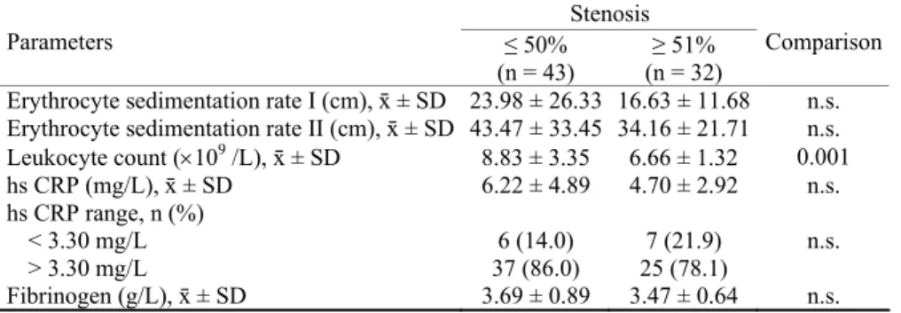
to the control group. The subjects with over 50% stenosis (2– 6.3%) belonged to the symptomatic group, and 30 (93.8%) to the asymptomatic group. The difference in the structure of belonging to certain groups among subjects with varying de-grees of stenosis was statistically significant (2 = 67,37; p < 0.001). The results showed no statistically significant sex and age distribution of the percentage of stenosis. The average number of leukocytes was significantly higher in the subjects with 50% stenosis (8.83 ± 3.35 : 6.66 ± 1.32 109/L, ANO-VA and post-hoc Dunnett’s test: p = 0.001) (Table 3). The values of other biochemical parameters were not notably dif-ferent among groups formed according to the percentage of stenosis (Table 3).
The average value of NIHSS score in the subjects of the symptomatic group was 5.10 ± 2.86, and the ischemic lesion diameter was 52.00 ± 30.83 mm (Figure 2). The correlation analysis showed a very high level of interdependence between the values of NIHSS score and diameter (r = 0.949, p < 0.001). These two characteristics also show the signifi-cant correlation with ESR I (NIHSS score: r = 0.445 and p = 0.014; diameter: r = 0.537 and p = 0.002), the number of leukocytes (NIHSS score: r = 0.822 and p < 0.001; diameter: r = 0.824 i p < 0.001) (Table 4, Figure 3).
The univariate logistic regression analysis as siginifi-cant predictors of CVI confirmed ESR I, ESR II, the number of leukocytes, hs-CRP, and fibrinogen. The increase in the
Table 2
Comparison of serum levels of inflammatory parameters and morphological characteristics Morphological characteristics
Characteristics Type I
(n = 35)
Type II (n = 27)
Type III (n = 13)
Comparison
Erythrocyte sedimentation rate I (cm) 25.97 ± 28.63 17.22 ± 11.37 14.54 ± 10.42 n.s. Erythrocyte sedimentation rate II (cm) 44.29 ± 35.43 37.41 ± 22.50 30.92 ± 21.33 n.s. Leukocyte count (109
/L) 8.82 ± 3.62 7.27 ± 1.73 6.76 ± 1.59 A*, B† hs CRP (mg/L) 6.92 ± 5.60 4.47 ± 2.05 4.21 ± 1.11 n.s. hs CRP range, n (%)
< 3.30 mg/L 7 (20.0) 5 (18.5) 1 (7.7)
> 3.30 mg/L 28 (80.0) 22 (81.5) 12 (92.3)
n.s.
Fibrinogen (g/L) 3.87 ± 0.75 3.35 ± 0.71 3.37 ± 0.86 A* hsCRP – high sensitivity C-reactive protein; A – symptomatic vs asymptomatic;
B – symptomatic vs control; ns – no significance; *p < 0.05; †p < 0.01; ‡p < 0.001.
Fig. 2 – Predominantly echolucent plaque with a thin echogenic cap (category I, Gray-Weale classification).
Table 3 Association of serum levels of inflammatory parameters and stenosis
Stenosis
Parameters ≤ 50%
(n = 43)
≥ 51% (n = 32)
Comparison
Erythrocyte sedimentation rate I (cm), ґ ± SD 23.98 ± 26.33 16.63 ± 11.68 n.s. Erythrocyte sedimentation rate II (cm), ґ ± SD 43.47 ± 33.45 34.16 ± 21.71 n.s. Leukocyte count (109
/L), ґ ± SD 8.83 ± 3.35 6.66 ± 1.32 0.001 hs CRP (mg/L), ґ ± SD 6.22 ± 4.89 4.70 ± 2.92 n.s. hs CRP range, n (%)
< 3.30 mg/L 6 (14.0) 7 (21.9)
> 3.30 mg/L 37 (86.0) 25 (78.1)
n.s.
Page 520 VOJNOSANITETSKI PREGLED Vol. 73, No. 6
ć ć
Table 4 Correlation between NIHSS score and serum levels of inflammatory
parameters in the symptomatic group
NIHSS scor Diameter Characteristcs
r p r p
Erythrocyte sedimentation rate I (cm) 0.445 0.014 0.537 0.002 Erythrocyte sedimentation rate II (cm) 0.254 0.176 0.338 0.068 Leukocyte count (x10-9 U/L) 0.822 0.000 0.824 0.000
hs CRP (mg/L) -0.256 0.172 -0.228 0.225
Fibrinogen (g/L) 0.141 0.458 0.178 0.346
hsCRP – high sensitivity C-reactive protein; NIHSS – National Institute of Health Stroke Scale.
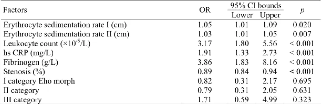
value of each of the following factors by a unit of measure-ment was associated with a significant increase in the risk of CVI as follows: ESR I by 5% (95% CI; 1 to 9%; p = 0.029),
ESR II by 3% (95% CI; 1 to 5%; p = 0.007), the number od leukocytes by 3.17 times (95% CI; 1.80–5.56 times; p = 0,001), hsCRP by 91% (95% CI; 33–173%; p = 0.001), fib-rinogen by 3.86 times (95% CI; 1.83–8.16 times; p < 0.001) (Table 5). On the contrary, the increase in stenosis percentage value by a unit of measurement was associated with a significant decrease in the risk of CVI by 10% (95% CI; 6–15%) (Table 5). In the subjects with category I of ec-homorphological characeristics the risk of CVI was notably higher than in the subjects with categories II and III by 4 times (95% CI; 1.50–10.66 times) (Table 5).
The multivariate logistic regression analysis as the most important predictor of CVI singled out the number of leukocytes. Each increase in the value of this characteristic by an unit of measurement was associated with a signifi-cant increase in the risk of CVI by 3.22 times (1.67– 6.22 times) (Table 6).
The regression model containing these two factors and the regression constant as independent variables, explained even 66% of the variability of the risk of CVI in subjects of the sample (determination coefficient - R2 = 0.66).
Fig. 3 – Ischemic cerebral lesion.
Table 5 Stroke risk factors stratified by inflammatory parameters (univariate analysis)
95% CI bounds
Factors OR Lower Upper p
Erythrocyte sedimentation rate I (cm) 1.05 1.01 1.09 0.020 Erythrocyte sedimentation rate II (cm) 1.03 1.01 1.05 0.007
Leukocyte count (×10-9/L) 3.17 1.80 5.56 < 0.001
hs CRP (mg/L) 1.91 1.33 2.73 < 0.001
Fibrinogen (g/L) 3.86 1.83 8.16 < 0.001
Stenosis (%) 0.89 0.84 0.94 < 0.001
I category Eho morph 0.82 0.31 2.17 0.695
II category 0.79 0.31 2.05 0.631
III category 1.71 0.59 4.99 0.323
OR – odds ratio; CI – confidence interval; NIHSS – National Institute of Health Stroke Scale. hsCRP –high sensitivity C-reactive protein.
Table 6 Stroke risk factors stratified by inflammatory parameters
(multivariate analysis)
95% CI bounds
Risk factor OR
ć ć Discussion
Contemporary knowledge about the potential reversibility of ischemic cerebral damage 12 influences the formation of a therapeutic approach in the prevention and treatment of acute cerebral infarction. The results of studies dealing with the morphology of atherosclerotic lesions have provided epidemiological support of the hypothesis that the vulnerability of carotid plaque is an etiopathogenic factor of acute cerebral infarction 15. Studies on the therapeutic effect of endarterectomy in symptomatic/asymptomatic patients emphasize stenosis as risk factor for cerebrovascular compli-cations 16 in clarifying the mechanisms which associate athe-roscletrosis with ischemic cerebral damage. Ultrasound eva-luation of the morphological and hemodynamic status of ca-rotid plaque 17, 18 influence the formation of the attitude that the size and structure of atheroma should be considered as separate risk factors of acute cerebral infarction in the asses-sment of embolic potential of atherosclerotic lesions.
Epidemiological support of the hypothesis that echolucency carries an increased risk of cerebrovascular complications 17–19, the definition of stable/unstable plaque and classification on the basis of echo-morphological charac-teristics 14 confirm that echomorphological characteristics are an indicator of the vulnerability and risk of future neurologi-cal symptomatology 17,18.
The traditional opinion that the size of atherosclerotic le-sion is the criterium in the assessment of high-risk changes 20 does not encompass the concept of inflammation as a basic mechanism of atherogenesis 21. Scientific research on experimental and human models provide theoretical knowledge and assumptions that inflammation can predispose distal embolization 22, 23. Even though we emphasize the im-portance of chronic subclinical inflammation in blood vessel wall, precise opinions on the proinflammatory response and the role of inflamatory mechanisms in destabilization of caro-tide plaque have not been formed. The connection of inflammatory, rheological and coagulation/fibrinolytic pro-cesses in the network of complex interactions 21, 22 gives the possibility of considering numerous parameters altered by the biochemical conditions of vulnerable carotid plaque.
By statistical analysis of biochemical markers characte-ristics it has been determined that the average values od the examined parameters are significantly higher in the subjects of the symptomatic group than in the subjects of the asymptomatic and control group.
The indepedent connection of Er dysfunction and caro-tide plaque incidence in the results of clinical studies repre-sents a rational basis of the assumption that inflammation and oxidative stress (OS) are the factors which change Er homeostasis through the alteration of the morphology and activity 22, 24. The influence of plasma proteins fibrinogen, immunoglobulin, lipoprotein, α2 macroglobulin on the ESR and increased aggregation potential under the influence of cytokines 22, 24 confirm that the altered activity of Er is a part of the chronic inflammatory processes of atherogenesis. In estimating the role of the ESR in the progression and stablilty of carotid plaque there were no statistically
signifi-cant differences in the values of the ESR I and ESR II in the subjects of the three compared categories of echomophological characteristics. The average value of fibrinogen was notably higher in the subjects with category I than in those with category II of echomorphological characteristics (p < 0.05). The values of the ESR I, ESR II and fibrinogen were not significantly diffe-rent among the two groups formed according to the hemodynamic significance of stenosis percentage (≥ 50%, < 50%). The obtained results were in accordance with the opinion formed after the publication of the Norwegian study TROMSO, that the size and structure of carotid plaque should be considered as separate factors in assessing the risk of the development of acute cerebral infarction 17, 18.
Fibrinogen as a reactant of the acute phase is an impor-tant determinant of the ESR 22, 24–26 which indicates that within a framework of the systemic response, the ESR can be the measure of response of brain alteration at an early stage of ischemia 27.
Significantly higher values of the ESR I and ESR II in the subjects of the symptomatic group compared to the sub-jects of the asymptomatic and control group represent a rati-onal basis of the conclusion that the ESR can be an acceptab-le test in the monitoring of chronic inflammatory processes related to atherosclerosis. In assessing the influence of the ESR as risk factor for cerebrovascular ischemic complicati-ons, its predictive significance was confirmed by the univari-ate logistic regression analysis, as well as that the increase in the value of this parameter by a unit of measurement was as-sociated with a significant risk growth. Even though the pro-gnostic significance of appearance of the clinical manifesta-tions of the progression and complicamanifesta-tions of carotid disease was not confirmed in multivariate logistic regression analysis, in the statistical processing of the interdependence between the values of NIHSS score (ischemic lesion diame-ter) and examined biochemical markers, a significant correla-tion during the first hour indicates that the ESR can carry important information for the early prognosis of acute cereb-ral infarction.
Page 522 VOJNOSANITETSKI PREGLED Vol. 73, No. 6
ć ć
increase in the value for an unit of measurement is associated with the risk increase. Similar results have been published by Atherosis Risk in Communites Study (ARIC) and TROMSO studies emphasizing the significance of pharmacological control of carotid atherosclerosis progression 15, 30. The mec-hanisms of the pleotropic effect of statin on multi-metabolic disorders encompass, within antiinflammatory effects, also the reduction of the concentration of fibrinogen in the primary and secondary prevention of acute cerebral infarcti-on 31. As a reactant of the acute phase, a part of the systemic response within the framework of cerebral ischemia can be a purposeful parameter in the assessment of carotid atheroscle-rosis progression and prediction at early stage of acute cereb-ral infarction.
Statistical analysis of biochemical parameters shows that the number of Le in the subjects of the symptomatic group is notably higher than in the subjects of the asymptomatic (p < 0.001) and the control group (p < 0.001). A connection of the number of Le and clinical progression of carotid disease and the influence of the increased number of Le on the risk of developing acute cerebral infarction is re-flected in the phenomena of inflammation and infection in the process of atherogenesis 32, 33. As the study on Risk Fac-tors in Impaired Glucosae Tolerance for Atherosclerosis and Diabetes (RAID) points out, the number of Le is an indepen-dent determinant of the initiation and progression of atheros-clerotic vascular disease 34. Low grade inflammation and subclinic infection, expressed through the number of Le, are a more informative indicator of the focal than general endothe-lial damage 35. The mechanisms of aggregation, adhesion and migration of Le 32, connection of the number of Le, thickness of intimomedia complexes and atherosclerotic plaque 31 indica-te that Le are a part of chronic subclinical inflammation. The rheological significance of Le stems from their size and deformability characteristics and the ability to release biologically active substances such as prostaglandins, leukotri-enes, cytokines make them a part of the inflammatory respon-se of the arterial wall 22.
In statistical analysis of the characteristics of biochemical markers, it was determined no statistically significant difference in the average values of Le among the asymptomatic and the control group. The connection of the number of Le and reducti-on in the blood vessel diameter, an insignificant difference of the number of Le in the subjects with hemodynamically significant carotid plaque (≥ 50%, asymptomatic group) and the subjects with hemodynamically insignificant carotid plaque (< 50%, con-trol group) confirm that the clinical neurologist should include both the size and structure of the atherosclerotic lesion when considering the appropriate treatment. In the conducted research the average value of Le in the subjects with category I of echo-morphological characteristics was notably higher than in the su-bjects with the categories II and III.
The results of an experimental research suggest the me-diating and modulatory role of Le in the acute inflammation inside the fibrous cap 34. The presence of inflammatory and immunocompetent cells in the human atheroma, synthesis and release of the numerous molecules with proinflammatory ef-fects 36 confirm that inflammation is a basic determinant of the
vulnerability of atherosclerotic lesions. In defining the hystological criteria of destabilization, we emphasized the ability of Le to dilute tissue by the secretion of protoeolitic enzymes and that they are rarely present in intact plaques 3. Starting from the premise that the presence of carotid plaque is the risk factor for cerebrovascular ischemic disorders, the re-sults of the Northern Manhattan Stroke Study (NOMASS) gi-ve an epidemiological confirmation of the association of the number of Le with the incidence of carotid plaque in persons who did not suffer from acute cerebral infraction 33.
The majority of studies on the connection of carotid dise-ase and acute cerebral ischemia 15, 37, 38 stemmed from the re-sults of the study Aortic Plaque and Risk of Ischemic Stroke (APRIS) suggesting no connection of the increased number of Le, appearance and size of atherosclerotic plaque and risk of acute cerebral infarction 33. The importance of clinical studies lies in the additional information which influences the forma-tion of attitudes that there is a connecforma-tion of the number of Le and subclinical atherosclerosis, independent on traditional risk factors 32 and that the connection of the number of Le, carotid ahterosclerosis and acute cerebral infarction confirm that the number of Le can be a significant predictor of cerebrovascular ischemic complications 17, 30. The altered permeability of the blood-brain barrier, accumulation of Le in the zone of acute ischemia, secretion of the proinflammatory cytokines, increa-sed endothelial permeability and production of reactive oxygen metabolities (ROM), increased expression of the potentially neurotoxic enzymes are a part of physiological changes within a framework of the inflammatory reaction which affects ischemic brain tissue 39. The reduced flexibility of Le under a reduced pressure in the zone of acute cerebral in-fraction, adhesion to endothelium, impaired hemodynamics, occlusion of capillaries and altered blood viscosity lead to an increase of the zone of ischemic brain damage 39. In the con-ducted research the average number of Le was statistically significanlty higher in the group of the subjects with acute ce-rebral infarction compared to the asymptomatic (p < 0.001) and the control group (p < 0.001). Correlation analysis estab-lished the values of the numerical characteristics of NIHSS score (ischemic lesion diameter) in a significant connection with the number of Le. The obtained results are in accordance with the attitude that the increased number of Le can be related to the risk of developing acute cerebral infarction 15 and that the degree of Le infiltration in the ischemic zone is in a positi-ve correlation with the size of tissue damage and the disease outcome 39.
Assessing the tested factors influence on the occurence of ischemic cerebral complications by the univariate logistic re-gression analysis showed that the values of the number of Le in the tested sample represented significant predictors of acute cerebral infarction. The multivariate logistic regression analysis singled out the number of Le as the most significant predictor of acute cerebral infarction.
ć ć
carotide plaques of lower echogenicity are a part of pathophysiological mechanisms of the change of atherosclerosis lesion phenotype from a structurally vulnerable to functionally unstable form and that there is an increase of the number of Le as a reactant of the acute phase, as well as a positive correlation with the lesion size 40 in the neuroinflammatory response and considering the results of the conducted research, it can be con-cluded that the number of Le is an informative parameter in the clinical assessment of the stability of carotide plaque and risk of developing acute cerebral infarction.
In the pathophysiological mechanisms of atherogenesis the penetration of hsCRP into the arterial wall at the sites of endothelial dysfunction and the presence of a deposit in the early atherosclerotic lesion, binding to Le, synthesis by monocytes and macrophages, increased platelet aggregation, proliferation of smooth muscle cells and reduced expression of endothelial nitric oxide synthase (e-NOS) 1, 38, 41 confirm the proinflammatory and proatherogenic effects of hsCRP in the initiation and progression of atherosclerotic vascular disease. By measuring numerous mediators of the inflammatory pro-cess, scientific studies on inflammation and atherosclerotic va-scular disease emphasized that hsCRP as indicator of the risk of cerebrovascular ischemic complications was in correalation with the incidence of acute cerebral infarction 35, 38, 39, 42, 43.
The association of the concentrations of hsCRP with hystological determinants of atherosclerotic lesion vulnerability in symptomatic and asymptomatic patients 23, 44 indicates that hs-CRP can be indicator of the change in atheroma phenotype from a stable to unstable form and a significant indicator of the risk of developing acute cerebral infarction 45.
Analysis of the connection of biochemical and ultraso-und parameters showed the average values of hsCRP were not significantly different among the groups formed accor-ding to the hemodynamic significance and structure of caro-tid plaque. In the atherogenic profile of subjects the concen-tration of hsCRP was considered a reactant of the acute pha-se, from the perspective of neurological practice as a part of the systemic response in acute cerebral ischemia without the presence of the extrahepatic synthesis factor of hsCRP 22.
The average value of hsCRP was notably higher in the symptomatic group compared to the asymptomatic and control
ones. All the subjects of the symptomatic group had the level of hsCRP above the reference values. Even though there was no correlation between the concentrations of hsCRP and severity of neurological deficit expressed through NIHSS sco-re, in assessing the influence of the concentrations of hsCRP on the development of acute cerebral infarction by univariate logistic regression analysis it was established that hsCRP was a significant indicator of the possible cerebrovascular ischemic complications. Multivariate logistic regression analysis con-firmed no predictive significance of hsCRP. The results of the conducted research and the results of other studies 44, 45 con-firm a connection of the increased concentrations of hsCRP, carotid atherosclerosis progression and the incidence of acute cerebral infarction and that hsCRP can be an indicator of the presence of unstable carotid plaque and the risk of ischemic cerebral complications. If we accept the attitude that hsCRP is a part of the process of inflammation in the pathophysiology of cerebral ischemia 18, 46–48 and that the increased concentration is related to the severity of neurological deficit 10, the fact that there are patients with normal values of hsCRP after acute ce-rebral infarction leads to the conclusion that the relation between hsCRP and brain damage is much more complex within a framework of the acute phase 10, 45, 46. Clinical trials in which the increase of the concentration of hsCRP in defined time intervals after acute cerebral infarction was compared to the values prior to the disease, confirmed that the significance of hsCRP in the pathogenesis of acute cerebral ischemia is the expression of inflammatory system individual response 10, 44, 45.
Conclusion
The predictive significance of the number of Le con-firms that the number of Le is associated with the phenome-non of atherosclerotic plaque vulnerability and may be a use-ful, additional marker in the clinical practice of neurologists in discovering new pharmacological approaches in the pre-vention of cerebrovascular complications. It is possible that the risk of developing acute cerebral infarction could be re-duced by controlling the carotid plaque stability through the continuous therapeutic influence on the pathogenic mecha-nisms of destabilization.
R E F E R E N C E S
1. Davignon J, Ganz P. Role of Endothelial Dysfunction in Atherosclerosis. Circulation 2004; 109(23 Suppl 1): 27−32. 2. Faxon DP, Fuster V, Libby P, Beckman JA, Hiatt WR, Thompson
RW, et al. Atherosclerotic Vascular Disease Conference: Writing Group III: pathophysiology. Circulation 2004; 109(21): 2617−25.
3. Morgan AR, Rerkasem K, Gallagher PJ, Zhang B, Morris GE, Calder PC, et al. Differences in matrix metalloproteinase-1 and matrix metalloproteinase-12 transcript levels among ca-rotid atherosclerotic plaques with different histopathological characteristics. Stroke 2004; 35(6): 1310−5.
4. Ministry of Health RS. National Guide. Belgrade: Ministry of Health of the Republic of Serbia 2004. (Serbian).
5. Grant EG, Benson CB, Moneta GL, Alexandrov AV, Baker JD, Bluth EI, et al. Carotid Artery Stenosis: Gray-Scale and
Dop-pler US Diagnosis—Society of Radiologists in Ultrasound Consensus Conference. Radiology 2003; 229(2): 340−6. 6. Touboul PJ, Hennerici MG, Meairs S, Adams H, Amarenco P,
Born-stein N, et al. Mannheim Intima-Media Thickness Consensus (2004-2006-2011). Crebrovasc Dis 2012; 34(4): 290−6. 7. Adams HP, Bendixen BH, Kappelle LJ, Biller J, Love BB, Gordon
DL, et al. Classification of subtype of acute ischemic stroke. Definitions for use in a multicenter clinical trial. TOAST. Trial of Org 10172 in Acute Stroke Treatment. Stroke 1993; 24(1): 35−41.
8. Special report from the National Institute of Neurological Disorders and Stroke. Classification of cerebrovascular dis-eases III. Stroke 1990; 21(4): 637−76.
Page 524 VOJNOSANITETSKI PREGLED Vol. 73, No. 6
ć ć
10. Ding S, Zhang M, Zhao Y, Chen W, Yao G, Zhang C, et al. The role of carotid plaque vulnerability and inflammation in the pathogenesis of acute ischemic stroke. Am J Med Sci 2008; 336(1): 27−31.
11. Faul F, Erdfelder E, Lang A, Buchner A. G*Power 3: a flexible statistical power analysis program for the social, behavioral, and biomedical sciences. Behav Res Methods 2007; 39(2): 175−91.
12. Uteyboogaart M, Schrijvers E, Vroomen P, Dekeyser J, Luijckx GJ. Routine Thrombolysis With Intravenous Tissue Plasminogen Activator in Acute Ischemic Stroke. Oxford J Med 2012; 36(5): 577−9.
13. Demarin V, Štikovac M, Thaller N. Blood-Vessel Doppler Ultra-sonography. Zagreb: Školska knjiga; 1990. (Croatian)
14. Gray-Weale AC, Graham JC, Burnett JR, Byrne K, Lusby RJ. Carotid Artery Atheroma: Comparison of Preoperative B-mode Ultra-sound Appearance With Carotid Endarterectomy Specimen Pa-thology. J Cardiovasc Surg 1988; 29(6): 676−81.
15. Chambless LE, Folsom AR, Davis V, Sharrett R, Heiss G, Sorlie P, et al. Risk factors for progression of common carotid atherosclero-sis: the Atherosclerosis Risk in Communities Study, 1987-1998. Am J Epidemiol 2002; 155(1): 38−47.
16. Spagnoli LG, Mauriello A, Sangiorgi G, Fratoni S, Bonanno E, Schwartz RS, et al. Extracranial thrombotically active carotid plaque as a risk factor for ischemic stroke. JAMA 2004; 292(15): 1845−52.
17. Mathiesen EB, Bønaa KH, Joakimsen O. Echolucent plaques are as-sociated with high risk of ischemic cerebrovascular events in ca-rotid stenosis: the tromsø study. Circulation 2001; 103(17): 2171−5.
18. AbuRahma AF, Wulu JT, Crotty B. Carotid plaque ultrasonic het-erogeneity and severity of stenosis. Stroke 2002; 33(7): 1772−5. 19. Yoshida K, Narumi O, Chin M, Inoue K, Tabuchi T, Oda K, et al.
Characterization of Carotid Atherosclerosis and Detection of Soft Plaque with Use of Black-Blood MR Imaging. Am J Neu-roradiol 2008; 29(5): 868−74.
20. Ohara T, Toyoda K, Otsubo R, Nagatsuka K, Kubota Y, Yasaka M, et al. Eccentric stenosis of the carotid artery associated with ipsilateral cerebrovascular events. Am J Neuroradiol 2008; 29(6): 1200−3.
21.Laskowitz DT, Kasner SE, Saver J, Remmel KS, Jauch EC. Clinical usefulness of a biomarker-based diagnostic test for acute stroke: the Biomarker Rapid Assessment in Ischemic Injury (BRAIN) study. Stroke 2009; 40(1): 77−85.
22.Packard RR, Libby P. Inflammation in atherosclerosis: from vascular biology to biomarker discovery and risk prediction. Clin Chem 2008; 54(1): 24−38.
23.Mallika V, Goswami B, Rajappa M. Atherosclerosis pathophysi-ology and the role of novel risk factors: a clinicobiochemical perspective. Angiology 2007; 58(5): 513−22.
24. Yang W. High Red Blood Cell Distribution Width is Closely Associated With Risk of Carotid Artery Atherosclerosis in Pa-tients With Hypertension. Exp Clin Cardiol 2010; 15(3): 37−40.
25. Zaremba J, Skrobanski P, Losy J. Acute Ischaemic Increases the Erytrocyte Sedimentation Rate, Which Correlates With Early Brain Damage. Folia Morphol (Warsz) 2004; 63(4): 373−6. 26.Mauriello A, Sangiorgi G, Palmieri G, Virmani R, Holmes DR,
Schwartz RS, et al. Hyperfibrinogenemia is associated with spe-cific histocytological composition and complications of athe-rosclerotic carotid plaques in patients affected by transient ischemic attacks. Circulation 2000; 101(7): 744−50.
27. Krupinski J, Tiru M, Font AM, Ahmed N, Sullivan M, Luque A, et al. Increased Tissue Factor, MMP-8 and D-dimer Expression in Diabetic Patients With Instable Advanced Carotid Athero-sclerosis. Vasc Health Risk Manag 2007; 3(4): 405−12.
28.Magyar MT, Szikszai Z, Balla J, Valikovics A, Kappelmayer J, Imre S, et al. Early-onset carotid atherosclerosis is associated with increased intima-media thickness and elevated serum levels of inflammatory markers. Stroke 2003; 34(1): 58−63.
29. Paximadas SA, Pagoni SN, Pitsavos CE, Skoumas JN, Karagianni ET, Nikitopoulou PD, et al. The changes of fibrinogen and lipoprotein a levels after six months treatment with statins. Atherosclerosis Suppl 2001; 2(2): 99.
30. Sen S, Hinderliter A, Sen PK, Simmons J, LeGrys VA, Beck J, et al. As-sociation of leukocyte count with progression of aortic atheroma in stroke/transient ischemic attack patients. Stroke 2007; 38(11): 2900−5.
31. Elkind MS, Sciacca R, Boden-Albala B, Homma S, di Tullio MR. Leu-kocyte count is associated with aortic arch plaque thickness. Stroke 2002; 33(11): 2587−92.
32. Temelkova-Kurktschiev T, Koehler C, Henkel E, Hanefeld M. Leukocyte count and fibrinogen are associated with carotid and femoral in-tima-media thickness in a risk population for diabetes. Cardiovasc Res 2002; 56(2): 277−83.
33. Libby P, Shi G. Mast cells as mediators and modulators of athero-genesis. Circulation 2007; 115(19): 2471−3.
34. Rost NS, Wolf PA, Kase CS, Kelly-Hayes M, Silbershatz H, Massaro JM, et al. Plasma concentration of C-reactive protein and risk of ischemic stroke and transient ischemic attack: the Framingham study. Stroke 2001; 32(11): 2575−9.
35. Jander S, Sitzer M, Schumann R, Schroeter M, Siebler M, Steinmetz H, et al. Inflammation in High-Grade Carotid Stenosis : A Possible Role for Macrophages and T Cells in Plaque Destabilization. Stroke 1998; 29(8): 1625−30.
36. Hashimoto H, Kitagawa K, Hougaku H, Etani H, Hori M. Relation-ship between C-reactive protein and progression of early carotid atherosclerosis in hypertensive subjects. Stroke 2004; 35(7): 1625−30.
37. Wardlaw JM, Farrall A, Armitage PA, Carpenter T, Chappell F, Doubal F, et al. Changes in background blood-brain barrier integrity be-tween lacunar and cortical ischemic stroke subtypes. Stroke 2008; 39(4): 1327−32.
38.Willeit J, Kiechl S, Oberhollenzer F, Rungger G, Egger G, Bonora E, et al. Distinct Risk Profiles of Early and Advanced Atheroscle-rosis : Prospective Results From the Bruneck Study. Athero-scler Thromb Vasc Biol 2000; 20(2): 529−37.
39.Hashimoto H, Kitagawa K, Hougaku H, Shimizu Y, Sakaguchi M, Nagai Y, et al. C-Reactive Protein Is an Independent Predictor of the Rate of Increase in Early Carotid Atherosclerosis. Circu-lation 2001; 104(1): 63−7.
40.Ceulemans A, Zgavc T, Kooijman R, Hachimi-Idrissi S, Sarre S, Mi-chotte Y. The dual role of the neuroinflammatory response after ischemic stroke: modulatory effects of hypothermia. J Neuro-inflammation 2010; 7(1): 74.
41. Libby P, Ridker PM, Maseri A. Inflammation and atherosclero-sis. Circulation 2002; 105(9): 1135−43.
42.Silva D, Albuquerque L, Narvaes L, Goldani M, Pereira G. C-Reactive Protein and Clinical Instability in Carotid Artery Ob-structive Disease. J Vasc Bras 2007; 6(2): 1−7.
43.Everett BM, Ridker PM. Using inflammatory biomarkers to guide lipid therapy. Curr Cardiovasc Risk Reports 2008; 2(1): 29−34.
44. Bos MJ, Schipper CM, Koudstaal PJ, Witteman JC, Hofman A, Bre-teler MM. High Serum C-Reactive Protein Level Is Not an In-dependent Predictor for Stroke: The Rotterdam Study. Circu-lation 2006; 114(15): 1591−8.
ć ć
46. Laterza O, Modur V, Crimmins D, Olander J, Landt Y, Lee JM. Identification of Novel Brain Biomarkers. Clin Chem 2006; 52(9): 1713−21.
47.Taylor A, Kent S, Flaherty P, Coyle L, Markwood T, Vernalis M. ARBITER: Arterial Biology for the Investigation of the Treatment Effects of Reducing Cholesterol: A Randomized Trial Comparing the Effects of Atorvastatin and Pravastatin on Carotid Intima Medial Thickness. Circulation 2002; 106(16): 2055−60.
48. Kettani F, Dragomir A, Côté R, Roy L, Bérard A, Blais L, et al. Impact of a better adherence to antihypertensive agents on ce-rebrovascular disease for primary prevention. Stroke 2009; 40(1): 213−20.