Iranian Journal of Basic Medical Sciences
ijbms.mums.ac.ir
The mitochondrial DNA mutations associated with cardiac
arrhythmia investigated in an LQTS family
Fatemeh Khatami
1, Mohammad Mehdi Heidari
1*, Massoud Houshmand
21 Department of Biology, Yazd University, Yazd, Iran
2 Department of Medical Genetics, National Institute for Genetic Engineering and Biotechnology, Tehran, Iran
A R T I C L E I N F O A B S T R A C T
Article type:
Original article Objective(s): As mitochondrial oxidative stress is probably entailed in ATP production, a candidate modifier factor for the long QT syndrome (LQTS) could be mitochondrial DNA (mtDNA). It has been notified that ion channels' activities in cardiomyocytes are sensitive to the ATP level. Materials and Methods: The sample of the research was an Iranian family with LQTS for mutations by PCR-SSCP and DNA sequencing. The study searched about 40% of the entire mitochondrial genome in the family.
Results: Four novel mutations that lead to an amino acid substitution and two mutations in mitochondrial tRNA have been informed in this study. A Statistically significant correlation (r = 0.737) between QTc (ms) and the age of LQTS patients has been reported.
Conclusion: The research data show that these mitochondrial mutations, in a family with LQTS, might be the responsible mitochondrial that defect and increase the gravity of LQTS.
Article history: Received: Nov 19, 2013 Accepted: Feb 24, 2014
Keywords:
Arrhythmia Long QT syndrome Mitochondrial DNA Mutation, SSCP
►
Please cite this paper as:Khatami F, Mehdi Heidari M, Houshmand M. The mitochondrial DNA mutations associated with cardiac arrhythmia investigated in an LQTS family. Iran J Basic Med Sci 2014; 17:656-661.
Introduction
The Long QT syndrome (LQTS) can be inherited or acquired. Patients are predisposed to the ventricular tachyarrhythmia torsade de pointes (TdP) which causes syncope and sudden death (1). This syndrome is a prototype ion channel disease (channelopathies) and leads to abnormal electrical properties of the heart (2, 3). It should be mentioned that cardiac ion channels contain protein complexes in the sarcolemma of cardiomyocytes, through very regular opening and closing of the gates, a selective and rapid flow of ions is conducted through a central pore. Abnormalities in ion channel function can have disastrous consequences that manifest themselves as ECG abnormalities and arrhythmias (4, 5). While doing some exercises or having some feelings, a repolarization disorder named LQTS develops. This kind of disorder is characterized by extension of the QT intervals on the ECG which is indicated by syncopal episodes. The cause in 50% to 60% of the patients that suffer from this type of syndrome, is gene mutations which encode the subunits of ion channels. It seems highly probable that the inheritance pattern of LQT is polygenic which embraces both mtDNA and nuclear DNA genes (6, 7). It is a fact that heart is greatly dependent on oxidative energy which is
generated in mitochondria, and ion channels are ATP-sensitive and exist in high density in the sarcolemma membrane (8). We have conducted our research on an Iranian family with LQTS and scrutinized mtDNA mutation among them. Although the exact mechanism of mitochondrial NNA and its impact on the syndrome is vague, it is probable that it might have a prominent etiological role.
Materials and Methods
Patients
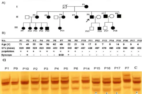
The study cases comprised a total of 18 members (11 females and 7 males) in a three-generation family (Figure 1A). The formula of Bazett was applied in order to assess the values of QTc (9). In the case study of the family, the patients were admitted
presenting symptomatic with QTc of 0 ms and
asymptomatic with QTc of 70 ms that were
tabulated according to the effect. All of the family members who were involved in the research had granted the informed consent, and blood samples and clinical evaluations were taken from them. The ethics committee of Yazd University ratified the under protocols. Thirty individuals (including 14 females and 16 males) who had no connection with definitive diagnoses of mitochondrial or cardiac
*Corresponding author: Mohammad Mehdi Heidari.Department of Biology, Yazd University, Yazd, Iran. Tel: +98-351-8122649, Fax: +98-351-8210644;
Figure 1. A) Pedigree structure of the LQT3 family. B) Clinical and ECG characteristics. C) Detection of maternally inherited 10463 T>C and 10530 G>A mutations in P1, P2, P3, P4, P5, P6, P7, P8, P9, P10 and P14 by PCR-SSCP method. These mutations did not appear in P15, P16, P17 or the controls (arrows indicate normal pattern bands)
disorders were assigned as the control group. All of the participants (both patients and control group) were notified of the research objectives and granted their informed consents to the genetic analysis.
Patient P6 (Proband) was a man aged 62 with a prolonged QT interval (QTc501 ms), his palpitation and echocardiography results were normal. P1 was a woman aged about 71, who had a 520 ms QT interval. Seven children of this woman suffered from LQT3. P7 was a man who had had a sudden cardiac arrest and a 533 ms QT interval, and the patients P2 and P8 had syncopes, in this pedigree. The rest of the patients' clinical manifestations are presented in Figure 1B.
Mutational analysis of mitochondrial genome
Through a DNA extraction kit the total DNA was extracted from peripheral blood lymphocytes of all patients and the controls (DNAfast Kit-Genfanavaran, Tehran, Iran). PCR of each sample was set in a 0.5 ml tube using 100 ng of total DNA, 10 pM of each primer, 200 µM of dNTPs, 1X PCR buffer containing 2.5 mM MgCl2 and 1 U Taq DNA polymerase (Roche
Diagnostics, Mannheim, Germany). After an initial 3
min denaturation at 94°C, 32 cycles were performed
(94°C for 30 sec, 57°C for 45 sec and 72°C for 50 sec)
followed by a final 5 min extension at 72°C. The
samples were mixed with 10 µl denaturing/loading solution containing 95% formamide, 20 mM EDTA, 0.05% bromophenol blue, 0.05% xylene cyanol and heated at 95°C for 10 min and immediately chilled on
ice. 5 µl of the PCR products were electrophoresed on an 8% nondenaturing acrylamide:bisacrylamide (62.5:1) gel in TAE buffer at 200 V; during electrophoresis, the temperature of the gel was constantly kept at 15°C for 16 hr. A strict control of
the gel temperature is of extreme importance to obtain a proper determination of polymorphisms, as no resolution was obtained by running the gels at temperatures other than 15°C. SSCP bands were visualized on the gels by silver staining (10).
If a band shift was detected by SSCP, it was confirmed with direct DNA sequencing by using an ABI 3700 capillary sequencer. Five segments, ranging in size from approximately 110 to 446 bp, were amplified by PCR and assayed by SSCP in both groups. Table 1, illustrates and defines respectively,
Table 1. SSCP primer sequences and PCR conditions
Primer sequence Nucleotide position Tm (°C) Size (bp) Fragment
F CTACGGTCAATGCTCTGAAA R AAATAGAATGATCAGTACTG
8161 - 8180
8605 - 8586 57
444
ATPase8
F TCTCTTATACTAGTATCCTT R CCAATTAGGTGCATGAGTAG
8731-8750
9050-9031 57 319 ATPase6-1
F ACAATTCTAATTCTACTGAC R TACTATATGATAGGCATGTGA
9106 - 9125
9239 - 9216 57 133 ATPase6-2
F TCTGGCCTATGAGTGACTAC R GAGCGATATACTAGTATTCC
10361-10380
10540- 10521 57 179 tRNAArg
F ACCACACCGCTAACAATCAG R TTCATCATGCGGAGATGTTG
14561-14580
the state of primers and PCR for SSCP analysis and sequencing, which were designed and optimized with Gene Runner version 3.05 (Hastings Software Inc. Hastings, NY, USA). These segments cover 40% of mtDNA, including the entire sequence of the 7 transfer RNA genes (tRNALys, Gly, Arg, His, Ser, Leu and Glu) and
the sequences of 9 protein-coding genes (CoxII, ATPase8, ATPase6, CoxIII, ND3, ND4L, ND4, ND5, and ND6). The results of DNA sequence analysis were compared with the revised Cambridge sequence (11, 12) and differences were recorded as mutations. Each sequence variation was then checked against the Mitomap database (13). Those that were not recorded in the database were categorized as novel mtDNA variations.
Pathogenicity analysis
The prediction of pathogenicity of amino acid changes was accomplished by the Sorting Intolerant from Tolerant (SIFT) database. SIFT score ranges from 0 to 1; the amino acid substitution is predicted damaging if the score is 0.0 and tolerated if the score is > 0.05. The median score ranges from 0 to 4.32. Protean (Protein Structure Prediction and Annotation) identified the secondary structural features and physiochemical characteristics including hydropathy index, flexibility index and antigenic index; the conservation of the amino acid changes was assayed using the MEGALIGN program that is part of the Lasergene V.6 software (DNASTAR, Inc. Madison, WI, USA). The hydropathy index was measured by Protean according to the Kyte-Doolitle method (14), which predicts the regional hydropathy of proteins from their amino acid sequence. All amino acids were designated by hydropathy values and they were averaged about a window size equal to 9. Results below 0 are hydrophobic and above 0 are hydrophilic. tRNA sequence and the two-dimensional (2D) structure comparison was accomplished according to the Mamit-tRNA database (http://mamit-tRNA.u-strasbg.fr).
Data analysis
Data are presented as Mean±SD values. Frequen- cy data betweennormal controlsand patientswere
Figure 2. Sequencing results: A) T8462C mutation, B) G10530A mutation
compared using Pearson's chi-square test. To carry out statistical analysis, we have applied the GraphPad Prism software; P presents values below 0.05 which is indicative of statistical significance.
Results
The age range of patients and controls were 4 to 71 (25.4±19.9, Mean±SD) and 12 to 61 (22.5±18.6, Mean±SD) years, respectively. The range of QTc of patients was 428 to 560 (489±39.2, Mean±SD). Statistics revealed that 10 patients had heart palpitation and 3 had syncopes.
SSCP analyses were carried out on 18 patients and 30 healthy controls. Normal controls of the same ethnicity were also genotyped to establish the frequency of mutations. DNA fragments showing abnormal banding patterns on SSCP analyses were sequenced for the identification of exact mutations (Figure 2).
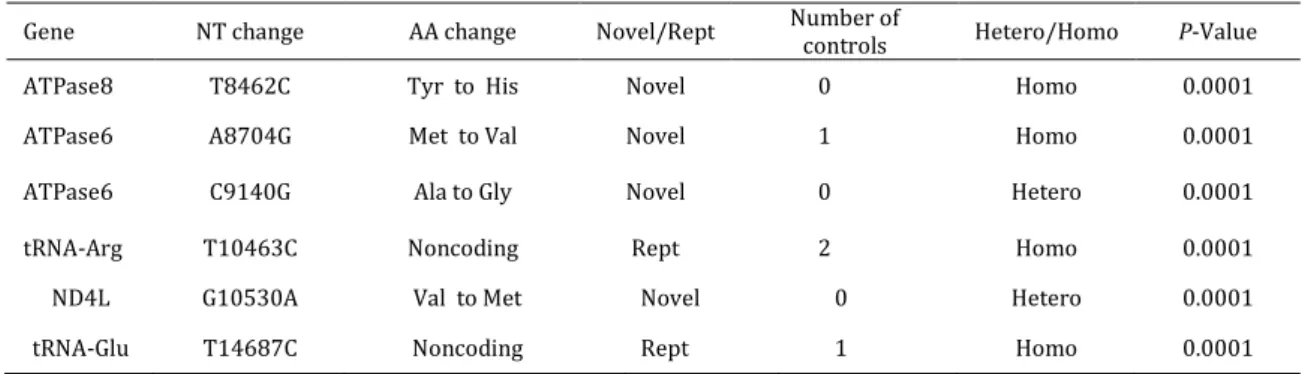
A total of six different point mutations were detected in 14 related patients (P1, P2, P3, P4, P5, P6, P7, P8, P9, P10, P11, P12, P13, and P14 patients) (Table 2). These mutations were not found in P15, P16, P17, and P18 patients. This familial history of mutations [4 novel mutations (m.8462 T>C, m.8704 A>C, m.9140 C>G and m.10530 G>A) and the indicator of maternal inheritance is 2 mutations in mitochondrial tRNA (m.10463 T>C and m.14687 T>C)]. These mutations were frequently detected in this family, although the difference relative to the controls was not statistically significant (P>0.05).
Table 2. Summery of mutations identified in the study subjects
Gene NT change AA change Novel/Rept Number of
controls Hetero/Homo P-Value
ATPase8 T8462C Tyr to His Novel 0 Homo 0.0001
ATPase6 A8704G Met to Val Novel 1 Homo 0.0001
ATPase6 C9140G Ala to Gly Novel 0 Hetero 0.0001
tRNA-Arg T10463C Noncoding Rept 2 Homo 0.0001
ND4L G10530A Val to Met Novel 0 Hetero 0.0001
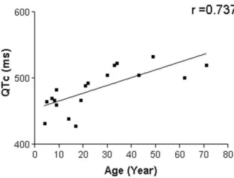
Figure 4. Correlation between QTc (ms) and age of LQTS patients
m.8462 T>C sequence change (homoplasmic state) is located at codon 33 in the outside transmembrane domain of the ATPase 8 gene, and changes a moderate conserved tyrosine, a hydrophilic amino acid, to histidine, a hydrophilic amino acid, modifying the hydropathy index from -0.867 to -1.08. SIFT projected that it tolerated with score of 0.08 and the median sequence conservation was 3.08. This sequence alteration is presented to 14 patients (77.7%) and compared with 2 normal subjects (6.6%) (P= 0.0001).
m.8704 A>C sequence variant (homoplasmic state) is located at amino acid position 60 in transmembrane domain of ATPase 6 gene. It changes a moderate conserved methionine, a hydrophobic amino acid, to valine, a hydrophobic amino acid, which changes the hydropathy index from -0.811 to -0.556. SIFT predicted that it affected protein function with score of 0.03 and median sequence conservation was 4.18. This sequence alteration was in 1 controls.
m.9140 C>G sequence change (heteroplasmic state) is located at amino acid position 205 in transmembrane domain of ATPase 6 gene. It changes a highly conserved alanine, a hydrophobic amino acid, to glycine, a hydrophobic amino acid, which changes the hydropathy index from 2.82 to 2.44. SIFT predicted that it affected protein function with score of 0.00 and median sequence conservation was 4.18. This sequence alteration was not observed in controls.
m.10530 G>A sequence variant (heteroplasmic state) is located at amino acid position 21 in transmembrane domain of ND4L gene. It reforms an average conserved valine and a hydrophobic amino acid, to methionine; a hydrophobic amino acid, which changes the hydropathy index from 0.389 to 0.133. SIFT predicted that it affected protein function with score of 0.00, and the median sequence conservation was 4.32. The
sequence fluctuation was seen in 1 control. Moreover, the results indicate that there are two different point mutations in tRNA genes, as explained earlier (15). These mutations were discovered among 14 patients (P1 to P14) and their mothers, as well.
m.10463 T>C sequence change is located at a moderate conserved region of the acceptor stem of tRNA arginine. This mutation was not observed in healthy control subjects but was previously reported as a polymorphism (16).
m.14687 T>C sequence change is located at a highly conserved region of the T-loop of tRNA glutamate. It was not detected in controls and was previously reported as pathologic (17).
Discussion
LQTS is a disorder resulting from a prolongation in ventricular repolarization and increases the risk of developing torsades de pointes and sudden cardiac death (6). The main criteria for clinical diagnosis of LQTS are a prolonged QTc value exceeding 450 ms and documented syncope episodes (18). On the condition that mitochondrial mutations arise in LQTS, it might be particularly noteworthy in under pressure states, which extend the rate of energy utilization (19).
Through examinations and detailed analysis of the mitochondrial genome of the family, 6
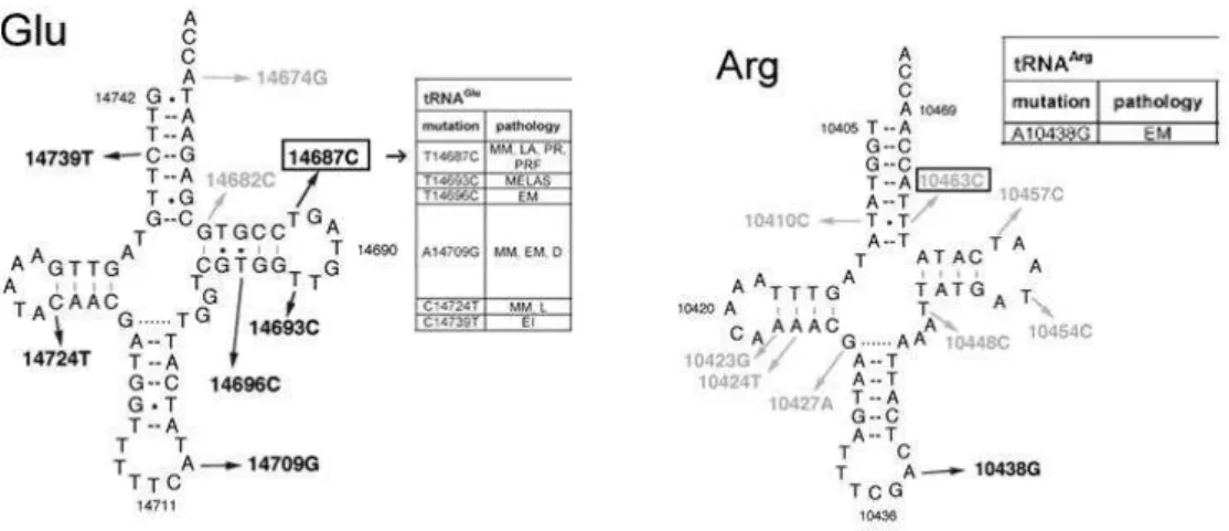
Figure 3. The position of pathogenic T14687C mutation in tRNAGlu and the position of polymorphic T10463C mutation in tRNAArg
By investigating seven tRNA genes, we found two mutations in tRNAArg (T10463C) and tRNAGlu
(T14687C) which have been discussed before (13, 20–22) (Figure 3). Among the control subjects the mutations were not noticed; T14687C is particularly located in a highly conserved region that may be one of several predisposing factors for LQTS.
Our result showed that the mitochondrial mutations in LQTS patients were higher than in control cases (P=0.0001). According to the statistics, the significant correlation between QTc (ms) and age of LQTS patients is (r= 0.737) (Figure 4). Hence, although the sample size (LQTS patients) is relatively small, the results of the research reveal that mitochondrial mutations might increase QTc as the patients grow older; moreover, they increase the gravity of LQTS.
Our data suggest that these mitochondrial mutations in a family with LQTS might not have caused the onset of the disease but had secondary effects. This phenomenon could be due to the sensitivity of the heart to defects in the mitochondrial respiratory chain (OXPHOS) and energy production (23). The substitutions might be due to either polymorphism or mutation, however, in order to validate further studies are necessary which consider other criteria with various sample sizes. It is possible that mutations in mtDNA could constitute a predisposing factor that in combination with nuclear mutations and environmental risk factors may trigger the sudden cardiac death.
Acknowledgment
This research was supported by Yazd University. We thank all patients for providing blood samples for scientific research as well as Special Medical Center (Tehran, Iran), whose cooperation and support was essential in our work. The study was approved by the Yazd University Human Research Ethics Committee. The authors declare that they have no conflicts of interest.
References
1. Vincent GM. The long QT syndrome. Indian Pac
Electrophysiol J 2002;2:127-142.
2. Chiang CE. Congenital and acquired long QT syndrome. Current concepts and management. Cardiol Rev 2004; 12:222-234.
3. Keating MT, Sanguinetti MC. Molecular and cellular mechanisms of cardiac arrhythmias. Cell 2001; 104:569-580.
4. Ackerman MJ, Clapham DE. Ion channels--basic science and clinical disease. N Engl J Med 1997; 336:1575-1586.
5. Grant AO, Carboni MP, Neplioueva V, Starmer CF,
Memmi M, Napolitano C, et al. Long QT syndrome,
Brugada syndrome, and conduction system disease are linked to a single sodium channel mutation. J Clin Invest 2002; 110:1201-1209.
6. Towbin JA, Vatta M. Molecular biology and the prolonged QT syndromes. Am J Med 2001; 110:385-398.
7. Opdal SH, Vege A, Egeland T, Musse MA, Rognum TO. Possible role of mtDNA mutations in sudden infant death. Pediatr Neurol 2002; 27:23-29.
8. Wilde AA, Bezzina CR. Genetics of cardiac arrhythmias. Heart 2005; 91:1352-1358.
9. Bazett HC. The time relations of the blood-pressure changes after excision of the adrenal glands, with some observations on blood volume changes. J Physiol 1920; 53:320-339.
10. Sambrook J, Russel DW. Molecular cloning: a laboratory manual. 3 th ed. New York: Cold Spring Harborn Laboratory Press; 2001.
11. Anderson S, Bankier AT, Barrell BG, de Bruijn MH,
Coulson AR, Drouin J, et al. Sequence and
organization of the human mitochondrial genome. Nature 1981; 290:457-465.
12. Andrews RM, Kubacka I, Chinnery PF, Lightowlers RN, Turnbull DM, Howell N. Reanalysis and revision of the Cambridge reference sequence for human mitochondrial DNA. Nat Genet 1999; 23:147.
13. Ruiz-Pesini E, Lott MT, Procaccio V, Poole JC,
Brandon MC, Mishmar D, et al. An enhanced
14. Kyte J, Doolittle RF. A simple method for displaying the hydropathic character of a protein. J Mol Biol 1982; 157:105-132.
15. Khatami M, Houshmand M, Sadeghizadeh M,
Eftekharzadeh M, Heidari MM, Saber S, et al.
Accumulation of mitochondrial genome variations in Persian LQTS patients: a possible risk factor? Cardiovasc Pathol 2010; 19:e21-27.
16. Uusimaa J, Finnila S, Remes AM, Rantala H,
Vainionpaa L, Hassinen IE, et al. Molecular
epidemiology of childhood mitochondrial
encephalomyopathies in a Finnish population: sequence analysis of entire mtDNA of 17 children reveals heteroplasmic mutations in tRNAArg, tRNAGlu, and tRNALeu(UUR) genes. Pediatrics 2004; 114:443-450.
17. Bruno C, Sacco O, Santorelli FM, Assereto S, Tonoli
E, Bado M, et al. Mitochondrial myopathy and
respiratory failure associated with a new mutation in the mitochondrial transfer ribonucleic acid glutamic acid gene. J Child Neurol 2003; 18:300-303.
18. Priori SG, Barhanin J, Hauer RN, Haverkamp W,
Jongsma HJ, Kleber AG, et al. Genetic and molecular
basis of cardiac arrhythmias: impact on clinical management part III. Circulation 1999; 99:674-681. 19. Arnestad M, Opdal SH, Vege A, Rognum TO. A mitochondrial DNA polymorphism associated with cardiac arrhythmia investigated in sudden infant death syndrome. Acta Paediatr 2007; 96:206-210. 20. Tang S, Batra A, Zhang Y, Ebenroth ES, Huang T. Left ventricular noncompaction is associated with
mutations in the mitochondrial genome.
Mitochondrion 2010; 10:350-357.
21. Gal A, Pentelenyi K, Remenyi V, Pal Z, Csanyi B,
Tomory G, et al. Novel heteroplasmic mutation in the
anticodon stem of mitochondrial tRNA(Lys)
associated with dystonia and stroke-like episodes. Acta Neurol Scand 2010; 122:252-256.
22. van Oven M, Kayser M. Updated comprehensive phylogenetic tree of global human mitochondrial DNA variation. Hum Mutat 2009; 30:E386-394.
23. Wallace DC. Mitochondrial defects in