INT. J. BIOAUTOMATION, 2013, 17(1), 5-16
5
Computer Modeling of Human Delta Opioid Receptor
Tatyana Dzimbova1*, Fatima Sapundzhi2, Nevena Pencheva3, Peter Milanov2,4
1
Institute of Molecular Biology “Roumen Tsanev” Bulgarian Academy of Sciences
1113 Sofia, Bulgaria
E-mail: [email protected]
2
Faculty of Mathematics and Natural Sciences South-West University “Neofit Rilski”
2700 Blagoevgrad, Bulgaria
3
Faculty of Public Health and Sports South-West University “Neofit Rilski” 2700 Blagoevgrad, Bulgaria
4
Institute of Mathematics and Informatics Bulgarian Academy of Sciences
1113 Sofia, Bulgaria
*
Corresponding author
Received: September 27, 2012 Accepted: April 5, 2013
Published: April 15, 2013
Abstract: The development of selective agonists of δ-opioid receptor as well as the model of interaction of ligands with this receptor is the subjects of increased interest. In the absence of crystal structures of opioid receptors, 3D homology models with different templates have been reported in the literature. The problem is that these models are not available for widespread use. The aims of our study are: (1) to choose within recently published crystallographic structures templates for homology modeling of the human δ-opioid receptor (DOR); (2) to evaluate the models with different computational tools; and (3) to precise the most reliable model basing on correlation between docking data and in vitro bioassay results.
The enkephalin analogues, as ligands used in this study, were previously synthesized by our group and their biological activity was evaluated. Several models of DOR were generated using different templates. All these models were evaluated by PROCHECK and MolProbity and relationship between docking data and in vitro results was determined. The best correlations received for the tested models of DOR were found between efficacy (erel) of the compounds, calculated from in vitro experiments and Fitness scoring function from docking studies.
New model of DOR was generated and evaluated by different approaches. This model has good GA341 value (0.99) from MODELLER, good values from PROCHECK (92.6% of most favored regions) and MolProbity (99.5% of favored regions). Scoring function correlates (Pearson r = -0.7368, p-value = 0.0097) with erel of a series of enkephalin analogues, calculated from in vitro experiments. So, this investigation allows suggesting a reliable model of DOR.
Newly generated model of DOR receptor could be used further for in silico experiments and it will give possibility for faster and more correct design of selective and effective ligands for
δ-opioid receptor.
Introduction
The opioid receptors are very important class of receptors that attract the attention of huge number of scientists. Development of the effective and selective ligands to each opioid receptor is a time consuming process, which involves knowledge and skills of different researchers: chemists, biologists, medics, pharmacologists, etc. Large number of compounds with opioid action were synthesized, characterized and biologically tested, but just some of them are with desired efficacy and selectivity to the respective opioid receptor.
On the other hand the isolation and crystallization of opioid receptors is very heavy task, due to their location. All of them are trans-membrane proteins and the isolation causes destruction of their tertiary structure. So, these are the reasons to search for different approaches for solving this problem. Many experiments, concerning mutation of the binding site of opioid receptors, were done in order to determine the key amino acid positions in the receptor, which are responsible for their action. Different theoretical models were proposed, but they are not available for the investigators in that field, because theoretical models have not been published in data base.
The aims of our study are: (1) to choose within recently published crystallographic structures templates for homology modeling of the human δ-opioid receptor (DOR); (2) to evaluate the models with different computational tools; and (3) to precise the most reliable model basing on correlation between docking data and in vitro bioassay results.
Materials and methods
Computational tools
In order to perform computational studies the different software is used in the present work. The protein sequence of δ-opioid receptor was obtained from UNIPROT. Homology modeling studies were carried out using Chimera, that provides a graphical interface to running the program Modeller via a Web service, hosted by the UCSF RBVI [1]. Ligand preparation was done with MOE (Molecular Operating Environment) [2]. Docking studies were performed by using GOLD 5.1 (Genetic Optimization for Ligand Docking) [3], run on Scientific LINUX 5.5 operating system. Molegro Molecular Viewer [4] was used for generating figures.
Sequence alignment
INT. J. BIOAUTOMATION, 2013, 17(1), 5-16
7
3D model generation and validation
Based on the MSA derived with CLUSTALW, 3D models of DOR were built using MODELLER of Chimera package with default parameters. The model with higher GA341 score (more than 95%) was chosen as a model for further evaluations. Validation of the structural quality of the generated models was done using PROCHECK [6] and MolProbity [7].
Docking of ligands
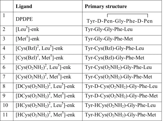
Eleven ligands, investigated for their potency, selectivity and efficacy to DOR with in vitro bioassay in our previous study [8], were selected for docking studies. Their primary structure, including that of selective ligand DPDPE and endogenous enkephalins, is presented in Table 1.
Table 1. Enkephalin analogues used as ligands in our study
Ligand Primary structure
1
DPDPE Tyr-D-Pen-Gly-Phe-D-Pen
2 [Leu5]-enk Tyr-Gly-Gly-Phe-Leu
3 [Met5]-enk Tyr-Gly-Gly-Phe-Met
4 [Cys(Bzl)2, Leu5]-enk Tyr-Cys(Bzl)-Gly-Phe-Leu
5 [Cys(Bzl)2, Met5]-enk Tyr-Cys(Bzl)-Gly-Phe-Met
6 [Cys(O2NH2)2, Leu5]-enk Tyr-Cys(O2NH2)-Gly-Phe-Leu
7 [Cys(O2NH2)2, Met5]-enk Tyr-Cys(O2NH2)-Gly-Phe-Met
8 [DCys(O2NH2)2, Leu5]-enk Tyr-D-Cys(O2NH2)-Gly-Phe-Leu
9 [DCys(O2NH2)2, Met5]-enk Tyr-D-Cys(O2NH2)-Gly-Phe-Met
10 [HCys(O2NH2)2, Leu5]-enk Tyr-HCys(O2NH2)-Gly-Phe-Leu
11 [HCys(O2NH2)2, Met5]-enk Tyr-HCys(O2NH2)-Gly-Phe-Met
3D structures of the ligands were modeled in MOE. Ligands were protonated at physiological pH 7.4.
Docking was carried out with GOLD 5.1 software. It uses generic algorithm and considers full ligand conformational flexibility and partial protein flexibility. The binding site for DOR, in literature [9], was defined as residues within 10 Å radius of aspartic acid of third TM domain, which is involved in the most crucial interaction. In the case of DOR this is Asp 128. All scoring functions of GOLD were used. The conformations of the ligands with best scoring functions were selected and parameters of the scoring functions were use in order to find correlations between them and previously obtained in vitro results. The following three, very important parameters were obtained for all compounds from in vitro bioassay: IC50 – potency, KA – affinity and erel – efficacy. Their relevance was well-argumented by Pencheva et al., [8]
Table 2. Values of IC50, KA and erel obtained in mouse vas deferens by in vitro assay [8]
Ligand IC50 (nM) KA (nM) erel
1 DPDPE 6.18±1.17 180±35 30.2±10.0
2 [Leu5]-enk 11.45±2.06 54.9±13.1 5.8±1.0
3 [Met5]-enk 18.91±2.15 48.4±7.5 3.6±0.3
4 [Cys(Bzl)2, Leu5]-enk 8.30±1.40 68.5±29.7 9.3±3.2
5 [Cys(Bzl)2, Met5]-enk 9.53±1.20 23.8±3.0 3.5±0.3
6 [Cys(O2NH2)2, Leu5]-enk 1.29±0.31 36.4±16.4 29.2±9.5
7 [Cys(O2NH2)2, Met5]-enk 2.22±0.45 14.1±5.4 7.3±2.0
8 [DCys(O2NH2)2, Leu5]-enk 11.40±2.01 73.4±12.7 7.4±1.9
9 [DCys(O2NH2)2, Met5]-enk 75.96±11.67 463±161 7.1±1.8
10 [HCys(O2NH2)2, Leu5]-enk 31.92±5.10 76.4±7.1 3.4±0.2
11 [HCys(O2NH2)2, Met5]-enk 16.09±1.90 55.7±6.1 4.5±0.3
Correlations
In order to find relationship between sets of data derived from in vitro assay and docking results, we tried to predict it with the help of Pearson's correlation, using GraphPad Prism 3.0.
Results and discussion
Sequence alignment
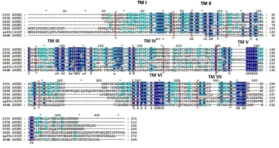
The final MSA for DOR is presented in Fig. 1. This result indicates that the sequences chosen are reasonably good to be used for homology modeling. The percentage of homologous residues in TM regions proved that these alignments are good (Table 3). It was found in MSA, that there was a sequence similarity greater than 50% in almost all TM regions. Thus, it could be expected that homology models built with this alignment would be more accurate.
Homology modelling
The 3D models with multiple alignments for DOR from respective templates, contain seven TM regions, as evident in all GPCRs. Homology models were generated with the focus to get suitable 3D models for docking and other in silico experiments in the absence of crystallographic structures.
I
NT
.
J
.
BIO
AU
TOMATI
ON
, 201
3,
17
(1)
, 5-16
9
Table 3. Percentage of residues identical or strongly similar with DOR sequence and different templates for each domain.
Percentage of residues identical or strongly similar for each domain Sequences
TM I TM II TM III TM IV TM V TM VI TM VII
2y00 68 67 80 60 65 73 75
2ycx 68 67 80 60 65 73 75
2r4s 68 67 80 60 61 73 75
3vg9 81 67 83 100 68 87 83
2ks9 100 87 83 67 73 93 33
4a4m 0 62 75 67 85 85 67
Fig. 2 Ramachandran plot of Model D of δ-opioid receptor, obtained by PROCHECK. Plot for the best model is presented.
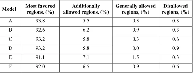
Table 4. PROCHECK results for selected DOR models
Model Most favored regions, (%)
Additionally allowed regions, (%)
Generally allowed regions, (%)
Disallowed regions, (%)
A 93.8 5.5 0.3 0.3
B 92.6 6.2 0.9 0.3
C 93.2 5.8 0.3 0.6
D 93.2 5.8 0.0 0.9
E 91.1 7.1 1.5 0.3
INT. J. BIOAUTOMATION, 2013, 17(1), 5-16
11
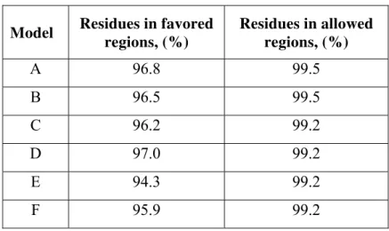
The results for the same Models A-F with the next tool, MolProbity score are shown in Table 5.
Table 5. MolProbity score for selected DOR models
Model Residues in favored regions, (%)
Residues in allowed regions, (%)
A 96.8 99.5
B 96.5 99.5
C 96.2 99.2
D 97.0 99.2
E 94.3 99.2
F 95.9 99.2
Docking results
An earlier study of Befort et al. [10] has shown that aspartic acid of TM III is important residue for ligand recognition. The binding site was defined as residues within 10 Å radius of Asp128 of the DOR. Docking was performed with all six models and 11 ligands. The results of docking studies of the ligands with the best model – Model B, are presented in Fig. 3, where the numbers correspond to the respective ligand form Table 1.
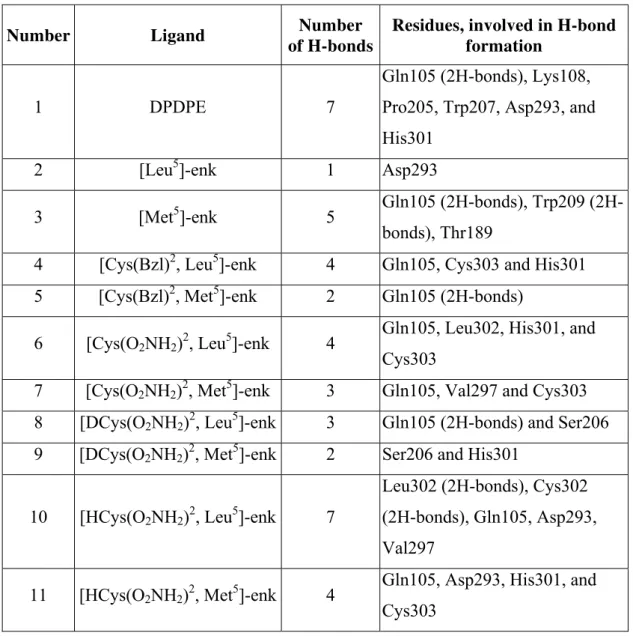
The best poses, including the interactions between target and ligands, obtained from docking with Model B of DOR, are presented in Table 6.
In all cases ligands interact with receptor in the cavity around Asp128 with 10 Å in radius. These results suggest the reliability of the selected model of DOR.
Correlations
Correlations of molecular docking data and in vitro studies are performed with GraphPad Prism 3.0. All parameters form in vitro studies such as IC50, KA and erel are compared with the
results from docking (fitness scoring function) in order to find correlation. It was found, that the parameter erel correlates with Fitness scoring function in five models. These data are
presented in the Table 7.
Table 6. Hydrogen bonds between ligands and receptor
Number Ligand Number
of H-bonds
Residues, involved in H-bond formation
1 DPDPE 7
Gln105 (2H-bonds), Lys108,
Pro205, Trp207, Asp293, and
His301
2 [Leu5]-enk 1 Asp293
3 [Met5]-enk 5 Gln105 bonds), Trp209
(2H-bonds), Thr189
4 [Cys(Bzl)2, Leu5]-enk 4 Gln105, Cys303 and His301
5 [Cys(Bzl)2, Met5]-enk 2 Gln105 (2H-bonds)
6 [Cys(O2NH2)2, Leu5]-enk 4
Gln105, Leu302, His301, and
Cys303
7 [Cys(O2NH2)2, Met5]-enk 3 Gln105, Val297 and Cys303
8 [DCys(O2NH2)2, Leu5]-enk 3 Gln105 (2H-bonds) and Ser206
9 [DCys(O2NH2)2, Met5]-enk 2 Ser206 and His301
10 [HCys(O2NH2)2, Leu5]-enk 7
Leu302 (2H-bonds), Cys302
(2H-bonds), Gln105, Asp293,
Val297
11 [HCys(O2NH2)2, Met5]-enk 4
Gln105, Asp293, His301, and
INT. J. BIOAUTOMATION, 2013, 17(1), 5-16
13
Table 7. Correlation between fitness scoring function from GOLD docking procedure and erel parameter of in vitro studies
Model Pearson's correlation, r p-value
A - 0.6491 0.0307
B - 0.7368 0.0097
C - 0.6479 0.0311
D - 0.6420 0.0332
E - 0.7495 0.0079
F - 0.5156 0.1045
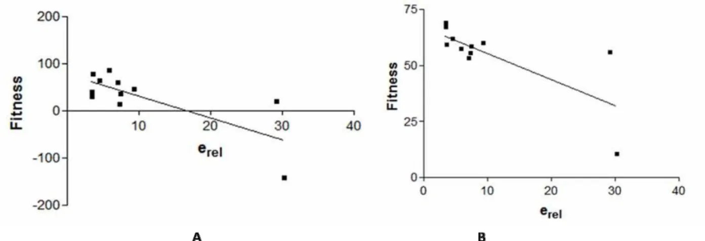
Best Pearson’s correlation was obtained for Model E of DOR, presented in Fig. 4.
Fig. 4 Pearson’s correlation of erel and fitness of ligands with Model E (A) and Model B (B) of DOR
The Model F can be excluded for further assessment, basing on comparative evaluation of all results obtained for DOR models. However, Model E with the best value of Pearson’s correlation is not satisfactory because according to of PROCHECK and MolProbity results the conformation of its 3D structure is inappropriate. So, the most acceptable model appears to be Model B. It shows good reliability in PROCHECK and MolProbity evaluation and reveals good correlation between in vitro results, concerning efficacy, and docking scoring function. 3D model of DOR – Model B is presented in Fig. 5.
Conclusion
New model of DOR was generated and evaluated by different approaches. This model has good GA341 value (0.99) from MODELLER, good values from PROCHECK (92.6% of most favored regions) and MolProbity (99.5% of favored regions). Scoring function correlates (Pearson’s correlation r = -0.7368, p-value = 0.0097) with erel of a series of enkephalin
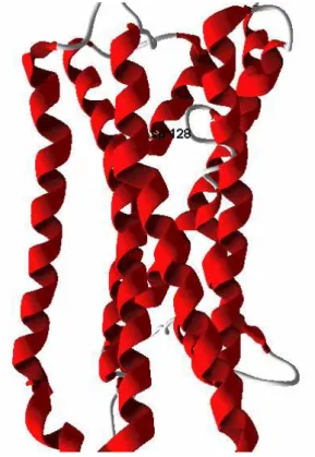
Fig. 5 3D model of DOR – Model B, generated by MODELLER. Colors of the backbone based on the secondary structure information:
α-helices are colored red, and coil is colored gray.
Newly generated model of DOR receptor could be used further for in silico experiments and it will give possibility for faster and more correct design of selective and effective ligands for DOR. Moreover, it could be compared in further studies with recently published data for crystal structure of DOR [11].
Acknowledgements
This work was supported by NFSR of Bulgaria project DVU 01/197/16.12.2008 and DO 02-135/31.07.2009.
References
1. Sali A., T. L. Blundell (1993). Comparative Protein Modeling by Satisfaction of Spatial Restraints, J Mol Biol, 234, 779-815.
2. Molecular Operating Environment (MOE), 2012.10; Chemical Computing Group Inc., 1010 Sherbooke St. West, Suite #910, Montreal, QC, Canada, H3A 2R7, 2012. Available at http://www.chemcomp.com.
3. Jones G., P. Wilett, R. C. Glen, A. R. Leach, R. Taylor (1997). Development and Validation of a Genetic Algorithm for Flexible Docking, J Mol Biol, 267, 727-748.
4. Molegro Molecular Viewer, http://molegro.com/index.php 5. RCSB Protein Data Base, www.rcsb.org
6. Laskowski R. A., M. W. MacArthur, D. Moss, J. M. Thornton (1993). PROCHECK: A Program to Check the Stereochemical Quality of Protein Structures, J Appl Cryst, 26, 283-291.
INT. J. BIOAUTOMATION, 2013, 17(1), 5-16
15
Validation for Macromolecular Crystallography, Acta Crystallogr D Biol Crystallogr, 66, 12-21.
8. Pencheva N., P. Milanov, L. Vezenkov, T. Pajpanova, E. Naydenova (2004). Opioid Profiles of Cys2-containing Enkephalin Analogues, Eur J Pharmacol, 498, 249-256.
9. Milanov P., Pencheva N. (2011). Hyperbolic Model of a Partial Agonism: Explicit Formulas for Affinity, Efficacy and Amplification of Partial Agonists, Serdica, Journal of Computing, 333-358.
10. Befort K., L. Tabbara, S. Bausch, C. Chavkin, C. Evans, B. Kieffer (1996). The Conserved Aspartate Residue in the Third Putative Transmembrane Domain of the Delta-opioid Receptor is Not the Anionic Counterpart for Cationic Opiate Binding but is a Constituent of the Receptor Binding Site, Mol Pharmacol, 49, 216-223.
11. Granier S., A. Manglik, A. C. Kruse, T. S. Kobilka, F. S. Thian, W. I. Weis, B. K. Kobilka (2012). Structure of the δ-opioid Receptor Bound to Naltrindole, Nature, 485, 400-404.
Tatyana Dzimbova, Ph.D.
E-mail: [email protected]
M.Sc. in Chemistry from South-West University “Neofit Rilski”, Blagoevgrad in 1995. Ph.D. from Institute of Molecular Biology, Bulgarian Academy of Sciences, Sofia in 2008. Presently Post-Doctoral position in Department “Molecular design and biochemical pharmacology”, Institute of Molecular Biology “Roumen Tsanev”, Bulgarian Academy of Sciences, Bulgaria. Main interests are in the field of Chemistry, Biochemistry, Pharmacology, Drug Design, Computer modeling.
Fatima Sapundzhi, Ph.D. student
E-mail: [email protected]
Prof. Nevena Pencheva, Ph.D.
E-mail: [email protected]
Prof. Pencheva obtained her Ph.D. in 1991 from the Institute of Physiology of Bulgarian Academy of Sciences, Sofia, where her research focuses on modulation of synaptic transmission, neuropharmacology of opioids and in vitro structure-activity relationships of enkephalin analogues. Currently, as a professor in South-West University in Blagoevgrad, she is a lecturer on Exercise physiology and works on opioid and cannabinoid modulation of exercise-induced hypoalgesia and modeling of opioid recepetors.
Prof. Peter Milanov, Ph.D.
E-mail: [email protected]
![Table 2. Values of IC 50 , K A and e rel obtained in mouse vas deferens by in vitro assay [8]](https://thumb-eu.123doks.com/thumbv2/123dok_br/16375709.191466/4.892.121.773.136.530/table-values-ic-obtained-mouse-deferens-vitro-assay.webp)