JSCS–4906 543.544–14+543.51
Original scientific paper
Chemical characterization of the photodegradation products of
midazolam complexes with randomly methylated-
β
-cyclodextrin
by HPLC and LC–MS/MS
SNEZANA AGATONOVIC-KUSTRIN1*, MOSIMOTSANA LEBETE2, MICHAEL E. BROWN2, DAVID W. MORTON3 and BEVERLEY D. GLASS4
1Faculty of Pharmacy, Universiti Teknologi MARA (UiTM), Selangor, Malaysia, 2Faculty of
Pharmacy, Rhodes University, Grahamstown, South Africa, 3The School of Pharmacy and
Applied Science, La Trobe Institute of Molecular Sciences, La Trobe University, Bendigo, Victoria, Australia and 4Pharmacy, College of Medicine and Dentistry,
James Cook University, Townsville, Queensland, Australia
(Received 2λ September, revised 20 December 2015, accepted 18 January 2016) Abstractμ Midazolam, a potent anxiolytic drug with sedative properties, is susceptible to degradation by both light and hydrolysis in aqueous solution. When formulated as an intranasal product, it was found to be effective in achieving seizure control in epileptic patients. In order to deliver an adequate therapeutic dose to a patient, a nasal formulation requires the concentration of midazolam to be higher than its aqueous solubility. One way to increase mida-zolam solubility to a therapeutic concentration is complexation with randomly methylated- -cyclodextrin. Thus, it is important to determine how complex-ation with cyclodextrin affects the rate of degradcomplex-ation and type of midazolam degradants that are formed. It was found that complexation with cyclodextrin decreases its photostability. More importantly, the degradation profile for midazolam is significantly altered when it is complexed with randomly methyl-ated- -cyclodextrin, which was partly confirmed in a previous work. By con-tinuing this study, degradation products, not found in the photodegradation of uncomplexed midazolam are observed in significant quantities when it was complexed with randomly methylated- -cyclodextrin. The decreased photo-stability was accompanied by the appearance of two new degradation products, an intermediate structure and a dimer. Photoproduct formation followed the same pattern as in the forced degradation studies, further confirming the pre-sence of an intermediate. The production of these new photodegradants, char-acterized with their MS spectra, and a proposed degradation mechanism of midazolam are discussed.
Keywordsμ benzodiazepine photostability; high performance liquid chromato-graphy; liquid chromatography-tandem mass spectrometry.
INTRODUCTION
The benzodiazepine, midazolam (MDZ), is a potent drug with anxiolytic, hypnotic, amnestic, anticonvulsant, skeletal muscle relaxant, and sedative pro-perties, and is commonly used as a preoperative anesthetic agent, especially for pediatric patients.1 A growing interest in alternative forms of drug administration has led to the development of nasal formulations of MDZ for sedation before surgical, dental or diagnostic procedures, and for treatment of seizures in children and adult patients as safe and inexpensive means to rapidly achieve seizure control.1 The nasal route has been explored widely for delivery of a large number of drug molecules, due to its rich vasculature and thin epithelial lining that enables drug to reach systemic circulation after administration via nasal route
directly and provides rapid onset of action. Since no nasal preparation is com-mercially available, midazolam solution for injection has been used.2,3 Since commercially available formulation for intravenous application contain low con-centration of MDZ, due to its low solubility, large application volumes exceeding the limited nasal capacity have to be used to achieve adequate dosing. In order to deliver therapeutic midazolam doses in smaller volumes, nasal preparations with MDZ concentration exceeding its solubility must be developed. MDZ is water-soluble at pH values lower than 4 and lipid-water-soluble at pH values above 5 and therefore lipid soluble at physiological pH.4 The water solubility of MDZs inc-reases at pH values less than 4.0 due to ring opening and ionization3 whereby MDZ is reversibly converted to the corresponding benzophenone, an open ring form with a highly ionizable primary amino group. Thus, at pH > 5, the drug is largely present in the lipid soluble, closed ring form. Although, solubility of MDZ increases at lower pH, the use of an intranasal acidic MDZ solution would result in severe irritation and swelling in the nasal cavity. In order to deliver an adequate therapeutic dose to a patient, a nasal formulation requires the concentra-tion of MDZ to be higher than its water solubility (i.e., <0.1 mg L–1 at the
phys-iological pH of 7.4).
hetero-cyclic molecules). Hence, -CD is commonly used as a complexing agent in many CD complex drug formulations.5
Formulations of a 0.2 % oral solution of MDZ containing -CD and citric acid were previously investigated.6 More recently, however, it was demonstrated that the use of randomly methylated- -CD (RM– -CD) as a solubilizer sig-nificantly reduces the rate of degradation of MDZ.7 RM– -CDs have replaced
-CD with partially methylated CDs in order to improve the solubility and stab-ility of MDZ oral formulations, with the aim of developing a palatable buccal-nasal formulation. Due to their high aqueous solubility (> 500 mg mL–1) and their ability to form inclusion complexes, methylated CDs are widely used in drug formulations to improve drug solubility8 and to reduce the rate of hydro-lysisλ and photodegradation.10 Substitution of the hydroxyl with methoxy groups imparts a slightly lipophilic character to the molecule, which also facilitates per-meation through the mucosa.11
Although the photostability of MDZ is well documented,4,12–14 the rate of photodegradation and the nature and distribution of photodegradants may change when MDZ forms an inclusion complex with CD.15–17 CDs have the potential to control chemical and photochemical reactions, due to the microplanarity of the host cavity and limited molecular mobility of the guest molecule, due to steric constraints. The nature of the lowest excited states of the guest molecule, deact-ivation pathways and the fate of the reaction may be modulated by the CD mic-roenvironment. Thus, structural changes in drug molecules that occur when they form a complex with CD may accelerate drug degradation.18 The rate of second-ary reactions may also be influenced and the chemical evolution of reaction intermediates controlled.18 Quantitative high performance liquid chromatography (HPLC) methods have been used to study the decomposition of MDZ in formul-ations1λ and to determine the amount of MDZ and its metabolites in blood plasma.20
Since the presence of RM– -CD results in different degradation from those observed in the absence of RM– -CD, the aim of present study was to investigate more deeply the obtained products. Based on results of the photostability, kinetic studies and MS characterization, a degradation mechanism of midazolam is proposed as well as a model of the RM– -CD–MDZ inclusion complex.
Phase solubility studies conducted according to the method of Higuchi and Connors21 confirmed the ability of RAMEB to solubilize the target concentration of 10 mg mL–1 of MDZ.
EXPERIMENTAL Chemicals
Limited (Cambridge). Water for chromatography was obtained from a Milli-Q® water purific-ation system (Millpore, MA, USA).
HPLC instrumentation
Analysis of MDZ and its degradants was performed using HPLC (Spectra series P100 pump, Thermo separation products, Virginia, USA) in an isocratic mode and a UV 100 vari-able wavelength ultraviolet (UV) detector. The flow rate was 1 mL min-1, the injection volume was 20 L, the column was used at room temperature (25±2 °C) and detection was performed spectrophotometrically at 240 nm. Methanol was used as the organic mobile phase component. The optimum organic/aqueous phase ration was found to be 70μ30 and the mobile phase was modified by addition of ammonium acetate as recommended for LC–MS studies to improve the performance of an LC–MS screening method. For very weak basic compounds, such as amides, the ammonium ions in the mobile phase promoted proton adduct responses.22
The retention times obtained with a Bondapak C18 10 m column were compared with those obtained using a Waters Spherisorb ODS2 5 m column.
Phase solubility studies
The phase solubility studies were performed in triplicate. A 75 mg of MDZ was placed in a conical flask and suspended in 5 ml of phosphate buffer solution (pH 5.8) containing 0, 5, 10, 20 and 30 % RM– -CD (i.e., 0.042, 0.084, 0.168, 0.252 mol L-1). The conical flaks were then stoppered, covered in foil, and shaken in a water bath at 25±1 °C. The samples were then analysed after 24 h using a UV method. The presence of photodegradation products was ruled out by covering the flasks with foil during the phase solubility studied. The procedure was repeated with phosphate buffer at pH 5.0.
UV method development
Midazolam (50 mg) was dissolved in 0.1 M HCl and diluted to 100 mL with the same solvent. The resultant solution was diluted to achieve concentrations of 0.005, 0.01, 0.015, 0.02 and 0.025 mg mL-1. The absorbances of these solutions were measured at 258 nm (λ
max) and a standard calibration curve was constructed by plotting absorbance versus concentration. Photostability kinetic studies
MDZ solutions (0.5 mg mL-1) in phosphate buffer (pH 5.0) were prepared in the pre-sence and abpre-sence of RM– -CD, in 2 mL clear glass (USP standard type 1 glass) ampoules (including dark controls) and irradiated at 550 W m-2 for 12 h (1.2 million lux h) in order to degrade the drug to approximately 10 % of its original concentration. An Atlas SUNTEST CPS+ (Atlas Material Testing Technology B.V, Germany), fitted with a Xenon lamp and Solar ID65 filter was used for the irradiation of the samples. The temperature in the Suntest cabinet was maintained at 40±2 °C.
Samples (2 mL) were removed at 1 h intervals over the 12 h period, diluted to a final volume of 10 mL and then analyzed. Control samples were covered in foil and treated in the same way as the exposed samples. Degradation was calculated as a percentage of the height of the drug peak, with respect to the peak height obtained from analysis of the original solution. The appearance of the major degradation products was also monitored and calculated as a percentage of the highest concentration achieved.
LC-MS/MS
degradation products. LC–MS/MS was then used to identify the photodegradants. For the MS studies, a Finnigan LCQ ion trap mass spectrometer (ITMS, Finnigan MAT, USA) equipped with an atmospheric pressure chemical ionization (APCI) ion source was used. Separation was performed on a Waters Spherisorb 5 m ODS2 column (250 mm×4.6 mm).
Thermal stability studies
To investigate the influence of temperature on the degradation of MDZ, 2 mL aliquots of MDZ solution (0.5 mg mL-1, with and without 30 % RM– -CD) were sealed in ampoules, covered in foil and placed in ovens at temperatures of 25, 30, 40, 50, 60 and 70 °C. The samples were removed after 12 h and analyzed.
RESULTS AND DISCUSSION
A simple UV method with 0.1 M HCl as the solvent was developed for use in midazolam–RM– -CD phase solubility studies. The method showed good linearity, with the linear regression equation y = –0.000051 + 0.0275A (y is the
midazolam concentration and A the absorbance) with a correlation coefficient of
0.λλ. It was accurate with λ8.λ–101.6 % percent recovery. Precision of the method was determined by calculating the relative standard deviation (RSD). The RSD for replicate measurement at three different concentration within the linear
range was less than 1 %, indicating adequate precision.22
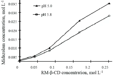
The highest concentration of MDZ of 8.4 mg mL-1 that was achieved in the
phosphate buffer pH 5.8, was below the target concentration. However, in the presence of 30 % RM-β-CD the concentration of 10.6 mg mL-1 was reached. In
both cases, the phase solubility curves obtained were of the Ap-type, suggesting the formation of higher order complexes (Fig. 1). The results of the phase sol-ubility studies indicated that desired solsol-ubility of 10 mg mL–1 was achieved with 30 % RM– -CD at pH 5.0.
The developed HPLC method was found to have sufficient accuracy with an average recovery of 100±2 % for replicate measurements at low, mid and high concentrations of MDZ within the working concentration range. The accuracy of the method was investigated by spiking a 30 % M– -CD solution with three known concentrations of MDZ (0.4, 0.5 and 0.6 mg mL–1). Linearity was con-firmed to be within the range 0.002 to 0.02 mg mL–1, with a high correlation coefficient of 1.00. The precision of the method, in terms of repeatability, was satisfactory with relative standard deviations of 0.62 and 0.40 % (n = 6) at low
and mid concentrations within the working range.
The limit of quantification (LOQ) was found to be 0.002 mg mL–1. The
robustness of the method was demonstrated during method development, when it was shown that the best peak resolution was reached when the pH of the mobile phase was between 6 and 8, the concentration of ammonium acetate in the mobile phase was between 20 and 40 mM, with the organic/aqueous phase ratio main-tained at 70μ30 methanol/water. The ruggedness of the method was confirmed by the robustness and intermediate precision results, which had a RSD of 0.λ and 0.5
%, respectively, thus ensuring that the method is precise and suitable for use for the analysis of MDZ. The LC–MS method was reliable in evaluating the specif-icity of the assay in the absence of RM– -CD, but for the solutions containing RM– -CD, a photodiode array detector was required in order to obtain a peak due to the presence of MDZ alone.
In the samples without RM– -CD, degradation products started appearing after 2 h of irradiation. A slight yellowish–brown color developed simultaneously that intensified on continued irradiation. After 6 h, a precipitate formed which was readily soluble in the dilution solvent used for analysis (methanol/water). The control samples, with and without RM– -CD, were clear and colorless, until the end of the experiment. Analysis of the exposed samples containing RM– -CD showed that degradation products begun to form during the first hour of exposure. No suspended particles were observed throughout the experiments, but a slight yellowish–brown color was observed.
The HPLC chromatograms of the degraded samples (i.e., MDZ with and
major degradants are marked as A, B and C (degradation product C being only present in the MDZ RM– -CD solution) on the HPLC chromatograms.
Time, min
(a)
Time, min
(b)
Fig. 2. HPLC chromatograms showing the peak of midazolam, M, after 8 h photodegradation (a) in the absence and (b) in the presence of RM– -CD and the peaks of degradants
A, B and C.
further degradants were evident at 8–λ min. For the purpose of this study, only identification of the major component, marked as a degradant C, was considered.
Production of degradant A follows degradation of the drug. The maximum concentration of photodegradant C was reached after 6 h, Production of the third degradant, degradant C, reached a maximum amount after about 7 h, when approximately λ0 % of the drug had been degraded. The amount of C then dec-reased over the next 3 h to about 15 % of its maximum value. At this total time of 10 h, the amount of degradant B reached a maximum. A similar trend was seen in the forced degradation studies. These results indicate that photodegradant C may be an intermediate that decomposes into degradant B.1
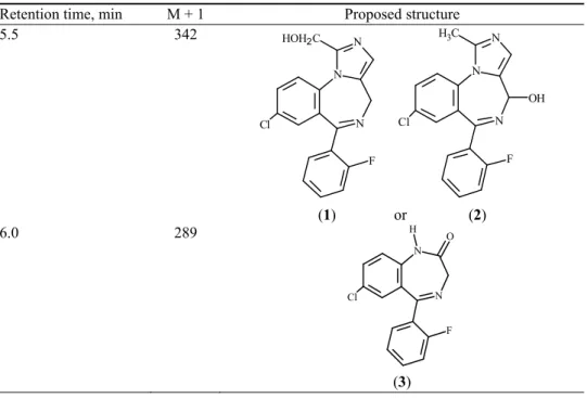
The results of LC–MS analysis including the M+1 peaks for the degradants, their retention times and proposed structures are listed in Table I. The degrad-ation products observed only in solutions of MDZ, at retention times 5.5 and 6.5 min, exhibited peaks at m/z 342 (14 %, relative abundance – RA) and 343 (27 %
RA), respectively. The structures proposed for the photoproduct with m/z 342 are
the hydrolysis products 1-hydroxymethylmidazolam (1) or 4-hydroxymidazolam (2), which are also the primary metabolites of MDZ. The photoproduct with m/z
343 has proposed structure (4).
TABLE I. Retention times, M + 1 peak values and proposed structures of photodegradants Retention time, min M + 1 Proposed structure
5.5 342
N
N N HOH2C
Cl
F
N
N N C H3
Cl
F OH
(1) or (2)
6.0 28λ
N
N O
Cl
F H
TABLE I. Continued
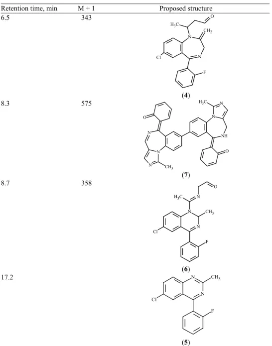
Retention time, min M + 1 Proposed structure
6.5 343
N
N CH2
Cl
F O C
H3
(4)
8.3 575
N N
N CH3
O N
NH N C H3
O
(7)
8.7 358
N N CH3
N O
F Cl
C H3
(6) 17.2
N N CH3
F Cl
(5)
Mass spectra of degradation products, with m/z values of 28λ, 273, 358 and
575 obtained by MS-MS analysis, are shown in Figs. 3a–d and 4a–d, respect-ively. In Fig. 3a, the molecular-ion peak (m/z 28λ, 23 % RA) is still visible, with
(a)
(b)
Fig. 3. Mass spectra ofμ a) degradant A with the M + 1 peak at m/z 28λ and b) degradant B with the M+1 peak at m/z 273.
pound is the same as one reported by Andersin et al.12 and, on this basis,
deg-radant A is proposed to be N-desalkylflurazepam, or
7-chloro-5-(2-fluorophe-nyl)-1,3-dihydro-2H-1,4-benzodiazepin-2-one (structure 3) and is one of the
starting compounds in the synthesis of MDZ. The base peak at m/z 261 (100 %
RA, M–H2O) and 260 (16 % RA, M–CO) were previously reported.23 Other peaks observed were at 226 (57 % RA), 208 (36 % RA) and 140 (56 % RA). Photodegradation of MDZ to degradant A follows the opening of the imidazole ring with subsequent substitution to form benzodiazepinone.
(a)
(b)
The M+1 peak m/z 273, which corresponds to the loss of Cl,21 for degradant
B was not detected (Fig. 3b). The base peak is at m/z 177 (100 % RA,
M–C6H4F) and other peaks were m/z 237 (44 % RA, M–Cl), 245 (21 % RA) and
253 (14 % RA, M–F). The molar mass of degradant B suggests that it is 6-chloro-2-methyl-4-(2-fluorophenyl)-quinazoline.12 Andersin et al. also rep-orted the formation of a precipitate in solutions of MDZ exposed to daylight. Their analysis of the purified precipitate revealed that the precipitate was 6-chloro-2-methyl-4-(2-fluorophenyl)-quinazoline. In this study, the precipitate appeared after 6 h of exposure and the amount of B was also approaching a maximum value. The proposed identification of B as structure 5, is consistent in that the compound is more hydrophobic than degradant A (structure 3). The ret-ention time of B was 17.2 min in contrast to the other degradants that had shorter retention times than MDZ (Rt = 10.5 min). Due to its non-polar nature, it is not
surprising that degradant B precipitated out of the aqueous solution as its con-centration increased. The absence of a precipitate in the RM– -CD solutions, in spite of the evidence of the presence of B as a major degradation product, could be explained by the ability of RM– -CD to improve the solubility of the hydro-phobic degradant compounds present.
The experimentally determined UV λmax values of degradants A and B were consistent with literature values (Table II) for their proposed structures.
TABLE II. UV max values of degradants A,B and C (nm)
Degradant Experimental Literature values13
A 22λ
318 230 318
B 230
327 206 230
326
C 215
333 –
The retention time (8.7 min) of degradant C suggests that it is more polar than MDZ. It possibly contains oxygen, resulting in a higher molecular mass. The mass spectrum of degradant C (Fig. 4a) shows an M+1 peak at m/z 358 (72
% RA). It is important to stress that a compound with this molecular mass has not been previously reported in photodegradation studies of MDZ. The base peak at
m/z 316 (100 % RA), and the peak at m/z 2λ7 (75 % RA) could be due to loss of
CH3, followed by subsequent loss of CH3CH2O. A fragment equivalent to the M+1 peak of degradant B, m/z 273 was observed. From the kinetic studies, it was
HPLC after a few weeks. After this time, the peak due to the drug had slightly decreased and the peak for degradant C had almost disappeared, while the peak for B had significantly increased, which is further indication that C may be an intermediate of B. The structure of C was therefore proposed to be structure 6. Degradation of MDZ through C to B is an additional reaction pathway that was observed in the presence of RM– -CD, which explains the slight increase in the degradation rate.
Another degradant caused by the presence of RM– -CD may be a dimer of one of the degradation products. The mass spectrum (Fig. 4b) shows a base peak at m/z 556 (100 % RA, M–H2O), and other peaks at 381 (52 % RA) and 367 (λ5
% RA). The proposed structure (structure 7) is a dimer of a major degradation product (6-(6-chloro-2-methyl-3H-quinazolin-4-ylidene)-cyclohexa-2,4-dienone)
of MDZ without CD in the aqueous solution.14 The MS data suggests the dimer is formed through loss of a Cl atom from each molecule, resulting in free rad-icals, which then combine with each other. Formation of this dimer and intermed-iate degradant C (structure 6) is unique to the CD environment. The reactions are made possible by the ability of CD to stabilize free radicals,24 and by conform-ational control which results in stabilization of certain reaction intermediates. Since hydroxylation is attributed to the escape of degradants from the CD cage, it is suggest that an important part of the degradation reaction pathway results from the recombination of radicals, which are not in the solvent but are trapped in the CD cavity.
The major degradation product (molar mass 323.1) in solutions exposed to a high pressure mercury lamp by Andersin and coworkers12 was not observed in the present work. Even the degradant that formed the dimer (m/z 575) was only
present in small quantities. Degradant B, one of the major degradation products in this study, occurred in the solutions exposed to daylight, where A was present in small amounts.
The results of this study indicate that MDZ undergoes a highly sequence- -selective photoreaction pathway following inclusion complexation with RM– - -CD. RM– -CD can thus alter the photobehavior of a MDZ molecule, by changing the ground state distribution of reactive and non-reactive conformers (“conform-ational control”) resulting in selectivity,25 and thus the reaction could be directed along one of the competing pathways.26
mole-cular size of MDZ, calculated using Molemole-cular Modeling pro 5.10 is estimated to be 14.67 Å, in molecular length (x), 11.5λ Å width (y), and 4.27 Å depth (z).
However, the RM– -CD diameter inside cavity is 6.0–6.5 Å, while its outside diameter is 15.4 Å and height is 7.λ Å, which corresponds approximately to the size of an aromatic ring (calculated size 6.18 Å in length, 6.82 Å in width, and 3.54 Å in depth). Therefore, RM– -CD is only able to accommodate slightly more than one aromatic ring within its cavity.
NMR studies have confirmed MDZ RM– -CD complex formation by the shift observed in the peaks for both MDZ and RM– -CD.271H- and 13C-NMR have provided an idea how the guest substrate is positioned in the CD cavity. The shielding of the CD cavity protons and associated shift changes in the signal for the MDZ protons in the mixtures of CD and MDZ are attributed to the aromatic ring penetrating into the CD cavity, thereby confirming the formation of an inc-lusion complex. The structure of the MDZ RM– -CD complex was established using two dimensional NMR (ROESY) spectral data.28 The signals for the pro-tons belonging to the aromatic ring containing fluorine exhibit strong cross cor-relation peaks with the CD cavity protons. Taking into account the 1μ1 stoichio-metry of the complex, it was concluded that the fluorine-containing ring pene-trates the -CD cavity resulting in the formation of a 1μ1 complex. The signals for the H-8 of the chlorine containing aromatic ring (next to the chlorine sub-stituent) also exhibited a strong interaction with the protons inside the CD cavity. However, H-λ and H-10 did not show any cross peaks with the CD cavity pro-tons (Fig. 5). Thus, the possibility of another 1μ1 complex involving penetration of the chlorine-containing ring was ruled out because interaction of the protons from the ring containing chlorine with the cavity protons was not evidenced. Therefore, it could be concluded that the fluorine-containing aromatic ring and the chlorine substituent and H-8 from the other aromatic ring are likely to be inside the RM– -CD cavity.
Fig. 5. Proposed model of the RM– -CD–MDZ inclusion complex.
medium. This occurs to a lesser extent in the solution outside the CD cavity due to more rapid diffusion.2λ
CONCLUSIONS
It has been shown that complexation with RM– -CD slightly decreased the photostability of MDZ. The decreased stability was accompanied by the appear-ance of two new degradation productsμ i) an intermediate [(E)-{1-[6-chloro-4-(2-
-fluorophenyl)-2-methylquinazolin-1(2H)-yl]ethylidene}amino]acetaldehyde
(structure 6/degradant C) that degrades to 6-chloro-2-methyl-4-(2-fluorophenyl)- -quinazoline (structure 5/degradant B); and ii) a dimer (structure 8) formed from
free radicals (structure 7) derived from previously reported photodegradant 6-(8- -chloro-1-methyl-4,5-dihydro-2,5,10b-triaza-benzo[e
]azulen-6-ylidene)-cyclo-hexa-2,4-dienone as a result of the loss of a Cl atom. Photodegradation product formation followed the same pattern as in the forced degradation studies, further confirming the presence of an intermediate. The photodegradation chemistry of a drug molecule is different when encapsulated because the interior of the cyclo-dextrin cavity constitutes an isolated environment where the included species is usually present as a single molecule. The photochemistry is therefore generally restricted to intramolecular events, except in cases of multiple occupancy.
While the presence of the RM– -CD improves the aqueous solubility of midazolam, it also alters its photostability. Introduction of new photo degradants must be taken into account when using RM– -CD to improve MDZ solubility and develop a feasible nasal formulation. Although photoinduced reactions might or might not be identical in vitro and in vivo, basic knowledge on the reaction
И З В О Д
ХЕМИЈСКАКАРАКТЕРИЗАЦИЈАПРОИЗВОДАФОТОРАЗГРАДЊЕКОМПЛЕКСА
МИДАЗОЛАМАСАНАСУМИЧНОМЕТИЛОВАНИМβ-ЦИКЛОДЕКСТРИНОМ
МЕТОДАМА HPLC И LC–MS/MS
SNEZANA AGATONOVIC-KUSTRIN1, MOSIMOTSANA LEBETE3, MICHAEL E. BROWN3, DAVID W. MORTON2
и BEVERLEY D. GLASS4 1
Faculty of Pharmacy, Universiti Teknologi MARA (UiTM), Selangor, Malaysia, 2The School of Pharmacy and Applied Science, La Trobe Institute of Molecular Sciences, La Trobe University, Bendigo, Victoria, Australia,
3
Faculty of Pharmacy, Rhodes University, Grahamstown, South Africa and 4Pharmacy, College of Medicine and Dentistry, James Cook University, Townsville, Queensland, Australia
Мидазолам, потенцијалнианксиолитичкилексаседативнимсвојствима, подложан
јефоторазградњи и хидролизиу воденимрастворима. Установљено је да су интрана
-залне формулације ефикасне у контроли епилептичних напада. Како би се постигла
адекватна терапијска доза, назалне водене формулације би морале да буду са већом
концентрацијом мидазолама негошто је његова растворљивост. Један од ачинада се
постигне терапијска доза јесте комплексирање мидазолама насумично метилованим
β-циклодекстрином. Стогајеважнодасеутврдебрзинаразграњемидазоламаиодгова
-рајући насталипроизводи уколико јекомплексиран. Установљено једа је комплекси
-рани мидазолам фотонестабилинији од некомплексираног. Оно штоје више значајно
јесте да се кодкомплексираног мидазолама јављајунови производи разградњеу зна
-чајнимконцентрацијама. Смањенафотостабилност јепраћенапојавомдвановапроиз
-водаразградње, једногсапрелазномструктуромиједногдимера. Настанакфотодегра
-дационихпроизводаодигравасепоистом механизмукојијеустановљениспитивањем
принудне разградње, штопотврђујеприсуство прелазнихструктура. Дата је дискусија
настанкафотодеградационихпроизвода, којисуокарактерисани MS подацима, ипред
-ложенјеодговарајућимеханизамразградњемидазолама.
(Примљено 29. септембра, ревидирано 20. децембра 2015, прихваћено 18. јануара 2016)
REFERENCES
1. S. Björkman, G. Rigemar, J. Idvall, Brit. J. Anaesth. 79 (1λλ7) 575 2. T. Mahmoudian, M. M. Zadeh, Epilepsy Behav. 5 (2004) 253
3. E. Lahat, M. Goldman, J. Barr, T. Bistritzer, M. Berkovitch, BMJ (Clin. Res. Ed.) 321 (2000) 83
4. R. Andersin, J. Pharm. Biomed. Anal. 9 (1λλ1) 451
5. G. Tiwari, R. Tiwari, A. K. Rai, J. Pharm. Bioallied Sci. 2 (2010) 72
6. F. Marçon, D. Mathiron, S. Pilard, A.-S. Lemaire-Hurtel, J.-M. Dubaele, F. Djedaini-Pilard, Int. J. Pharm. 379 (200λ) 244
7. D. Mathiron, F. Marcon, J. M. Dubaele, D. Cailleu, S. Pilard, F. Djedaini-Pilard, J. Pharm. Sci. 102 (2013) 2102
8. J. Mannila, T. Jarvinen, K. Jarvinen, M. Tarvainen, P. Jarho, Eur. J. Pharm. Sci. 26 (2005) 71
λ. T. Loftsson, M. E. Brewster, J. Pharm. Sci. 85 (1λλ6) 1017
10. S. Scalia, R. Tursilli, V. Iannuccelli, J. Pharm. Biomed. Anal. 44 (2007) 2λ
11. T. Loftsson, S. B. Vogensen, M. E. Brewster, F. Konradsdottir, J. Pharm. Sci. 96 (2007) 2532
14. B. D. Glass, M. E. Brown, S. Daya, M. S. Worthington, P. Drummond, E. Antunes, M. Lebete, S. Anoopkumar-Dukie, D. Maharaj, Int. J. Photoenergy 3 (2001) 205
15. P. Bortolus, G. Grabner, G. Kohler, S. Monti, Coord. Chem. Rev. 125 (1λλ3) 261 16. J. Mielcarek, J. Pharm. Biomed. Anal. 15 (1λλ7) 681
17. U. Tadanobu, I. Keiko, H. Fumitoshi, U. Kaneto, Eur. J. Pharm. Sci. 1 (1λλ3) 81
18. S. Sortino, S. Giuffrida, G. De Guldi, R. Chillemi, S. Petralia, G. Marconi, G. Condorelli, S. Sciuto, Photochem, Photobiol. 73 (2001) 6
1λ. R. Andersin, S. Tammilehto, Int. J. Pharm. 123 (1λλ5) 22λ
20. J. A. Carrillo, S. I. Ramos, J. A. Agundez, C. Martinez, J. Benitez, Ther. Drug Monit. 20 (1λλ8) 31λ
21. T. Higuchi, K. A. Connors, in Advances in Analytical Chemistry and Instrumentation, C. N. Reilley, F. W. McLafferty, Eds., Interscience, New York, 1λ65; pp. 117
22. H. G. Brittain, Pharm. Tech. 22 (1λλ8) 82
23. R. Selkämaa, S. Tammilehto, Int. J. Pharm. 49 (1λ8λ) 83
24. A. V. Veglia, A. M. Sanchez, R. H. De Rossi, J. Org. Chem. 55 (1λλ0) 4083 25. B. N. Rao, N. J. Turro, V. Ramamurthy, J. Org. Chem. 51 (1λ86) 460 26. G. D. Reddy, V. Ramamurthy, J. Org. Chem. 52 (1λ87) 5521 27. A. R. Hedges, Chem. Rev. 98 (1λλ8) 2035
28. S. M. Ali, S. K. Upadhyay, Magn. Reson. Chem. 46 (2008) 676