ORIGINAL ARTICLE
Fuelling of TCA cycle in hepatic cells
Marwari
goat during
ambi-ent temperature associated stress
Kataria N.
1*, Kataria A.K.
2, Joshi A.
11Department of Veterinary physiology, College of Veterinary and Animal Science, Rajasthan Uni-versity of Veterinary and Animal Sciences, Bikaner – 334 001, Rajasthan, India
2Apex Centre for Animal Disease Investigation, Monitoring and Surveillance, College of Veterin-ary and Animal Science, Rajasthan University of VeterinVeterin-ary and Animal Sciences, Bikaner – 334 001, Rajasthan, India
*
Phone , 0091 – 151- 2546399*Email- nalinikataria@rediffmail.com
Received July 16, 2010
The present study was launched to assess the effect of extreme ambient temperature associated stress on
fuelling of TCA cycle in hepatic cells of Marwari goat. Based on the fact that whenever a hepatocyte needs fuel
for TCA cycle, the activity of enzyme glutamate dehydrogenase (GD) increases making alpha-ketoglutarate
available for TCA cycle, 600 apparently healthy Marwari goats of either sex, between 6 months to 3 years of age
were screened and blood samples were collected during moderate, cold and hot ambient temperature periods to
determine the serum glutamate dehydrogenase enzyme and glucose concentration. The mean value of serum GD
was significantly (p≤0.05) higher during cold and hot ambient temperature periods in comparison to overall
moderate mean value. However, the rise was greater in cold (2.20 times) than hot ambient temperature (1.19
times). The serum GD activity was higher in male and younger animals. Serum glucose concentration showed a
reverse trend as compared to serum GD activity. The results indicated that in cold condition associated stress the
fuelling to TCA cycle was more than moderate and hot ambient temperature periods. Serum GD activity was
also found related with glucose homeostasis. Further the study has shown that variations in the enzyme levels are
not always pathological and while interpreting clinical data, a clinician must consider these variations.
ORIGINAL ARTICLE
Fuelling of TCA cycle in hepatic cells
Marwari
goat during
ambi-ent temperature associated stress
Kataria N.
1*, Kataria A.K.
2, Joshi A.
11Department of Veterinary physiology, College of Veterinary and Animal Science, Rajasthan Uni-versity of Veterinary and Animal Sciences, Bikaner – 334 001, Rajasthan, India
2Apex Centre for Animal Disease Investigation, Monitoring and Surveillance, College of Veterin-ary and Animal Science, Rajasthan University of VeterinVeterin-ary and Animal Sciences, Bikaner – 334 001, Rajasthan, India
*
Phone , 0091 – 151- 2546399 *Email- nalinikataria@rediffmail.comReceived July 16, 2010
The present study was launched to assess the effect of extreme ambient temperature associated stress on
fuelling of TCA cycle in hepatic cells of Marwari goat. Based on the fact that whenever a hepatocyte needs fuel
for TCA cycle, the activity of enzyme glutamate dehydrogenase (GD) increases making alpha-ketoglutarate
available for TCA cycle, 600 apparently healthy Marwari goats of either sex, between 6 months to 3 years of age
were screened and blood samples were collected during moderate, cold and hot ambient temperature periods to
determine the serum glutamate dehydrogenase enzyme and glucose concentration. The mean value of serum GD
was significantly (p≤0.05) higher during cold and hot ambient temperature periods in comparison to overall
moderate mean value. However, the rise was greater in cold (2.20 times) than hot ambient temperature (1.19
times). The serum GD activity was higher in male and younger animals. Serum glucose concentration showed a
reverse trend as compared to serum GD activity. The results indicated that in cold condition associated stress the
fuelling to TCA cycle was more than moderate and hot ambient temperature periods. Serum GD activity was
also found related with glucose homeostasis. Further the study has shown that variations in the enzyme levels are
not always pathological and while interpreting clinical data, a clinician must consider these variations.
key words: Ambient temperature /cold/ glucose/glutamate Dehydrogenase/ hot/Marwari goat
To determine the energy balance of the animals
at cellular levels, various tests are performed. One
simple way is to determine the levels of enzyme
regulators involved in these reactions like glutamate
dehydrogenase enzyme. Glutamate dehydrogenase
important branch-point enzyme between carbon and
nitrogen metabolism (Stillman et al., 1993). It
catalyses the reversible NAD (P)+
-linked oxidative
deamination of L-glutamate into alpha ketoglutarate
and ammonia, making alpha-ketoglutarate available
to tricarboxylic acid cycle (TCA cycle).
The TCA cycle is the central metabolic hub of
the cell. It is the gateway to the aerobic metabolism
of any molecule that can be transformed into an
acetyl group or dicarboxylic acid.
It is involved in the chemical conversion of
carbohydrates, fats and proteins into carbon dioxide
and water to generate a form of usable energy. One
source of oxidative energy is through catabolism of
amino acids which are derived from the normal
breakdown of cellular proteins, ingested proteins or
break down of body proteins in lieu of other fuel
sources. Whenever a hepatocyte needs fuel for TCA
cycle, the activity of enzyme glutamate
dehydrogenase (GD) increases making
alpha-ketoglutarate available for TCA cycle (Lehninger et
al., 1993). The mechanism involves conservation of
energy by Krebs reactions which can be illustrated
by looking at the fate of glutamic acid, or
glutamate. Glutamate enters the intermembrane
space through the porins. A transport mechanism in
the inner membrane called the glutamate-aspartate
exchange carrier takes the glutamate molecule into
the matrix. The enzyme complex known as
glutamate dehydrogenase binds the glutamate
molecule, a molecule of oxidized nicotine adenine
dinucleotide (NAD), and a water molecule. Off
comes the amino group, and glutamate is partially
oxidized to alpha-ketoglutarate.
Glutamate dehydrogenase enzyme is also
important in normal glucose homeostasis. It
enhances oxidation of glutamate which is
transformed to alpha ketogluterate, a substrate of
TCA cycle, which thereby stimulates insulin
secretion (Tanizawa et al., 2002). In this way
higher serum GD activity helps in the peripheral
utilization of glucose under the influence of insulin.
Marwari breed of goat has a significant status in
the economy of arid tracts in India. Vast variations
in the ambient temperatures ranging from very low
to high produce stress to these animals. Therefore
physiological mechanisms are modulated to sustain
the life. Despite of immense quality characteristics
of Marwari goat very little scientific has been given
to explore these mechanisms. Looking towards the
significance of glutamate dehydrogenase enzyme in
energy metabolism and paucity of work on this
aspect in Marwari
goat, the present study was
launched to assess the effect of extreme
ambient temperature stress on fuelling of
TCA cycle.
MATERIALS AND METHODS
To carry out the study, 600 apparently healthy
Marwari goats of either sex, between 6 months to 3
years of age were screened during various ambient
temperature periods.
Blood samples were collected during
slaughtering (jugular vein) from private slaughter
houses (Bikaner, Rajasthan). Sampling was carried
out in morning hours during moderate (mean
maximum ambient temperature was 29.08±0.12o C),
hot (mean maximum ambient temperature was
44.50±0.05o
C), and cold (mean minimum ambient
temperature was 3.01 ± 0.05o
C),ambient
temperature periods. Blood was collected directly
into the clean, dry test tubes without any
anticoagulant and serum was harvested.
Supernatant clear serum was collected and stored at
4oC in the refrigerator until analysis (King, 1965).
In each ambient temperature period
200 animals were taken and grouped into male
divided according to age as 6-9 months (100) and
1-3 years (100).Serum glutamate dehydrogenase
activity was determined by spectrophotometric
method (King,1965) and serum glucose was
determined by Folin-Wu method as described by
Oser (1976).
The mean value obtained during moderate
ambient temperature period was considered as
control value and in each category the mean values
were compared with the respective moderate mean
value as per Snedecor and Cochran (1967).
RESULTS AND DISCUSSION
Mean ±SEM values of serum GD and glucose
during different ambient temperature periods, sex
and age groups are presented in table 1.
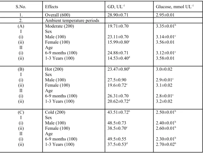
Table 1. Mean ± SEM values of serum glutamate dehydrogenase (GD) and glucose in Marwari goats
S.No. Effects GD, UL-1 Glucose, mmol UL-1
1. Overall (600) 28.90±0.71 2.95±0.01 2. Ambient temperature periods
(A) Moderate (200) 19.71±0.70 3.35±0.01b
I Sex
(i) Male (100) 23.11±0.70 3.14±0.01c (ii) Female (100) 15.99±0.80c 3.56±0.01
II Age
(i) 6-9 months (100) 24.88±0.71 3.12±0.01c (ii) 1-3 Years (100) 14.53±0.40d 3.58±0.01
(B) Hot (200) 23.47±0.80b
3.0±0.02
I Sex
(i) Male (100) 27.5±0.90 2.9±0.01c (ii) Female (100) 19.6±0.72c 3.1±0.02
II Age
(i) 6-9 months (100) 26.31±0.70 2.8±0.01c (ii) 1-3 Years (100) 20.62±0.72d 3.2±0.02
(C) Cold (200) 43.51±0.72b
2.50±0.01b
I Sex
(i) Male (100) 48.5±0.73 2.40±0.01b (ii) Female (100) 38.5±0.70c
2.60±0.01b
II Age
(i) 6-9 months (100) 49.5±0.55 2.30±0.01b (ii) 1-3 Years (100) 37.5±0.53d
2.70±0.02b
(i) Figures in the parenthesis indicate number of animals.
(ii) Superscript ‘b’ indicates a significant difference (p≤0.05) between moderate and hot and moderate and cold mean values.
(iv) Superscript ‘c’ indicates a significant (p≤0.05) difference between male and female mean values within an ambient temperature period.
(vi) Superscript ‘d’ indicates a significant (p≤0.05) difference between 6-9 months and 1-3 years old animals within an ambient temperature period.
The mean value of GD was significantly
(p≤0.05) higher during cold and hot ambient
temperature periods in comparison to overall
greater in cold (2.2 times) than hot ambient
temperature (1.19 times).This clearly showed that
GD activity during cold increased in otherwise
healthy animals to meet out energy requirements of
the body and for thermoregulation. Its activity
increased to provide more fuel for TCA cycle by
making alpha-ketoglutarate available (Lehninger et
al., 1993). Therefore blood levels of this enzyme
can be used to assess fuelling of TCA cycle.
Simultaneously the changes in glucose
concentrations also showed a link with serum GD
activity.
Variation in the serum GD levels becomes more
important from energy stand point of view as GD is
allosterically regulated by the cell’s energy state.
During the formation of alpha ketoglutarate, GDP
and ADP positively regulate GD in mammals, and
GTP, ATP, leucine, and coenzyme inhibit the
enzyme. Therefore, when the level of ATP is high,
conversion of glutamate to alpha ketoglutarate is
limited; however when the cellular energy charge is
low, glutamate is converted to ammonia and alpha
ketoglutarate (Banerjee et al., 2003). This happens
in cold conditions when energy requirement is high
to meet the demand for thermoregulation and
metabolism. GD plays a central role in amino group
metabolism. The effect of varying ambient
temperatures on metabolism is well documented
(Locher et al., 2008). Increased GD activity was
perhaps to meet the fuelling requirement of TCA
cycle in stress periods as extreme ambient
temperature periods are considered as stressors for
the animals. Increased glucocorticoids due to
ambient temperature related stress (Kataria et al.,
2000) could be the possible cause of higher GD
activity.
In each ambient temperature period, sex and age
wise variations was also observed in GD activity
and glucose concentration. The mean value of
serum GD was higher in male animals as compared
to female animals. Probably the higher cortisol
levels in males could be the proper explanation for
higher GD levels, modulating the energy processes
(Lehninger et al., 1993). In each ambient
temperature period, the mean value of serum GD
decreased significantly (p≤0.05) with the
advancement of age. Higher GD activity probably
helped the young animals in maintaining high BMR
(Locher et al., 2008).The variations in serum
glucose showed a reverse trend as compared to
serum GD activity.
In present study the value of the serum GD was
higher (range was from 15 to 43 UL-1) than the
reported values in other animals (Boyd,1962). It is
because of the fact that GD is highly concentrated
in liver of goat (Tennant, 1999) and is associated
with microsomal function of hepatocytes (El
Samani et al.,1985).Due to its higher concentration
in the goat, the estimation of GD as a liver function
test is being emphasised in goat (Tennant, 1999).
Sener and Malaisse (1980) suggested that oxidative
deamination reaction of GD feeds the tricarboxylic
acid (TCA) cycle by converting L-glutamate
to-ketoglutarate, whereas the reductive amination
reaction supplies nitrogen for several biosynthetic
pathways.
An increased serum GD activity was related
with a decrease in serum glucose concentration.
This showed the association of serum GD with
glucose homeostasis. Many earlier researchers
(Maechler and Wollheim, 1999) have correlated the
serum GD activity with insulin stimulation. In
pancreatic-cells, GD is involved in the regulation of
insulin secretion, especially amino acid–stimulated
insulin secretion and in this way it is also important
in glucose homeostasis. Glutamate was suggested
to be a mediator of glucose-stimulated insulin
granules (Maechler and Wollheim, 1999).In present
study, during cold and hot ambient temperature
periods, when serum GD activity increased, the
serum glucose concentration decreased. The pattern
of change in glucose concentration substantiated the
fact that GD functions as an enzyme to regulate
glucose concentration also through hormone
insulin. Therefore lower serum glucose
concentration in hot and cold ambient temperatures
indicated towards its greater peripheral utilisation to
meet out the energy requirement of the body.
These results indicated that in cold condition
fuelling to TCA cycle was higher than moderate
and hot ambient temperature periods. Serum GD
values showed variations due to changes in ambient
temperatures, sex and age, each having the relevant
physiological significance. Low glucose
concentration during hot and cold ambient
temperature periods indicated a high rate of
peripheral utilization. Further the study has shown
that variations in the enzyme levels are not always
pathological and while interpreting clinical data, a
clinician must consider these variations.
REFERENCES
Banerjee, S., Schmidt, T., Fang, J., Stanley, C.A.,
Smith, T.J.(2003). Structural Studies on
ADP Activation of Mammalian Glutamate
Dehydrogenase and the Evolution of
Regulation. Biochem. 42, 3446-3456.
Boyd, J. W. (1962). The comparative activity of
some enzymes in sheep, cattle and
rats-normal serum and tissue levels and
changes during experimental liver
necrosis. Res.Vet.Sci. 3,256-268.
El Samani, F., Mahmoud O.M., Fawi, M.T.,
Gameel, A.A., Haroun, E.M. (1985).
Serum enzyme activity and bilirubin
concentration in sheep experimentally
infected with Fasciola gigantica. J. Comp.
Pathol. 95(4), 499-503.
Kataria, N., Kataria, A.K., Agarwal, V.K., Garg,
S.L., Sahani, M.S. and Singh, R. (2000).
Effect of water restriction on serum
aldosterone and cortisol in dromedary
camel during winter and summer. J.
Camel Prac. Res. 7,1-7.
King, J. (1965). In, Practical clinical enzymology.
D. Van Nostrand Company Ltd., London.
pp 25-301.
Lehninger, A.L., Nelson,D.L. and Cox, M.M.
(1993). In, Principles of Biochemistry. 2nd
edn. Worth publishers, New York. pp
400-787.
Locher,L., Rudovsky, A. and Fürll, M. (2008).
Investigations on the antioxidative
metabolism during the periparturient
period in dairy goats with special regard to
seasonal differences in feeding and
management. Beri Munch Tierarzti
Wochenschr. 121 (9-10), 341-348.
Maechler, P. and Wollheim, C.B. (1999).
Mitochondrial glutamate acts as a
messenger in glucose-induced insulin
exocytosis. Nature. 402,685–689.
Oser, B.L. (1976). In, Hawk’s physiological
chemistry. 14th ed. Tata McGraw Hill
Publishing Co. Ltd., New Delhi. Pp
975-1152.
Sener, A. and Malaisse, W.J. (1980).L-leucine and
a nonmetabolized analogue activate
pancreatic islet glutamate dehydrogenase.
Nature. 288,187–189.
Snedecor, G. W. and Cochran, W. G. (1967).In,
Stillman, T.J., Baker, P. J., Britton, K.L. and Rice,
D.W. (1993). Conformational flexibility in
glutamate dehydrogenase: role of water in
substrate recognition and catalysis. J. Mol.
Biol. 234(4), 1131-1139.
Tanizawa, Y., Nakai, K., Sasaki,T., Anno,T.,
Ohta,Y., Inoue,H., Matsuo,K. , Koga,M. ,
Furukawa,S. and Oka,Y. (2002).
Unregulated elevation of glutamate
dehydrogenase activity induces
glutamine-stimulated insulin secretion identification
and characterization of a GLUD1 gene
mutation and insulin secretion studies with
MIN6 cells overexpressing the mutant
glutamate dehydrogenase. Diabetes.
51,712-717.
Tennant, B.C. (1999). Hepatic function. In, Clinical
Biochemistry of Domestic Animals. 5th
edn. Harcourt Brace & Company, Asia