vol. 40, n. 1, jan./mar., 2004
Analysis of the molecular association of rifampicin with
hydroxypropyl-
βββββ
-cyclodextrin
Denise Alves Ferreira
1, Antonio Gilberto Ferreira
2, Lucinéia Vizzotto
2, Alberto Federman Neto
3,
Anselmo Gomes de Oliveira
1*
1Departamento de Fármacos e Medicamentos, Faculdade de Ciências Farmacêuticas, Universidade Estadual Paulista
Júlio de Mesquita Filho, Araraquara, 2Laboratório de RMN, Departamento de Química, Universidade Federal de
São Carlos, 3Laboratório de Química Farmacêutica, Faculdade de Ciências Farmacêuticas de Ribeirão Preto,
Universidade de São Paulo
Inclusion complexes of rifampicin (RP) were prepared with hydroxypropyl-β-cyclodextrin (HPβCD). The aqueous solubility of RP increased linearly with cyclodextrin concentration in all range of the solubility diagram. The data was analyzed using the framework of Higuchi and Connors. The stability constant (K) values for RP/HPβCD complex at pH 6.9 were 18 and 120-125 M-1
for ionic strength 0.01 and 0.18M, respectively.The analysis of the chemical shift data of 1H and 15N for free RP and RP/HPβCD
inclusion complex reveal that only peaks of the side chain related to the piperazine ring of RP change substantially, probably due to interaction of this region of RP molecule with the hydrophobic core of HPβCD. We also postulated the optimized structure of RP/ HPβCD inclusion complex using molecular modelling study. We found that the postulated structure was in agreement with 1H and 13C-NMR and 15N-NMR spectra.
* Correspondence:
A.G. Oliveira
Departamento de Fármacos e Medicamentos.
Faculdade de Ciências Farmacêuticas UNESP
Rodovia Araraquara-Jaú km 01 14801-902 - Araraquara - SP - Brasil E-mail: [email protected]
Uniterms
• Rifampicin • Hydroxypropyl-ß-• cyclodextrin • Inclusion complex • Molecular Modelling • Proton Nuclear Magnetic • Resonance
INTRODUCTION
Rifampicin (RP) is a wide-spectrum antibiotic active against the most Gram-positive bacteria and variable active against Gram-negative microrganisms. Due to the development of resistance to rifampicin by many bacteria its clinical use is mainly in the treatment of tuberculosis (Reynolds, 1993). RP (Figure 1) is a potent inductor of the cytochrome P-450 of the hepatic and intes-tinal enzymatic system and of P-glycoprotein of the transport system, which results in numerous drug interactions and side effects (Watcher et al., 1998; Achira
et al., 1999). Thus, the interaction of RP with drug carriers to improve effectiveness and minimize side effects is of obvious relevance.
hydrogen bonds and hydrophobic effects contribute to the formation of a stable complex of drugs in the apolar cavity of cyclodextrins (Bibby et al., 2001). Thus, a large number of drug molecules can be complexed to CD modifying solubility, bioavaila bility and stability (Loftsson et al., 1991; Vianna et al., 1998; Dalmora, Oliveira, 1999; Dalmora et al., 2001; Loftsson et al., 2001; Másson et al., 1998). Important reviews on cyclodextrin applications in the pharmaceutical field were published with details (Bibby et al., 2001; Hirayama, Uekama, 1999; Szejtli, 1994; Loftsson, Másson, 2001).
Several experimental procedures have been used to analyze the structural and thermodynamic parameters related to cyclodextrin inclusion complex formation (Goyenechea, et al., 2001; Connors, 1997; Bobec et al., 2000; Díaz et al., 1999; Myake et al., 1999). Experimen-tal procedures using NMR (Másson et al., 1998; Díaz et al., 1999; Loukas, 1997; Myake et al., 1999), solubility analysis and UV-Vis spectroscopy (Dalmora, Oliveira, 1999; Oh et al., 1998), molecular mechanical calculations (Montassier et al., 1997; Vianna et al., 1998; Bobek et al., 2000; Madri, et al., 1997; Myles et al., 1994), DSC study (Bermúdez et al., 1999) and X-ray diffraction (Montassier
et al., 1997; Bobek et al., 2000) are frequently used to elucidate the structure of molecular complexes.
In this work we have studied the increase of the solubility of rifampicin by molecular complexation with hydroxypropyl-β-cyclodextrin. Molecular Modelling study of the structure of HPβCD, rifampicin and RP/ HPβCD inclusion complexes were performed. The molecular complexes were also analysed by 1H, 13C and 15N-NMR spectroscopy in order to characterize the
structure of the inclusion complex.
MATERIAL AND METHODS
Material
Hydroxypropyl-β-cyclodextrin (HPßCD), D.S. 4.6, was purchased from CTD Inc., Florida, USA. Rifampicin (RP) [3-(4-methylpiperazinyl-iminomethyl]rifamycin and deuterium oxide (D2O), were obtained from Sigma Chemical Company, St. Louis, MO, USA. Reagents used for the preparations of citrate and Tris-HCl buffer, were analytical grade. Double distillate and deionised water were used in all experiments. All others reagents were analytical grade.
Methods
Stability Constant Determination
Amounts of RP (about 300 mg) that exceeded its solubility were weighed in a 25 mL plastic flask, to which 10 mL of buffer solution (pH 6.9) containing HPβCD with concentrations in the range of 0-100 mM was added. The mixture was kept under agitation by 24 hours at controlled temperature of 25 °C to reach steady state. Then, the ma-terial was filtered through 0.22 µm membrane. Afterwards appropriate dilution with buffer solution was performed and the concentration of rifampicin was measured by spectrophotometry at 331 nm. The solubility diagram was plotted, relating the absorbance as a function of dissolved rifampicin. The stability constant (K) was calculated using the framework of Higuchi and Connors model (Higuchi, Connors, 1965):
K
RPf + HPβCD RPb (1)
Where K is the stability constant for RP/HPbCD 1:1 complex and the subscripts f and b refer to the free and bound RP, respectively.
K = slope/So (1 – slope) (2)
Where slope is calculated from of the linear phase solubility diagram, and So is the intrinsic apparent solubility of RP in the absence of cyclodextrin.
The effect of the buffer and ionic strength on the formation of RP complexes were carried out in citrate buffer pH 6.9, µ =180 mM and in Tris-HCl buffer pH 6.9, µ =10 mM and 180 mM. The ionic strength was adjusted using sodium chloride.
Molecular Modelling Studies
A model of inclusion complex was investigated
through molecular graphic software using the Chem 3D Ultra software, version 6.0, serial number 553923 with use authorized by Cambridge Software Corporation (http:// www.cambridgesoft.com/products/ family.cfm?FID=3, 2004). The structures of RP and HPβCD were built separately, and the conformational energy was minimized using MM2 method and force field (Schupfer, Gulacar, 2001). A set of structures of minimum-energy was chosen, and the molecule of RP was introduced into cavity of HPβCD using docking method. Steric and electrostatic interactions between the molecules were calculated by moving the atoms of the molecules back and forth. For the simulation solvents were not considered. Following, the atoms of RP and HPßCD were allowed to move freely, and the most stable docking position of the two molecules was obtained.
Nuclear Magnetic Resonance (NMR) Measurements NMR measurements of liquid HPßCD inclusion complex were taken at 298°K on a Bruker 9.4 Tesla DRX 400 (400 MHz for hydrogen frequency), using as external reference TSPAd4 to 1H and 13C and CH
3NO2 to 15N
experiments. A 5 mm probe with inverse detection, gradient coil in z direction and D2O as a solvent were used. The 13C and 15N chemical experiments were performed using gHSQC and gHMBC techniques. For gHMBC experiments the coupling constants were optimised to 4, 8 and 16 Hz for 13C and also 15N.
The complex was prepared in D2O buffer, as well as the free rifampicin and CD’s. The spectra were recorded at the following conditions:
Hydrogen: The spectrum was recorded using a pulprog zgpr, acquisition time 5.1 s, 90o pulse width 8.3 µs, spectral width 6377 Hz, relaxation delay 1s and acquired 128 scans. They were processed using zero filling and without any window function.
COSY: The spectrum was obtained using a pulse sequence cosygs, with acquisition time 0.16 s, 90o pulse width 8.3 µs, spectral width 6377 Hz, relaxation delay 1.2 s, acquired 16 scans and 256 experiments. It was processed using SINE apodization function and zero filling in both dimensions.
gHSQC: The spectrum was recorded using a pulse sequence inviestgssi, acquisition time 0.16 s, 90o pulse width 8.3 and 12.0 µs for 1H and 13C respectively, relaxation delay 1.2 s, spectral widths 6377 and 18000 Hz for F2 and F1 dimension, 64 scans and 256 experiments. It was processed using QSINE apodization function (SSB=3 for booths dimensions) a zero filling in F2 and F1 dimension. gHMBC 1H-13C: The spectra were recorded using a pulse sequence inv4gslplrnd, acquisition time 0.16 s, 90o
pulse widths 8.3 and 12.0µs for 1H and 13C respectively, relaxation delay 1.2 s, spectral widths 6377 and 18000 Hz for F2 and F1 dimension, 64 scans and 256 experiments. It was processed using QSINE apodization function (SSB=2 for both dimensions) a zero filling in F2 and F1 dimension.
gHMBC 1H-15N: The spectra were recorded using a pulse sequence inv4gslrnd, acquisition time 0.26 s, 90o pulse widths 8.3 and 20.4 µs for 1H and 15N respectively, relaxation delay 2 s, spectral widths 8012 and 20249 Hz for F2 and F1 dimension, 1024 scans and 34 experiments. It was processed using QSINE apodization function (SSB=2 for booths dimensions) a zero filling in F2 and F1 dimension
RESULTS
The analysis of the drug inclusion complex with cyclodextrin in solution can be achieved by several methods. The analysis of the drug solubilization associated to spectral changes can be used to measure the transference of the drug from the aqueous phase to the apolar cavity of cyclodextrin. If the drug has limited aqueous solubility, the concentration of solubilized drug can be increased by the inclusion in cyclodextrin.
In our results, linear responses of rifampicin concentrations dissolved against HPβCD concentrations can be observed (Figure 2).
The phase solubility behaviors show that rifampicin followed the AL type phase solubility diagram (Másson et al., 1998) indicating that the stoichiometry of molecular complex between RP/HPβCD was probably 1:1.
Using the equation de Higuchi and Connors, the calculated results for K were 18 M-1 for 10 mM Tris-HCl buffer ionic strength 10 mM. For 30 mM citrate and 10 mM Tris-HCl buffer ionic strength 0.18 M, the values obtained for K were 120 M-1 and 125 M-1, respectively.
Nuclear magnetic resonance study at high field has been used to investigate the stoichiometries of important drug inclusion complexes. This technique provides evidence for the existence of true inclusion complexes and allows unequivocal determination of the stoichiometry for complexes obtained in the solid state, as well as in solution (Djedaini, Perly, 1991). For the analysis of the spectra of the figures 4-7, we have used the RP structure of figure 1.
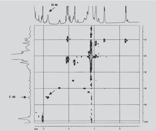
We can verify in the Figure 4 that for pure RP the signal of hydrogen 46 at δ = 2.91 ppm is a singlet coupled to carbon 46 (δ = 45.8 ppm). The arrow indicates the correlation signal for 1H-13C.
The spectra of Figure 5 characterizes the identification of the multiplet of hydrogen 46 coupled to carbon 46 which suggests that this region of the RP molecule is one site of interaction between RP and CD.
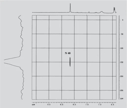
Figure 6 shows the RP spectra with the correlations values obtained by HMBC for the bounds 1H-15N. It is possible to see the hydrogen 38 (δ = 3.23 ppm) coupled to N39 (δ = 350 ppm) and with N40 of the piperazine ring (δ = 114 ppm).
The variation of the chemical displacement of the N40 (δ =150 ppm) coupled to hydrogen 38 for the RP complex with CD can be seen in Figure 7. This chemical displacement suggests that this region of RP molecule interacts with the cyclodextrin in the inclusion complex.
DISCUSSION
Although rifampicin has a bulky structure, the values of stability constants for the antibiotic (18 M-1 for µ=0.01 and 120-125 M-1 for µ=0.18) show that the inclusion complexes are formed and that the solubility of antibiotic in the presence of hydroxypropyl-β -cyclodextrin increase about 6 fold in high ionic strength independently of the buffer system used. This
FIGURE 2 - Solubility diagram of rifampicin with HPβCD at 25 °C. Key: ({) 30 mM citrate buffer, pH 6.9, µ=180 mM;
(z) 10 mM Tris-HCl buffer, pH 6.9, µ=10 mM; (T) 10 mM Tris-HCl buffer, pH 6.9, µ=180 mM. The ionic strength was adjusted with sodium chloride.
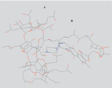
FIGURE 3 - Molecular Model of RP inclusion complex
FIGURE 4 - gHSQC 1H-13C spectrum of the pure RP with magnification of the region of H46. The arrow indicates the correlation signal for 1H-13C.
FIGURE 6 - gHMBC 1H-15N spectrum of pure rifampicin.
phenomenon can be understood since at pH 6.9 the antibiotic is a zwiterion and binds to cyclodextrin cavity by hydrophobic effect. Thus, the activity coefficient of neutral organic compounds increases with ionic strength of medium and therefore favours the transfer of the drug from hydrophilic medium to hydrophobic cavity of cyclodextrin increasing the value of the stability constant (Oliveira et al., 1997).
We have shown that MM2 Molecular Modelling calculations for RP/CD complex using computational tools demonstrate the experimental evidence that the complex at molar ratio 1:1 are thermodynamically stable. For RP/HPβCD complex of minimum energy it was possible to predict that the piperazine tail of RP penetrates into cyclodextrin cavity through hydrophobic interactions. However, the low values obtained for K indicate a weak bond of RP into HPβCD molecule, probably due to steric hindrance of the substitutive hydroxypropyl group of cyclodextrin, which do not allow the approach of the bulkiest region of RP molecule.
NMR studies were used to compare the chemical shift of 1H and 13C of rifampicin with a similar compound from literature (Fuhrer von, 1973). However, since the D2O solubility of rifampicin was not good to obtain a direct detection of 13C chemical shift and the data of literature were obtained in deuterated methanol, we have adopted the inverse detection approach.
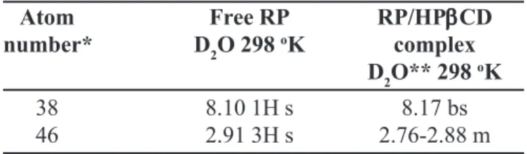
Comparing the chemical shift data of 1H and 15N for free RP and RP/HPßCD complex it was verified that only peaks of the side chain related to the piperazine ring change substantially. In fact, the data of table I show that the RP hydrogen in the positions 38 (δ 8.10 1H s) and 46 (δ 2.91 3H s) change to (8,17 bs) and (2.76-2.88 m), respectively (Table I).
In addition, it is possible to verify that not only occur the chemical shift but also a clear multiplicity in the signal of hydrogen 46 coupled to carbon 46. On the other hand, the 15N chemical shift for N40 changes from d 114 for free
RP to 150 ppm for RP/HPßCD complex. All the carbons and hydrogens attributions are in agreement with the data founded in the literature (Fuhrer von, 1973; Gallo, et al., 1974) although the authors have used different solvents. Thus, this group of data obtained by 2D-NMR allow suggesting that the RP was complexed with the HPßCD through the lateral chain containing piperazine ring and that the geometry of the complex is in agreement with the predictions of the Molecular Modelling Calculations.
CONCLUSIONS
The increase in the aqueous solubility of RP caused by the presence of HPbCD was dependent on the ionic strength and is one of experimental evidence of the molecular complexation. The spectroscopy analysis by 1H and 15N NMR showed that the signals of the hydrogen at C38 and C46 of RP were shifted upfield due to interaction with HPßCD. The postulated structure of complex RP/ HPbCD was in agreement with 1H and 13C-NMR and 15 N-NMR spectra.
RESUMO
Análise da associação molecular da rifampicina com hidroxipropil-β-ciclodextrina
Foram preparados complexos de inclusão de rifampicina (RP) com hidroxipropil-β-ciclodextrina (HPβCD). A so-lubilidade da RP em água aumentou linearmente com a concentração de ciclodextrina na faixa de concentração utilizada no diagrama de solubilidade. Os resultados fo-ram analisados quantitativamente através do modelo de Higuchi e Connors. Os valores da constante de estabilida-de (K) para o complexo RP/HPβCD em pH 6,9 foram 18 e 120-125 M-1 para as forças iônicas 0,01 e 0,18 M,
res-pectivamente. A análise dos dados de deslocamento quí-mico de 1H e 15N-NMR para a RP livre e do complexo de
inclusão RP/HPβCD revelou que somente os picos da cadeia lateral relacionada com o anel piperazina modifi-caram substancialmente, provavelmente devido à interação desta região da molécula da RP com a cavida-de hidrofóbica da HPβCD. Com base no estudo de mode-lagem molecular foi proposta a estrutura otimizada para o complexo de inclusão RP/HPβCD. A estrutura propos-ta está de acordo com os resulpropos-tados dos espectros de 1H e 13C-NMR e 15N-NMR .
UNITERMOS: Rifampicina. Hidroxipropil-ß-ciclodextrina. Complexo de inclusão. Modelagem molecular. Ressonância Nuclear Magnética.
TABLE I - Chemical shift δ (ppm) of 1H NMR for RP/
HPβCD inclusion complex
Atom Free RP RP/HPβββββCD
number* D2O 298 oK complex
D2O** 298 oK
38 8.10 1H s 8.17 bs
46 2.91 3H s 2.76-2.88 m
ACKNOWLEDGMENTS
The authors wish to thanks to FAPESP and CNPq (Anselmo Gomes de Oliveira) for financial support and CAPES (Denise Alves Ferreira) for a student fellowship.
REFERENCES
ACHIRA, M.; ITO, K.; SUZUKI, H.; SUGIYAMA, Y. Comparative studies to determine the selective inhibitors for P-glycoprotein and cytochrome P-450 3A4. AAPS PharmSciTech., Arlington,v.1, p.1-5, 1999.
BERMÚDEZ, M.; MARTINEZ, E.; MORA, M.; SAGRISTÁ; M. L.; MADARIAGA, DE M.A. Molecular and physicochemical aspects of the interactions of the tuberculostatics ofloxacin and rifampicin with liposomal bilayers: a P-31-NMR and DSC study. Colloid Surf. A.,
Amsterdam,v.158, p.59-66, 1999.
BIBBY, D. C.; DAVIES, N.M.;TUCKER, I. G.; Mechanisms by which cyclodextrins modify drug release from polymeric drug delivery systems. Int. J. Pharm.,
Amsterdam, v.197, p.1-11, 2001.
BOBEK, M. M.; GIESTER, G.; KÄHLIG, H.; BRINKER, U. H. A ‘sugar-coated’ carbene precursor: a single crystal X-ray diffraction and NMR study. Tetrahedron Lett.,
Amsterdam, v.41, p.5663-5667, 2000.
CAMRIDGE SOFTWARE CORPORATION. Disponível em: <http://www.cambridgesoft.com/products/ family.cfm?FID=3>. Acesso em: abril. 2004.
CONNORS, K. A.The stability of cyclodextrin complexes in solution. Chem. Rev., Washington, v.97, p.1325-1357, 1997.
DALMORA, M. E.; DALMORA, S. L.; OLIVEIRA, A. G. Inclusion complex of piroxicam with β-ciclodextrin and incorporation in cationic microemulsion. In vitro release and in vivo topical anti-inflammatory effect. Int. J. Pharm., Amsterdam, v.222, p.45-55, 2001.
DALMORA, M. E.; OLIVEIRA, A. G. Inclusion complex of piroxicam with β-ciclodextrin and incorporation in hexadecyltrimethylammonium bromide based microemulsion. Int. J. Pharm., Amsterdam, v.184, p.157-164, 1999.
DÍAZ, D.; BERNARD, M. J. B.; GRACIA-MORA, J.; LLANOS, C. M. E. Solubility, H-1-NMR, and molecular mechanics of mebendazole with different cyclodextrins. Drug Dev. Ind. Pharm., New York, v.25, p.111-115, 1999.
DJEDAINI, F.; PERLY, B. Nuclear magnetic resonance investigation of the stoichiometries in beta-cyclodextrin:steroid inclusion complexes. J. Pharm. Sci., Washington, v.80, p.1157-61, 1991.
FUHRER VON, H. Zuordnung des 13C-NMR. Helv. Chim.
Acta., Zürich, v.56, p.244-245, 1973.
GALLO, G.G.; MARTINELLI, E.; PAGANI, V.; SENSI, P. The conformation of rifamycin S in solution by H NMR spectroscopy. Tetrahedron, Amsterdam, v.30, p.3093-3097, 1974.
GOYENECHEA, N.; SANCHEZ, M.; VELAZ, I.; MARTIN, C.; MARTINEZ-OHARRIZ, M. C.; GONZALEZ-GAITANO, G. Inclusion complexes of nabumetone with beta-cyclodextrins: thermodynamics and molecular modeling studies. Influence of sodium perchlorate. Luminescence, Chichester,v.16, p.117-27, 2001.
HIGUCHI, T.; CONNORS, K. A. Phase-solubility techniques. Adv. Anal. Chem. Instrum., New York, v.4, p.117-212, 1965.
HIRAYAMA, F.; UEKAMA, K. Cyclodextrin-based controlled drug release system. Adv. Drug Deliver. Rev.,
Amsterdam, v.36, p.125-141, 1999.
LOFTSSON, T.; BREWSTER, M. E. Pharmaceutical applications of cyclodextrins. 1.Drug solubilization and stabilization. J. Pharm. Sci.,Washington, v.85, p.1017-1025, 1996.
LOFTSSON, T.; FRIDRIKSDÓTTIR, H.; ÓLAFSDÓTTIR, B. J. Solubilization and stabilization of drugs through cyclodextrin complexation. Acta Pharm. Nord.,
Stockholm, v.3, p.215-217, 1991.
LOFTSSON, T.; GUÕMUNDSDÓTTIR, H.; SIGURJÓTTIR, J. F.; SIGURÕSSON, H. H.; SIGFÚSSON, S. D.; MÁSSON, M.; STEFÁNSSON, E. Cyclodextrin solubilization of benzodiazepines: formulation on kidazolam nasal spray. Int. J. Pharm.,
LOFTSSON, T.; MASSON, M. Cyclodextrin in topical formulations: theory and pratice. Int. J. Pharm.,
Amsterdam, v.225, p.15-30, 2001.
LOUKAS, Y.L. Measurement of molecular association in drug: cyclodextrin inclusion complexes with improved 1H-NMR studies. J. Pharm. Biom. Anal., Arlington, v.49, p.944-948, 1997.
MADRI, J.M.; POZUELO, J.; MENDICUTI, F.; MATTICE, W.L. Molecular Mechanics Study of the Inclusion Complexes of 2-Methyl Naphthoate with a and ß-Cyclodextrins. J. Colloid Interface Sci., London, v.193, p.112-120, 1997.
MÁSSON, M.; LOFTSSON, T.; JÓNSDÓTTIR, S.; FRIDRIKSDÓTTIR, H.; PERTERSEN, D.S. Stabilization of ionic drugs through conplexation with non-ionic cyclodextrins. Int. J. Pharm., Amsterdam, v.164, v.45-55, 1998.
MONTASSIER, P.; DUCHENE, D.; POELMAN, M.C. Inclusion complexes of tretinoin with cyclodextrins. Int. J. Pharm., Amsterdam, v.153, p.199-209, 1997.
MYAKE, K.; IRIE, T.; ARIMA, H.; HIRAYAMA, F.; UEKAMA, K.; HIRANO, M.; OKAMOTO, Y. Characterization of itraconazole/2-hydroxypropyl-beta-cyclodextrin inclusion complex in aqueous propylene glycol solution. Int. J. Pharm., Amsterdam, v.179, p.237-245, 1999.
MYLES, A.M.C.; BARLOW, D.J.; FRANCE, G.; LAWRENCE, M.J. Analysis and modelling of the structures of beta-cyclodextrin complexes. Biochem. Biophys. Acta., Amsterdam, v.1199, p.27-36, 1994.
OH, I.; LEE, M.; SHIN, S.; PARK, I. Spectroscopy characterization of ibuprofen/2-hydroxypropyl-β -cyclodextrin inclusion complex. Int. J. Pharm.,
Amsterdam, v.175, p.215-223, 1998.
OLIVEIRA, A.G.; SCARPA, M.V.; CHAIMOVICH, H. Effect of hexadecyltrimethylammmonium bromide-based microemulsions on the rate of decomposition of the β-lactam antibiotic cephaclor. J. Pharm. Sci.,
Washington, v.86, p.616-620, 1997.
REYNOLDS, J.E.F., ed. Martindale The Extra Pharmacopoeia. 30 ed. London: Pharmaceutical Press, 1993, p.197-200.
SCHUPFER, P.Y.; GULACAR, F.O. Relative stabilities of cholestadienes calculated by molecular mechanics and semi-empirical methods: application to the acid-catalyzed rearrangement reactions of cholesta-3, 5-diene.
Org. Geochem., Oxford, v.31, p.1589-1596, 2001.
SZEJTLI, J. Medicinal applications of cyclodextrins. Med. Res. Rev., Chichester, v.14, p.353-86, 1994.
VIANNA, R.F.L.; BENTLEY, M.V.L.B.; RIBEIRO, G.; CARVALHO, F.S.; NETO, A.F.; OLIVEIRA, D.C.R.; COLLET, J.H. Formation of cyclodextrin inclusion complexes with corticosteroids: their characterization and stability. Int. J. Pharm., Amsterdam, v.167, p.205-213, 1998.
WACHER, V.J.; SILVERMAN, J.A.; ZANG, Y.; BENET, L.Z. Role of P-glycoprotein and cytochrome P450 3A in limiting oral absorption of peptides and peptidomimetics.
J. Pharm. Sci., Washington, v.87, p.1322-1330, 1998.