Structure-Based Network Analysis of
Activation Mechanisms in the ErbB Family
of Receptor Tyrosine Kinases: The
Regulatory Spine Residues Are Global
Mediators of Structural Stability and
Allosteric Interactions
Kevin A. James1, Gennady M. Verkhivker1,2*
1.School of Computational Sciences and Crean School of Health and Life Sciences, Schmid College of Science and Technology, Chapman University, Orange, California, United States of America,2.Department of Pharmacology, University of California San Diego, La Jolla, California, United States of America
Abstract
The ErbB protein tyrosine kinases are among the most important cell signaling families and mutation-induced modulation of their activity is associated with diverse functions in biological networks and human disease. We have combined molecular dynamics simulations of the ErbB kinases with the protein structure network modeling to characterize the reorganization of the residue interaction networks during conformational equilibrium changes in the normal and oncogenic forms. Structural stability and network analyses have identified local communities integrated around high centrality sites that correspond to the regulatory spine residues. This analysis has provided a quantitative insight to the mechanism of mutation-induced ‘‘superacceptor’’ activity in oncogenic EGFR dimers. We have found that kinase activation may be determined by allosteric interactions between modules of structurally stable residues that synchronize the dynamics in the nucleotide binding site and theaC-helix with the collective motions of the
integrating aF-helix and the substrate binding site. The results of this study have pointed to a central role of the conserved His-Arg-Asp (HRD) motif in the catalytic loop and the Asp-Phe-Gly (DFG) motif as key mediators of structural stability and allosteric communications in the ErbB kinases. We have determined that residues that are indispensable for kinase regulation and catalysis often corresponded to the high centrality nodes within the protein structure network and could be
distinguished by their unique network signatures. The optimal communication
OPEN ACCESS Citation:James KA, Verkhivker
GM (2014) Structure-Based Network Analysis of Activation Mechanisms in the ErbB Family of Receptor Tyrosine Kinases: The Regulatory Spine Residues Are Global Mediators of Structural Stability and Allosteric Interactions. PLoS ONE 9(11): e113488. doi:10.1371/journal.pone. 0113488
Editor:Natarajan Kannan, University of Georgia, United States of America
Received:April 12, 2014
Accepted:October 27, 2014
Published:November 26, 2014
Copyright:ß2014 James, Verkhivker. This is an open-access article distributed under the terms of theCreative Commons Attribution License, which permits unrestricted use, distribution, and repro-duction in any medium, provided the original author and source are credited.
Data Availability:The authors confirm that all data underlying the findings are fully available without restriction. All crystal strctures used in this study are available from the Protein Data Bank.
Funding:This work is supported by funding from Chapman University. No additional external was funding received for this study. The funders had no role in study design, data collection and analysis, decision to publish, or preparation of the manu-script.
pathways are also controlled by these nodes and may ensure efficient allosteric signaling in the functional kinase state. Structure-based network analysis has quantified subtle effects of ATP binding on conformational dynamics and stability of the EGFR structures. Consistent with the NMR studies, we have found that nucleotide-induced modulation of the residue interaction networks is not limited to the ATP site, and may enhance allosteric cooperativity with the substrate binding region by increasing communication capabilities of mediating residues.
Introduction
The human protein kinases play a fundamental regulatory role in orchestrating
functional processes in complex cellular networks [1–3]. The mechanisms that
regulate catalytic activities of protein kinases include phosphorylation,
auto-inhibition and allosteric activation by binding partners [4]. The diversity of
structural mechanisms that regulate a dynamic switch between inactive and active kinase forms may involve several layers of allosteric control that enable various
kinase functions [5–16]. The crystal structures of protein kinases in different
functional states have underscored the role of specific regions in the catalytic
domain whose structural variations can determine regulatory preferences [17,18].
The main regulatory elements within the kinase catalytic domain include thea
C-helix, the DFG-Asp motif (DFG-Asp in, active; DFG-Asp out, inactive), and the
activation loop (A-loop open, active; A-loop closed, inactive) (Figure 1,Table 1).
Structural coupling of the DFG motif and the regulatory aC-helix has been long
recognized as central in controlling a dynamic equilibrium between major
functional forms that include an inactive state (DFG-out/aC-helix-in), a Cdk/Src
inactive conformation (DFG-in/aC-helix-out) and an active state (DFG-in/a
C-helix-in). Protein kinase regulation is also governed by a dynamic coupling of two spatially distributed networks of mostly hydrophobic residues that form a
regulatory spine (R-spine) and a catalytic spine (C-spine) [19–21]. The analysis of
protein kinase crystal structures has identified that the R-spine and the hydrogen bond networks that connect the N-terminal and the C-terminal kinase lobes may be perturbed and often disrupted in the inactive conformations, whereas a cooperative assembly and stabilization of the spine motifs along with the characteristic salt bridges constitute critical features of activation kinase
mechanisms [22].
The ErbB protein tyrosine kinases are among the most important cell signaling families and mutation-induced modulation of their activity is associated with
diverse functions in biological networks and human disease [23,24]. A common
regulatory signature of the ErbB kinases is based on sharing a Cdk/Src inactive structure with a characteristically low catalytic activity. Crystal structures of the
EGFR catalytic domain in the wild-type (WT) [25–27] and mutant forms [28–30]
active conformations (DFG-in/aC-helix-in) (Figure 1), demonstrating that oncogenic mutants stabilize the active form of EGFR. The crystal structures of the inhibitory complexes between the EGFR kinase domain and a fragment of the
cytoplasmic protein MIG6 [31] have unveiled an alternative Cdk/Src inactive
form with DFG-out/aC-helix-out (Cdk/Src-IF2) (Figure 1), in which the DFG
motif is in the inactive DFG-out position, but the interactions constraining the
aC-helix in the inactive position are removed, and the A-loop is in a fully
extended conformation (A-loop open) as in the active EGFR structures. Another
Cdk/Src inactive conformation (Cdk/Src-IF3) was detected in the crystal structure
of the ErbB2 kinase where the aC-helix and the DFG motif conform to their
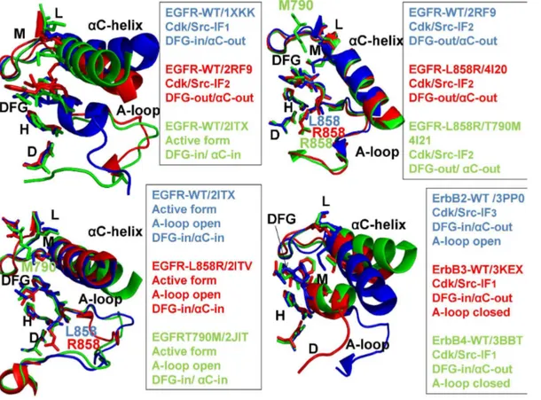
Figure 1. Structural Characteristics of the ErbB Kinases.The crystal structures of the ErbB kinase family in different functional states are depicted using a comparison of key regulatory regions in the catalytic domain. The three regulatory elements of the kinase domain shown are theaC-helix, the DFG-Asp motif (DFG-Asp in, active; DFG-Asp out, inactive), and the activation loop (A-loop open, active; A-loop closed, inactive). In Cdk/Src inactive structures the
aC-helix is displaced outwards the N-terminal lobe adopting aaC-out (swung-out) conformation that inhibits the formation of the active enzyme form. The R-spine residues (M766, L777, H835, F856, and D896) and the DFG motif are shown in colored sticks. Note that the R-R-spine residues in a different sequence numbering of the EGFR kinase domain correspond to M742, L753, H811, F832, and D872 residues. Left Upper Panel. Structural differences in the functional regions of the EGFR-WT crystal structures: Cdk/Src-IF1 state (in blue), DFG-in/aC-helix-out (pdb id 1XKK, 2GS7); Cdk/Src-IF2 conformation (in red), DFG-out/aC-helix-out (pdb id 2RF9); and the active conformation (in green), DFG-in/aC-helix-in (pdb id 2ITX, 2J6M). Right Upper Panel. Structural similarities in the functional regions of the Cdk/Src-IF2 EGFR-WT conformation (in blue), DFG-out/aC-helix-out (pdb id 2RF9); Cdk/Src-IF2 EGFR-L858R conformation (in red), DFG-out/aC-helix-out (pdb id 4I20); and Cdk/Src-IF2 EGFR-L858R/T790M double mutant conformation (in green), DFG-out/aC-helix-out (pdb id 4I21). Left Lower Panel. Structural similarities in the functional regions of the active EGFR-WT conformation (in blue), DFG-in/aC-helix-in (pdb id 2ITX, 2J6M); the active EGFR-L858R conformation (in red), DFG-in/aC-helix-in (pdb id 2ITV); and the active EGFR-T790M conformation (in green), DFG-in/aC-helix-in (pdb id 2JIT). Right Lower Panel. Structural differences in the functional regions of Cdk/Src-IF3 ErbB2-WT conformation (in blue), DFG-in/aC-helix-out, A-loop open (pdb id 3PP0); Cdk/Src-IF1 ErbB3-WT conformation (in red), DFG-in/aC-helix-out, A-loop closed (pdb id 3KEX, 3LMG); and Cdk/Src-IF1 ErbB4-WT conformation (in green), DFG-in/aC-helix-out, A-loop closed (pdb id 3BBT).
DFG-in/aC-helix-out positions, but the A-loop adopts an active, open
conformation [32] (Figure 1). The ErbB3 kinase has long been considered as
inactive, and classified as a pseudokinase, since the key catalytic residues are conspicuously missing in ErbB3. However, recent crystallographic studies have indicated that the catalytically inactive ErbB3 kinase domain can bind ATP and
serve as an activator of the EGFR kinase domain [33]. The crystal structure of the
catalytically inactive ErbB3 kinase domain has revealed a Cdk/Src-IF1
con-formation that is similar to that of EGFR and ErbB4 kinases, albeit with a
shortened aC-helix [33]. Subsequent studies have reported a crystal structure of
the ErbB3 kinase domain bound to an ATP analogue and have demonstrated that human ErbB3 kinase can bind ATP and retain sufficient kinase activity, though
,1000-fold less than the canonical ErbB kinases [34]. Crystal structures of the
ErbB4 kinase domain in the active and inhibited Cdk/Src-IF1forms [35,36] have
suggested that structural determinants of kinase activation may be conserved among the EGFR and ErbB4 catalytic domains. Crystallographic studies of the ErbB kinase domains have also discovered that the formation of an asymmetric dimer between the C-lobe of a ‘‘donor’’ (activator) monomer and the N-lobe of an adjacent ‘‘acceptor’’ (receiver) monomer is a common structural mechanism
required to achieve full kinase activation [37–40].
A number of human cancers are associated with mutations causing the increased expression of the ErbB kinases. More than 200 activating and drug
resistance EGFR mutations have been reported [41], and molecular mechanisms
of mutation-induced kinase activation have been extensively discussed [42,43].
Oncogenic kinase mutants have been long linked with their ability to lock the catalytic domain in a constitutively active state - a functional form whose
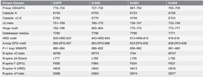
Table 1.The Functional Regions of the ErbB Kinases.
Kinase Domain EGFR ErbB2 ErbB3 ErbB4
P-loop GSGAFG 719–724 727–732 697–702 700–705
Catalytic K K745 K753 K723 K726
CatalyticaC-E E762 E770 H740 E743
aC-helix 751–769 760–775 738–747 733–749
Hinge motif 792–796 800–804 770–774 773–777
Gatekeeper residue T790 T798 T768 T771
HRD motif 835-HRD-837 843-HRD-845 813-HRN-815 816-818
A-loop DFG motif 855-DFG-857 863-DFG-888 833-DFG-835 836-DFG-838
P+1 loop WMAPE 880–884 888–892 858–862 861–865
R-spineaC-helix M766 M774 I744 M747
R-spineb4-Strand L777 L785 L755 L758
R-spine F (DFG) F856 F864 F834 F837
R-spine H (HRD) H835 H843 H813 H816
R-spineaF-helix D896 D904 D874 D877
The residue ranges of functional regions in the ErbB kinases are based on the crystal structures of EGFR (pdb id 2ITX), ErbB2 (pdb id 3PP0), ErbB3 (pdb id 3LMG), and ErbB4 (pdb id 3BCE).
uncontrollable activity may contribute to the initiation or progression of cancer
[44,45]. Recent crystallographic studies [46,47] have discovered that the catalytic
domains of the EGFR-L858R and EGFR-L858R/T790M mutants in the inactive
form can adopt a mobile Cdk/Src-IF2conformation (DFG-out/aC-helix-out) that
may facilitate conformational release from the inactive dormant state, resulting in an accumulation of a constitutively active form and elevated enzyme activities. Biochemical reconstitution analysis in combination with the crystal structure of
an asymmetric dimer of the L858R/T790M mutant [48,49] have revealed a new
mechanism of mutant-specific kinase regulation in which oncogenic EGFR mutants can preferentially assume the acceptor role in the regulatory dimers.
Structural and computational approaches have been instrumental in revealing the atomic details of protein kinase dynamics at different levels of complexity: from detailed analyses of the catalytic domain to simulations of the regulatory dimer assemblies. A significant body of computational studies has focused on
elucidating molecular mechanisms of the ErbB kinases [50–59]. Molecular
dynamics (MD) simulations and the energy landscape analysis have investigated the structural and energetic basis of mutation-induced changes in the EGFR
kinase domain [60,61]. These studies have determined that the inactive
EGFR-WT state is more stable than the active state, and the L858R mutation could differentially perturb both active and inactive conformations to shift thermo-dynamic preferences towards the activated form. Recently, simulation boundaries have been pushed to new unprecedented levels of multiple microsecond
simulations, revealing that the catalytic domain of EGFR may sample a locally disordered state and that oncogenic mutations could reduce the disorder in the
aC-helix region of the dimerization interface, thus promoting acquisition of an
active asymmetric dimer and stabilization of a constitutively active form [62].
Subsequent multi-scale simulations have witnessed spontaneous conformational transitions between the inactive and active states via locally disordered
intermediate conformations, whose functional relevance was independently confirmed by hydrogen exchange mass spectrometry (HX-MS) experiments
[63,64]. The effects of oncogenic mutations on the conformational landscape of
the EGFR kinase have been also quantified in another series of large-scale
computer simulations [65]. These studies have similarly concluded that
mutation-induced alterations in the relative stability of the kinase states and the reduction of disorder at the dimer interface may serve as catalysts of kinase activation by oncogenic mutations. Recent investigations have combined multi-scale molecular simulations with structure-functional approaches to demonstrate that the activation mechanism may involve a cooperative effect of the external, internal,
and transmembrane segments of the complex EGFR assembly [66,67].
modulated by activation mutations and controlled by a small number of
‘‘privileged’’ functional residues [68]. Understanding how conformational
equilibrium between functional kinase states can be altered and redistributed upon ligand binding and/or mutations is critical for quantifying molecular basis
of allosteric regulation. Structural studies [69,70], NMR spectroscopy
investiga-tions [71–76] and computer simulations [77,78] of cAMP-dependent protein
kinase A (PKA-C) have confirmed the existence of multiple functional forms and allosteric interaction networks that can regulate conformational equilibrium between dynamically committed, uncommitted, and quenched states. ATP binding can redistribute the relative populations of these states and activate a nucleotide-bound functional form of PKA-C that is structurally and dynamically
committed to catalysis [71–76]. These NMR studies have also discovered the effect
of positive allosteric cooperativity in PKA-C, according to which ATP binding in the nucleotide binding site can enhance the substrate affinity in the allosteric site, thus confirming that the interaction networks and long-range communication between distal kinase regions may control catalytic reaction and mediate substrate recognition.
The residue interaction networks can be described as weighted graphs providing a convenient and robust framework for understanding allosteric
communications in protein systems [79,80]. Structure-based network models
often employ common measures of node centrality (degree, closeness, and
betweenness) to characterize local and global connectivity of residues [81–83].
Integration of molecular dynamics simulations and protein structure network analysis has been successfully used to identify functionally important regulatory sites and model allosteric communication pathways for a variety of protein
systems [84–88]. These studies have shown that the residue interaction networks
in protein structures can be characterized by small-world organization, in which local interactions and long-range coupling between mediating nodes are properly balanced to achieve an optimal trade-off between network resilience and efficiency
[89–91]. These networks are efficient in transmitting long-range signal due short
paths between any pair of nodes, but may become vulnerable to targeted attacks on a small number of central nodes.
In this work, atomistic simulations were combined with the ensemble-based network analysis to characterize evolution of the residue interaction networks in the ErbB kinases during conformational equilibrium changes. Conformational dynamics of the ErbB kinases was analyzed in different functional states by simulating multiple crystal structures of the catalytic domain and regulatory dimer complexes. We also investigated the allosteric effect of ATP binding on conformational dynamics and structural stability of the EGFR structures. This study shows that structural stability and allosteric interactions in the ErbB kinase family can be mediated by a small number of sparsely distributed high centrality nodes that correspond to the conserved functional residues in the R-spine, the
regulatory HRD and DFG motifs, and the substrate binding P+1 loop. We
states. This study reveals that the residue interaction networks in the kinase structures may exhibit elements of modularity that may have evolved to achieve a trade-off between structural stability, the efficiency of allosteric communications and resilience against perturbations in the protein environment.
Results
Conformational Dynamics of the ErbB Kinases: Structural Stability
of Regulatory Regions
MD simulations of the ErbB kinases were performed using multiple crystal structures of the catalytic domain and regulatory dimer complexes in the normal and oncogenic forms. We analyzed equilibrium simulations of the EGFR
structures and asserted that conformational dynamics of the regulatory regions is conserved in the active kinase forms, but may vary significantly depending on the inactive kinase state. Equilibrium fluctuations in the inactive and active EGFR structures displayed characteristic differences in the conformational mobility and
coordinated motions of the P-loop, aC-helix, and the A-loop (Figure 2A). These
regions displayed larger thermal fluctuations and the increased conformational flexibility in the active kinase form as evident from the root mean square fluctuation (RMSF) of the backbone residues and computed B-factors. However,
the inactive Cdk/Src-IF1 form showed smaller thermal variations in the key
functional regions, mainly due to the autoinhibitory interactions between the
P-loop, a helical motif within the A-P-loop, and the aC-helix. The greater structural
rigidity of the Cdk/Src-IF1structure may lock EGFR in the autoinhibited dormant
state, thereby decreasing the probability of inadvertent activation. In contrast, a
markedly greater flexibility could be seen in the Cdk/Src-IF2 structure, where
thermal motions were especially pronounced in the aC-helix, the aC-b4-loop,
and the A-loop (Figure 2A). In this structure, the autoinhibitory constraints are
removed, leading to the increased flexibility and decoupling of the aC-helix and
the A-loop movements. A more uniform pattern of small thermal fluctuations was
detected across all regions in the active EGFR structures (Figure 2B). The
atom-based fluctuation profiles of the EGFR structures depicted a more detailed view of
variations experienced by the P-loop, aC-helix, and the A-loop regions (Figure
S1). These equilibrium profiles similarly highlighted the increased flexibility of the
inactive Cdk/Src-IF2 form of EGFR as compared to the autoinhibited and active
EGFR structures (Figure S1). Simulations of the L858R and the
EGFR-L858R/T790M mutants in the inactive Cdk/Src-IF2 structure revealed not only
local adjustments near the mutational sites, but also long-range changes in the
aC-helix and the N-terminal lobe (Figure 2C). In this case, we also observed
additional intermediate conformations that were similar to the Cdk/Src-IF2 form
(DFG-out/aC-helix-out, A-loop open), but in which the aC-helix drifted away
from the inactive conformation towards an active aC-helix-in position. At the
(Figure 2B). As a result, oncogenic EGFR mutants may escape from the
autoinhibitory trap and induce the increased mobility of the inactive conforma-tions by populating a rapidly interconverting region of the conformational landscape. This could facilitate fast conformational transitions between the
inactive Cdk/Src-IF2 state and the active EGFR form, ultimately leading to
stimulated activities of oncogenic EGFR mutants. MD simulations of the inactive
ErbB2 crystal structure (Cdk/Src-IF3conformation with DFG-in/aC-helix-out,
A-loop open) (Figure 2D) revealed a significant conformational mobility in the
P-loop, aC-helix and A-loop regions. Unlike other members of the ErbB family, the
Figure 2. Residue-Based Equilibrium Fluctuations of the ErbB Kinases.(A) The computed B-factors describe time-averaged residue fluctuations obtained from simulations of Cdk/Src-IF1 EGFR-WT (pdb id 1XKK, in blue), Cdk/Src-IF2 EGFR-WT structure (pdb id 2RF9, in red), and the active EGFR-WT structure (pdb id 2ITX, in green). (B) The computed B-factors obtained from simulations of the active structures of WT (pdb id 2ITX, in blue), EGFR-L858R (pdb id 2ITV, in red), and EGFR-T790M (pdb id 2JIT, in green). C) The computed B-factors for Cdk/Src-IF2 structures of EGFR-WT (pdb id 2RF9, in blue), EGFR-L858R (pdb id 4I20, in red), and EGFR-L858R/T790M (pdb id 4I21, in green). (D) The computed B-factors obtained from simulations of Cdk/ Src-IF3 ErbB2-WT structure (pdb id 3PP0, in blue), Cdk/Src-IF1 ErbB3-WT structure (pdb id 3LMG, in red), Cdk/Src-IF1 ErbB4-WT structure (pdb id 3BBT, in green), and the active ErbB4-WT structure (pdb id 3BCE, in maroon).
increased conformational flexibility in the ErbB2 structure was spread beyond the
aC-helix region suggesting a partial weakening of the entire catalytic core.
The collective motions of the ErbB kinase structures were evaluated using
principal component analysis (PCA) [92,93]. According to a comprehensive
account of PCA applications in protein dynamics, MD simulations at the nanosecond time scale may be sufficient for an effective separation of time scales
and an adequate characterization of essential dynamics [94]. This study has also
noted that PCA of protein conformational dynamics based on the heavy atoms, as
opposed to Ca atoms only, can provide a better description of slow degrees of
freedom and yield a more accurate view of global collective motions. Consistent with these arguments, we utilized the extended set of backbone heavy atoms (N,
Ca, Cb, C, O) in the PCA modeling of the ErbB kinase structures. We observed
that almost the same shapes can be obtained for the slowest modes of motion when using between 3 and 10 lowest frequency modes. Based on our
computations, the 10 lowest eigenvectors captured between 85% and 90% of the total variance in the collective kinase motions, while the essential subspace covered by the first three lowest PCA modes typically accounted for at least 75% of atomic fluctuations in each trajectory and approached 80% in simulations of
the active kinase structures. These results are in line with earlier findings [94]
indicating that the heavy atom-based PCA may provide a sufficient convergence of high frequency subspaces and thus allow for a better separation of orthogonal low frequency modes. Indeed, we found that a relatively small number of low frequency modes may describe most of the slow conformational motions. Based on these results and to streamline functional dynamics analysis, we characterized conformational dynamics and collective movements of the ErbB kinases in the essential space of the three low frequency modes. For all catalytic domain structures, the first principal mode typically corresponded to the opening and closing movements of the N-terminal and C-terminal lobes with respect to each other. In the second principal mode, the kinase lobes displayed a shear motion between the N-terminal and C-terminal lobes, in which a sliding movement of the lobe interface corresponded to the forward displacement of one lobe and inward displacement of the other lobe. In another principal mode, the C-terminal and N-terminal tails rotated in opposite directions with respect to the C-N-terminal lobe. The observed pattern of principal motions is conserved among protein kinase
folds [95] and consistent with the NMR studies of conformational dynamics in
protein kinases [71–77].
Conformational dynamics profiles were mapped onto the ErbB structures where the residue mobility referred to an average B-factor value computed over
backbone atoms in each given residue (Figures 3, 4). The distributions revealed
that structural rigidity of theaC-b4 loop can be linked to the positional variability
of the aC-helix. The ‘‘boundary’’ between the rigid aC-b4 loop and a more
flexibleaC-helix can define a functional hinge connecting regions of high and low
structure (Figure 4) demonstrated the increased conformational mobility in all
regions of the catalytic domain. Noticeably, structural stability of theaC-b4-loop,
aC-helix, and the R-spine residues was compromised in the inactive ErbB2
structure. The more restricted thermal fluctuations in the inactive ErbB3 kinase were reminiscent of those in the autoinhibitory form of EGFR. Despite a
shortened aC-helix in the crystal structures of ErbB3, the catalytic core and the
aC-b4/aC-helix region were rigid. The obtained dynamic profile of the ErbB3
kinase corroborates with structural studies [33,34] that attributed the lack of the
ErbB3 catalytic activity to its overly stable inactive form. To characterize patterns of structurally stable and flexible regions in the functional kinase forms, we analyzed conformational dynamics of the R-spine residues. The EGFR R-spine
includes L777 from theb4-strand, M766 from the C-terminal end of theaC-helix,
F856 of the DFG motif in the activation segment, H835 of the HRD motif of the
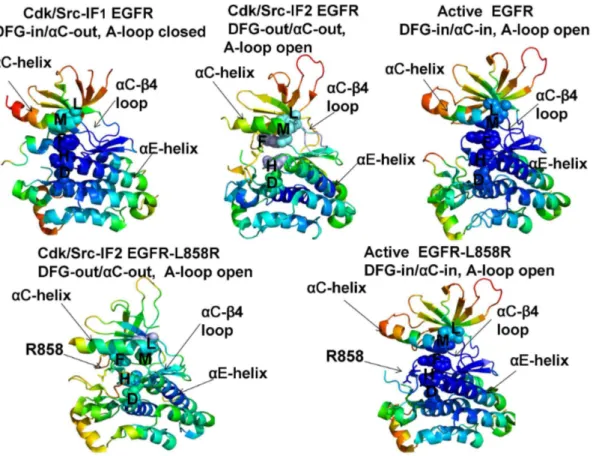
Figure 3. Conformational Mobility Analysis of the EGFR-WT and EGFR-L858R Kinases.Conformational mobility profiles of EGFR-WT are shown for the inactive Cdk/Src-IF1 form (pdb id 1XKK, left upper panel), the inactive Cdk/Src-IF2 state (pdb id 2RF9, middle upper panel) and the active conformation (pdb id 2ITX, right upper panel). Conformational mobility of EGFR-L858R is shown for the Cdk/Src-IF2 form (left lower panel) and the active conformation (right lower panel). The backbone heavy atoms (N,Ca,Cb,C,O) were employed for the PCA computations. Conformational dynamics profiles were computed by averaging protein motions in the space of three lowest frequency modes. The color gradient from blue to red indicates the decreasing structural rigidity (or increasing conformational mobility) of the protein residues and refers to an average value over the backbone atoms in each residue. The functional kinase regionsaC-helix,aC-b4-loop, andaE-helix as well as the R-spine residues are annotated and their positions are indicated by arrows. The R-spine residues are also highlighted in spheres and colored according to their degree of structural stability. Conformational mobility profiles were obtained from simulations of complete structures, where unresolved segments and disordered loops were modeled with the ModLoop server [127,128]. These profiles were mapped onto the original crystal structures of EGFR for clarity of presentation.
catalytic loop, and D896 of the aF-helix (Table 1). The backbone of H835 is
anchored to the aF-helix via hydrogen bonding to D896. The substrate binding P
+1 loop, the A-loop, and the aH-aI loop bind to theaF-helix forming a dense
interaction network. While the R-spine residues of active EGFR are linearly
connected, the aC-helix position in the inactive EGFR confirmation leads to a
dislocation between M766 and L777 residues and partly disassembled spine architecture. A common dynamics signature of the active structures was a uniform structural stability acquired by all spine residues that integrate
coordinated movements of the aC-b4-loop, aC-helix,aE-helix, and aF-helix in
their active positions. A partially assembled architecture of the hydrophobic spine in the autoinhibitory structures of EGFR, Erbb3 and ErbB4 is fairly constrained due to structural rigidity of the spine residues, thus increasing the energetic cost of inducing the active conformation. In contrast, the regulatory regions are fairly dynamic and the R-spine structure is loose in the mobile Cdk/Src-IF2
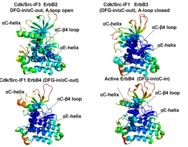
Figure 4. Conformational Mobility Analysis of the ErbB Kinases.Conformational mobility mapping of ErbB2-WT in the inactive Cdk/Src-IF3 form (left upper panel), ErbB3-WT in the inactive Cdk/Src-IF1 conformation (right upper panel), ErbB4-WT in the Cdk/Src-IF1 form (left lower panel) and the active form (right lower panel). The backbone heavy atoms (N, Ca, Cb, C, O) were employed for the PCA calculations. Conformational dynamics profiles were computed by averaging protein motions in the space of three lowest frequency modes. The color gradient from blue to red indicates the decreasing structural rigidity (or increasing conformational mobility) of the protein residues and refers to an average value over the backbone atoms in each residue. The key functional regionsaC-helix,aC-b4-loop, andaE-helix as well as the R-spine residues are annotated and their positions are indicated by arrows as in
Figure 3. The conformational mobility profiles were mapped onto the original crystal structures of ErbB kinases.
conformations that are adopted by the EGFR mutants. Among interesting findings of this analysis was a striking similarity between functional dynamics
profiles of the catalytic domains of EGFR (Figure 3) and ErbB4 (Figure 4). The
R-spine residues in EGFR (M766, L777, H835, F856, D896) and ErbB4 (M747, L758, H816, F837, D877) have a very similar profile, revealing structural stability of the
HRD and DFG motifs, whereas the aC-b4/aC-helix interface residues (M766,
L777 in EGFR and M747, L758 in ErbB4) mark the border between regions of high and low structural stability. The dynamics profile of the EGFR dimer (Figure 5) revealed the increased stability of the acceptor monomer that extended beyond the interface, suggesting that the formation of an asymmetric complex may allosterically strengthen structural integrity of the active kinase form. The R-spine residues in the acceptor monomer become structurally stable and effectively immobilized in their active positions. This is consistent with the notion that EGFR
and ErbB4 kinases employ the same autoinhibitory mechanisms [39,40]. Hence,
conformational dynamics of the ErbB kinases underscored structural stability of
the inactive Cdk/Src-IF1 structure (DFG-in/aC-helix-out, A-loop closed) that
could be contrasted with the conformationally mobile Cdk/Src-IF2state
(DFG-out/aC-helix-out, A-loop open) and Cdk/Src-IF3conformations (DFG-in/a
C-helix-out, A-loop open).
Structural Stability Profiles of the Kinase Catalytic Doman and
Active Regulatory Dimer: The Force Constant Analysis
In the previous section we asserted that conformational dynamics and functional motions of the ErbB kinases may be associated with allosteric interactions between regulatory regions. Here, we analyzed structural stability of the regulatory regions in different functional states of the ErbB kinases and characterized mutation-induced changes in stability profiles that may be relevant for activation mechanisms. For this analysis, we employed a number of complementary approaches, including the force constant profiling of residue connectivity, the contact network analysis of residue closeness, the relative solvent accessibility (RSA) evaluation of local residue environment, and the network-based analysis of local contact density. In the ensemble-based force constant analysis, the
equilibrium fluctuations of the mean distance between each residue and the rest of the protein were converted into force constants that measure the energy cost of
the residue displacement during equilibrium simulations [96,97]. The high force
constants are typically associated with structurally stable residues that display small fluctuations in their distances to other residues and often correspond to highly connect and effectively communicating rigid sites. Previous studies have linked structural stability of functionally important residues with their high connectivity, particularly indicating that catalytic and binding site residues typically have high force constant values, which reflects functional constraints
imposed on their movement [98,99]. Abrupt changes between maxima and
hypothesis tested in our analysis is that the R-spine residues could effectively mediate structural stability and allosteric interactions via regulatory regions. The analysis revealed that high force constant residues in the catalytic domain are
assembled near theaC-helix,aE-helix andaF-helix regions (Figure 6), suggesting
that structural stability of these structural elements may be critical for allosteric coupling between regulatory regions. In addition, in all functional states, we detected a clear maximum corresponding to the conserved regulatory motif
WMAPE (substrate binding P+1 loop) that is anchored to theaF-helix. We also
observed abrupt changes in the force constant profiles of the EGFR catalytic
domain (Figure 6A) that corresponded to the border between the more flexible
aC-helix (residues 751–769) and the more rigid aC-b4 loop (residues 770–777).
The force constant values and structural stability of the aC-helix residues were
considerably higher in the active EGFR form than in the inactive EGFR
conformation. Similar changes were also detected in the force constant profile of
the ErbB4 kinase domain (Figure 6D) in which theaC-helix region (residues 733–
749) displayed appreciable differences between the inactive and active ErbB4
conformations. At the same time, the force constant values for the aC-b4 loop
(EGFR residues 770–777 in Figure 6Aand ErbB4 residues 750–758 inFigure 6D)
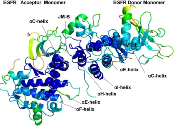
Figure 5. Conformational Mobility Profile of the Active EGFR Dimer.Structural distribution of conformational mobility in the asymmetric active dimer of EGFR-WT. In an asymmetric dimer arrangement a donor monomer interacts with an acceptor through interactions involving theaH-helix andaI-helix of the donor as well as the JM-B segment and theaC-helix of the acceptor. The key functional regions are annotated and pointed to by arrows as inFigures 3,4. Note the increased stability of the acceptor monomer, particularly a uniform stabilization of the R-spine residues in the acceptor subunit. The conformational mobility profiles were mapped onto the original crystal structure of the active EGFR dimer.
were similar in the inactive and active kinase forms. Accordingly, the hinge site
located at the border between the aC-b4 loop (higher structural stability region)
and the aC-helix (lower structural stability region) may be involved in
modulating large conformational transitions between the inactive and active kinase states. These findings corroborate with the experimental studies in which
theaC-b4 loop/aC-helix region was recognized as an intramolecular switch of the
ErbB kinase activity [100]. Notably, the aC-b4 loop and the R-spine residues in
the inactive form of ErbB2-WT displayed lower force constant values and did not
correspond to the distribution peaks (Figure 6B). As a result, structural stability of
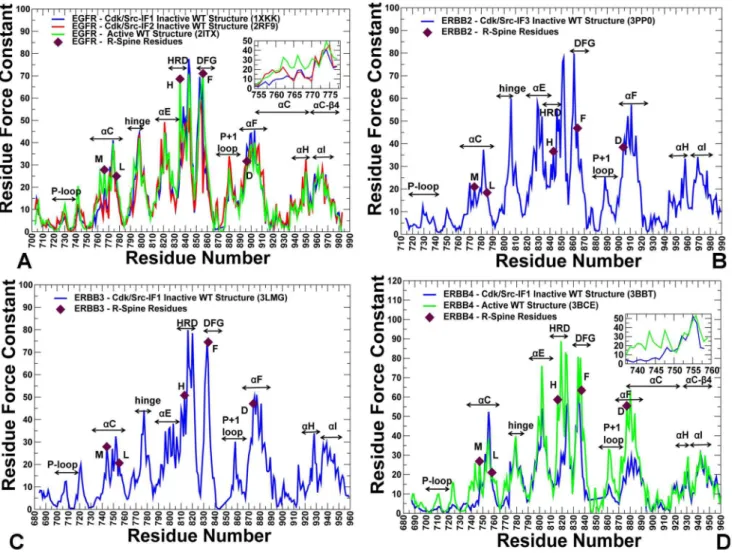
Figure 6. The Force Constant Profiles of the Kinase Catalytic Domain.Dynamics-based analysis of structural stability in the ErbB crystal structures. (A) The residue-based force constant profiles of the EGFR-WT crystal structures: Cdk/Src-IF1 conformation (in blue), Cdk/Src-IF2 conformation (in red), and the active conformation (in green). A close-up view of the EGFR force constant profile in theaC-helix (residues 752–768) and the adjacentaC-b4-loop regions (residues 769–777) is provided as an inset. (B) The force constant profile of Cdk/Src-IF3 ErbB2 structure. (C) The force constant profile of Cdk/Src-IF1 ErbB3 structure. (D) The force constant profiles of Cdk/Src-IF1 ErbB4 conformation (in blue) and the active ErbB4 conformation (in green). A close-up view of the ErbB4 force constant profile in theaC-helix (residues 735–749) and the adjacentaC-b4-loop (residues 750–758) is provided as an inset. The annotated functional regions included P-loop,aC-helix, hinge,aE-helix, HRD motif, DFG motif, substrate binding P+1 loop,aF-helix,aH, andaI helix. The R-spine residues are indicated by filled maroon-colored diamond symbols. Note that the R-R-spine residues corresponded to the peaks in the distributions.
the regulatory regions a may be compromised in the inactive ErbB2 structure. These factors may contribute to the experimentally observed low catalytic activity
of ErbB2 [32]. Hence, the force constant profiles highlighted the conserved
features and differences in structural stability of the inactive and active kinase forms.
Structural stability of the regulatory residues tends to become considerably
more pronounced in the force constant profiles of the EGFR dimer (Figure 7).
The noticeable peaks in the acceptor monomer (Figure 7A) corresponded to L680
from the juxtamembrane segment (JM-B segment includes residues 664 to 682)
and prominently included the R-spine residues M742 (aC-helix), H811 (HRD
motif), F832 (DFG motif), and D872 (aE-helix). Strikingly, the R-spine residues
coincided precisely with the highest peaks in the force constant profile of the acceptor monomer, suggesting that these functional residues may serve as global mediators of structural stability in the active EGFR dimer. The stability profile of
the donor monomer revealed the important contribution of the aF-helix and the
aH-helix, owing to a stable dimer interface that rigidified the position of thea
H-helix (Figure 7B). The crystal structure of the L858R/T790M dimer is essentially
identical to the EGFR-WT, and structural stability profiles of the acceptor (Figure 7C) and donor monomers (Figure 7D) in the mutant were similar to the respective distributions in EGFR-WT. However, we noticed the emergence of wider peaks in the mutant form of the EGFR dimer. In particular, a broader peak
was seen in theaC-helix of the acceptor molecule (Figure 7C), while in the donor
molecule the individual peaks corresponding to the aE-helix, HRD and DFG
motifs tend to aggregate into a broader maximum (Figure 7D). Similarly, single
peaks corresponding to W880 (P+1 substrate site) and D896 (aF-helix) seem to
consolidate into a wider maximum (Figure 7D). In our interpretation, this may
reflect the integration of structurally stable residues into consolidated modules, pointing to the reorganization of the interaction network and the enhanced structural stability of the mutant dimer. These findings corroborate with the
biochemical experiments [48,49] and provide a useful insight to the mechanism
of mutation-induced ‘‘superacceptor’’ activity, which may result from the lower energetic cost of inducing the active conformation in the EGFR mutant relative to EGFR-WT. Because of functional dependency for dimerization it is possible that only the acceptor subunit should be catalytically fully active.
Probing Residue Environment in the Regulatory Kinase Regions:
Local and Global Network Analysis of Residue Connectivity
shortest path between a given residue and all other residues in the protein network
and represents a global measure of residue connectivity [81–83]. The residues with
high closeness can interact directly or indirectly with all other residues of the protein. The degree of a node and closeness are radial measures of network centrality that tend to be highly correlated with each other because they are both based on direct connections. According to our conjecture, high connectivity residues, determined by the consensus of local and global metrics, may
correspond to structurally stable sites that are important for kinase function. We first analyzed the relationship between global residue connectivity measures that
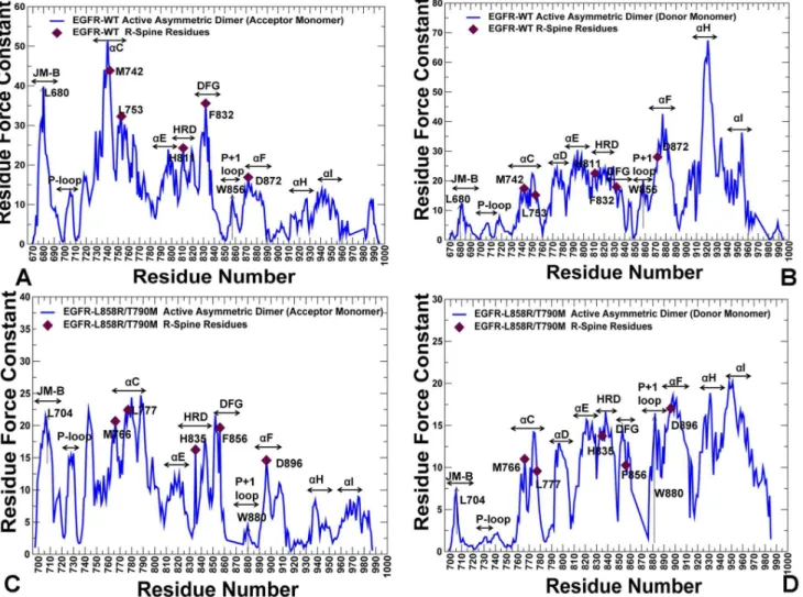
Figure 7. The Force Constant Profiles of the Active EGFR Dimers.Dynamics-based analysis of structural stability in the active asymmetric dimers of EGFR-WT (A, B) and EGFR-L858R/T790M double mutant (C, D). Note that a different EGFR sequence numbering was adopted in these crystal structures and we adhered to the original numbering to streamline the discussion and comparison with the experimental data. The force constant profiles are shown separately for the acceptor monomer (A, C) and donor monomers (B, D). The annotated functional regions included JM-B region, P-loop,aC-helix, hinge,
aE-helix, HRD motif, DFG motif, substrate binding P+1 loop,aF-helix,aH, andaI helix. The annotated peaks in the profiles reflecting structural stability of the EGFR-WT dimer included L680 (JM-B region), M742, L753, H811, F832, D872 (R-spine residues), and W856 (P+1 substrate loop). The respective peaks in the profile of the EGFR-L858R/T790M dimer corresponded to L704 (JM-B region), M766, L777, H835, F856, and D896 (R-spine residues), and W880 (P+1 substrate loop). The R-spine residues are indicated by filled maroon-colored diamond symbols. The position of JM-B peaks (L680 in EGFR-WT, L704 in EGFR- L858R/T790M) and P+1 loop peaks (W856 in EGFR-WT, W880 in EGFR- L858R/T790M) are indicated by arrows.
are represented by the force constant and residue closeness. These parameters are derived from the mean distance of a residue node to all other nodes and thus integrate the effect of the entire protein on a given single residue. By correlating these parameters in different kinase states, we tested whether the R-spine residues could correspond to similar high peaks in these distributions. We found a
significant correlation (R,0.85) between the force constant and the residue
closeness values for both the inactive (Figure 8A, B) and active EGFR-WT
structures (Figure 8C). A similar level of correlation was also evident in the
analysis of the inactive and active forms of ErbB4 (Figure 8D, E). Noteworthy,
these global connectivity parameters revealed a significant correlation and cooperativity of the R-spine residues in the active kinase forms, while this relationship was weaker in the inactive structures. In the inactive EGFR form, the
aC-helix spine residues (M766, L777) were more flexible, while the HRD and
DFG motifs (H835, F856, D896) remained structurally stable. In the active EGFR conformation, we observed the synchronously increased force constant and
residue closeness values for the aC-helix and all R-spine residues. This analysis
underscored that a uniform structural stabilization of all spine residues could be
achieved only in the active dimer (Figure 8 F, G). In this case, both the force
constant and residue closeness values of the R-spine residues were generally higher as compared to the other core residues.
We complemented the global analysis of residue connectivity by probing local residue environment using an energetics-based evaluation of relative solvent accessibility (RSA). This approach is rooted in thermodynamic principles of protein stability and demonstrates a strong correspondence with computational
and experimental measures of conformational flexibility [101–103]. The global
RSA values can be used as a simple proxy for predicting intrinsic flexibility and stability of monomeric proteins and the extent of conformational changes that
would occur upon complex formation or disassembly [101,102]. A
residue-specific local RSA measure employed here is defined as the ratio of the observed solvent-accessible surface area for a residue to the expected unfolded state value
for that amino acid type [104]. According to this model, residues are considered
to be solvent exposed if the ratio value exceeds 50% and to be buried if the ratio is less than 20%. As expected, we found that residues with low force constant values
are mainly highly exposed (RSA >50%), while unexposed amino acids (core,
RSA,0–10%) have high force constant values (Figure S2). It is evident that the
solvent-exposed residues have on average lower force constant values and are more flexible than the buried residues. Notably, the force constant and RSA values are only partially related, with a moderate correlation for the EGFR structures
(R,0.5). As a result, these measures of residue connectivity may describe
complementary features related to structural stability. Nonetheless, the R-spine residues in the EGFR structures were characterized by both low RSA values
(RSA,10%) and high force constants values (Figure S2). The correlation between
the force constants and RSA values improved in the active EGFR form (R,0.55)
where the R-spine residues became completely buried (RSA,3%) and attained
Cdk/Src-IF2 structures (Figure S2), reflecting the reduced local contact density and the increased solvent exposure of functional residues in this flexible inactive form. In the original hypothesis, we argued that structurally stable regulatory residues could be distinguished by the consensus of both local and global measures of high residue connectivity. To further test this proposal, we
determined the local contact density defined by the residue degree in the protein structure network. In particular, we evaluated the distribution of local contacts in the EGFR and ErbB4 structures by specifically focusing on ‘‘well-connected’’ residues with the number of directly interacting neighbors exceeding the selected threshold of four. This analysis indicated that the R-spine residues had on average high local connectivity in both inactive and active EGFR structures (Figure S3). In
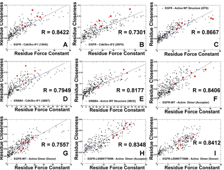
Figure 8. A Comparative Analysis of Residue Connectivity Parameters in the Functional States of the Kinase Domains and Active EGFR Dimers. The scatter graphs between the force constant (a dynamics-based residue connectivity measure) and residue closeness (a network-based residue connectivity measure) values are shown for Cdk/Src-IF1 EGFR-WT (A), Cdk/Src-IF2 EGFR-WT (B), active EGFR-WT (C), Cdk/Src-IF1 ErbB4-WT (D), active ErbB4-WT (E), acceptor monomer of the EGFR-WT dimer (F), donor monomer of the EGFR-WT dimer (G), acceptor monomer of the EGFR-L858R/ T790M dimer (H), and donor monomer of the EGFR-L858R/T790M dimer (I). The positions of the R-spine residues are indicated by filled squares colored in red.
the inactive conformations, theaC-helix residues (L777 in EGFR, M747 and L758 in ErbB4) had fewer local neighbors and appeared to be more flexible as compared to the rest of the spine. However, all spine residues displayed higher local connectivity and greater stability in the active kinase conformations. The local contact density was generally higher in the regulatory dimers (Figure S4),
especially in the aC-helix,aE-helix, andaF-helix of the acceptor monomer. The
interaction network of the active dimers is strongly influenced by the JM-B
segment and could lead to a denser network near the mediating aC-helix. A
number of highly connected residues in the donor monomer corresponded to the
aH-helix residues involved in the intermonomer interface (Figure S4). These
results indicated that high connectivity of the regulatory residues in the active structures could be amplified by the presence of well-connected neighboring residues with a similar local contact density. The interactions between high connectivity residues with similar node degree are often referred to as the
‘‘rich-club’’ phenomenon [105] which is recognized as a signal of network robustness
against random perturbations and mutations. To this end, we explored various structural and energetic measures of residue connectivity in the kinase structures. The global and local measures produced a certain consensus by identifying the R-spine residues as important functional sites that could mediate structural stability of the regulatory regions in the ErbB kinases.
The Interaction Communities Differentiate between Inactive and
Active Forms of the ErbB Kinases
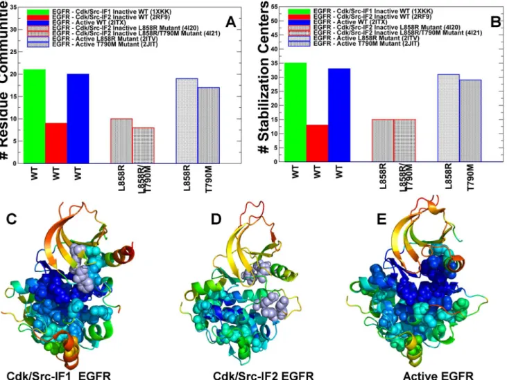
Using protein structure network analysis we also characterized the evolution of the residue interaction networks during conformational equilibrium changes in the ErbB kinases. The distribution and the aggregate number of stable interaction
communities (Figure 9A) and stabilization centers (Figure 9B) were computed for
different functional forms of EGFR. We observed that the number of communities
in the inactive EGFR state (Cdk/Src-IF1) is greater than in the alternative
Cdk/Src-IF2conformation and in the active EGFR-WT state. The interaction communities
in the inactive Cdk/Src-IF1 state (Figure 9C) have well-defined boundaries and
consist of a significant number of residues. Accordingly, strong residue
interactions formed within partly overlapping local communities may contribute to the intrinsic rigidity of the autoinhibited EGFR structure and reduce the probability of inadvertent kinase activation. The lower number of small disjointed
communities was encountered in the inactive Cdk/Src-IF2conformation, which is
consistent with the intrinsic flexibility of this EGFR form (Figure 9D). We
observed similarities in the number of communities and stabilization centers for
the inactive and active states of EGFR (Figure 9) and ErbB4 (Figure 10). The
results also revealed the reduced number of communities in the flexible inactive
form of ErbB2-WT (Cdk/Src-IF3), whereas a larger number of stable interaction
networks were observed for the rigid inactive form of ErbB3-WT (Cdk/Src-IF1)
structures onto functional dynamics profiles. A critical interaction network conserved in the autoinhibited EGFR conformation was formed by the residues F723-K745-D855-L858 (Figure S5). These interactions ensure the stability of the
rigid cluster formed between the aC-helix and a short a-helix of the A-loop,
which is a common structural feature of the inactive autoinhibitory form shared by the ErbB kinases. Indeed, a similar interaction community was detected in the inactive ErbB3 (F701-K723-D833-V836) and ErbB4 structures (F704-K726-D836-L839) (Figure S5). These interactions that determine structural stability of the autoinhibitory state involve a contribution of a conserved hydrophobic residue from the A-loop that is targeted by oncogenic mutations in the ErbB kinases
Figure 9. Community Analysis of the EGFR Kinase.The distribution of residue interaction communities (A) and stabilization centers (B) in different functional states of the EGFR kinase. The analysis is based on structurally stable residue interaction networks that were maintained in more than 75% of the simulation samples. The principal interaction communities were mapped onto conformational dynamics profiles of Cdk/Src-IF1 EGFR conformation (C), Cdk/Src-IF2 EGFR conformation (D) and the active EGFR conformation. The communities that are characteristic of different functional states are highlighted in spheres and colored according to structural stability of protein residues. A larger number of stable communities were observed in Cdk/Src-IF1 (C) and active EGFR forms (E).
(EGFR-L858, ErbB3-V836, and ErbB4-L839). Activating mutations targeting a weak link in the critical interaction network of the autoinhibitory structure may be sufficient to promote destabilization of the inactive state and shift the
thermodynamic equilibrium towards the active conformation.
We also observed that that stabilizing communities could be anchored by the R-spine residues from the HRD and DFG motifs. For instance, the local
interaction networks in the inactive EGFR included (L828-V774-F856-H835), (V769-M766-F856), (M766-L858-F856), (M825-H835-D837-D896), and (D837-R841-P877) communities (Figure S5). Collectively, these interactions contribute
to structural stabilization of the autoinhibitory state by engaging the aC-helix
(V765, M766, and V769), the aC-b4-loop (L774), the HRD motif (H835, D837)
and DFG motif (D855, F856) in the global interaction network. Of particular
Figure 10. Community Analysis of the ErbB Kinases.The distribution of residue interaction communities (A) and stabilization centers (B) is shown for different functional states of ErbB2, ErbB3, and ErbB4 kinases. The principal interaction communities were mapped onto conformational dynamics profiles of the inactive Cdk/Src-IF3 ErbB2 (C), inactive Cdk/Src-IF1 ErbB3 (D), inactive Cdk/Srdc-IF1 ErbB4 (E), and active ErbB4 (F). The communities that are characteristic of different functional states are highlighted in spheres and colored according to structural stability of protein residues. A larger number of stable communities were observed in the functional states of ErbB4.
interest was the distribution of local communities near the hinge site formed by
the aC-helix and the aC-b4-loop that could be involved in coordinating
conformational changes during activation. A structurally stable community in this region (V769-M766-F856) is proximal to a weaker interaction cluster formed
between the aC-helix residue V769 andaE-helix residues Y827 and R831. The
interactions between these residues may represent another ‘‘weak’’ link in the structurally rigid autoinhibited form of EGFR that may be targeted by known
activation mutation EGFR-V769L [41].
In the active EGFR form, the emergence of larger communities (F856-L828-V774-V853-I853-M825-H835), (V765-M766-V769-L777-F856), and
(V774-Y827-V769-L777) could stabilize the active positions of theaC-helix (V765, M766, and
V769) and the aC-b4-loop (V774, L777) (Figure S5). These N-terminal
communities are also linked to the C-terminal communities (R841-D837-P877-L858), (R836-L858-V876-D837), (R836-Y869-Y891), and (Y891-M881-W880). Collectively, these stable interaction modules couple the nucleotide and substrate binding sites by linking R836 of the HRD motif with Y869 in the A-loop (primary phosphorylation site), Y891 and W880 from the conserved WMAPE motif in the
substrate P+1 loop. Similar interaction networks were observed in the active
ErbB4 structure (Figure S5). In this case, interaction communities link the a
C-helix (I746, M747, M750), theaC-b4-loop (L755), and theaE-helix (M806, Y808,
L809, R812) thus stabilizing the active position of the regulatory aC-helix. The
conserved arginine R817 within the HRD motif tethers the A-loop residue L839 (L858 in EGFR) to Y850 (phosphorylation site) via communities (R817-L839-M857-D818) and (R817-Y850-F872). These interaction networks in ErbB4 are highly similar to the respective EGFR communities (R841-D837-P877-L858), (R836-Y869-Y891). A similar reorganization of stabilizing interaction networks during conformational changes is reflective of the conserved activation
mechanisms in EGFR and ErbB4 kinases. The community analyses suggested that the R-spine residues could form critical bottlenecks in the interaction networks and correspond to bridging nodes connecting local communities, which may be indicative of their important mediating role in allosteric interaction networks.
Structure-Based Network Analysis of Allosteric Communications:
The Global Centrality of the R-spine Residues
would likely have a significantly higher betweenness as compared to the network average. The ensemble-derived centrality profiles supported this conjecture and revealed key differences between the inactive and active kinase forms. The characteristic feature of the active kinase states is the higher average betweenness, as compared to the inactive structures, and the emergence of sharper peaks
corresponding to the R-spine residues (Figure 11). Furthermore, similar peaks in
the force constant profiles and centrality distributions of active kinase forms often pointed to the same residues. Some of the characteristic peaks for the inactive EGFR structures corresponded to F723 and L858 residues that are involved in the local community (F723-K745-D855-L858) critical for stability of the autoinhibi-tory EGFR state. However, these residues are no longer among mediating sites in the active state, as the disintegration of the autoinhibitory lock dissolves the
P-loop/A-loop interactions holding the aC-helix in the inactive position.
The characteristic centrality features of the active EGFR structure also included the emergence of strong peaks corresponding to the catalytic residue pair (K745, E762) and ATP-binding site residues. These functional sites are highly central not only compared to the peripheral solvent-exposed residues, but also relative to the core of the catalytic domain. We also detected clear peaks in the EGFR centrality profiles that corresponded to H835 from the HRD catalytic motif, F856 from the
DFG motif, W880 from the conserved WMAPE motif in the substrate P+1 loop,
and Y869 of the primary phosphorylation site in the A-loop (Figure 11A–C).
Similar peaks were seen in the ErbB4 profiles and included respectively functional
residues H816, F837, W861, and Y850 (Figure 11D). Notably, the phosporylatable
residues Y869 (in EGFR) and Y850 (in ErbB4) are hydrogen-bonded to the mediating HRD-Arg from the catalytic loop, thus stabilizing the active
conformation of the A-loop. In the active EGFR and ErbB4 conformations, the
high centrality WMAPE motif anchors the substrate binding P+1 loop to thea
F-helix, providing a plausible route for communication between allosteric sites.
Interestingly, the high centrality residues in theaF-helix included T903 and L907
that contribute to stabilization of the C-spine (V726, A743, L844, V843, V845, L798, T903, and L907) in the EGFR kinase domain. Noteworthy, both the R-spine
and C-spine are anchored to the aF-helix, which is a highly stable integrating
component of the kinase core. The oncogenic mutations reduced the average
residue betweenness in the inactive form (Figure 11B), while this effect was
moderately stimulating in the active forms (Figure 11C). The enhanced centrality
of the R-spine residues in the oncogenic mutants corroborates with the notion that activating mutations may enhance structural integrity of the hydrophobic spine.
The important contribution of the R-spine residues becomes even more
apparent from the centrality analysis of the regulatory dimers (Figure 12). A
strong correspondence between the R-spine residues and the highest peaks of the centrality distribution could be seen in the acceptor monomer of EGFR-WT (Figure 12A). The aC-b4-loop spine residues (M742, L753) along with H811
(HRD motif), F832 (DFG motif), and W856 (WMAPE motif in the P+1 loop)
segment was seen in the emerging peak corresponding to L680 in the acceptor
monomer (Figure 12A). We observed a number of similar peaks in the donor
monomer (Figure 12B), but the high centrality sites shifted to the aH-helix and
aI-helix regions that are involved in the extensive intermonomer contacts. The
profile of the L858R/T790M dimer revealed interesting peculiarities as we observed considerable changes in mediating capabilities of the mutated residues (Figure 12 C, D). Indeed, the relatively moderate betweenness values for the
Figure 11. Centrality Analysis of the EGFR and ErbB4 Kinase Domains.(A) The residue-based betweenness profiles of the EGFR-WT structures are shown for Cdk/Src-IF1 (in blue), Cdk/Src-IF2 (in red) and the active conformation (in green). (B) The betweenness profiles of Cdk/Src-IF2 EGFR structures (WT in blue, L858R in red, and L858R/T790M in green). (C) The betweenness profiles of the active EGFR structures (WT in blue, L858R in red, and T790M in green). In (A–C) a close-up view of the EGFR force constant profile in theaC-helix (residues 752–768) and the adjacentaC-b4-loop regions (residues 769–777) is provided as an inset. (D) The betweenness profiles of Cdk/Src-IF1 and active ErbB4 structures are shown in blue and green respectively. A close-up view of the EGFR force constant profile in theaC-helix (residues 752–768) and the adjacentaC-b4-loop regions (residues 769–777) is provided as an inset. The annotated EGFR residues and respective functional regions corresponding to the peaks in the profiles (A–C) included: F723 (P-loop), catalytic pair K745 and E762, M766, L777 (aC-helix), hinge,aE-helix, H835(HRD motif), F856 (DFG motif), W880 (P+1 substrate loop), D896 (aF-helix),aH, andaI helix. The R-spine EGFR residues (M766, L777, H835, F856, D896) are shown by filled maroon-colored diamond symbols. The annotated ErbB4 residues and functional regions in (D) included F704 (P-loop), catalytic pair K726 and E743, M747, L758 (aC-helix), hinge,aE-helix, H816 (HRD motif), F837 (DFG motif), W861 (P+1 substrate loop), D877 (aF-helix),aH, andaI helix. The R-spine ErbB4 residues (M747, L758, H816, F837, and D877) are shown by filled maroon-colored diamond symbols.
EGFR-WT residues T766 and L834 (this corresponds to the original residue numbering in the crystal structure) were seen in both acceptor and donor monomers. In the mutant, these sites (T790M and L858R respectively) experienced a significant increase in their centrality level that was especially
pronounced in the acceptor monomer (Figure 12C). According to this analysis,
mutated sites L858R and T790M may become global mediators of allosteric communications in the oncogenic dimer, and this effect may be especially pronounced in the acceptor monomer. Hence, mutation-induced structural
Figure 12. Centrality Analysis of the Active EGFR Dimers.The residue-based betweenness profiles of the active EGFR dimer are shown for EGFR-WT (A, B) and L858R/T790M (C, D). The profiles are shown for the acceptor (left panels A, C) and donor monomers (right panels B, D). The annotated functional regions included JM-B region, P-loop,aC-helix, hinge,aE-helix, HRD motif, DFG motif, substrate binding P+1 loop,aF-helix,aH, andaI helix. The annotated peaks in the profiles reflecting structural stability of the EGFR-WT dimer included L680 (JM-B region), M742, L753, H811, F832, D872 (R-spine residues), and W856 (P+1 substrate loop). The respective peaks in the profile of the EGFR-L858R/T790M dimer corresponded to L704 (JM-B region), M766, L777, H835, F856, and D896 (R-spine residues), and W880 (P+1 substrate loop). The R-spine residues are annotated as maroon-colored diamond symbols and the oncogenic mutation sites are indicated as orange-colored diamond symbols. Note that a different EGFR sequence numbering was adopted in the original crystal structures of the EGFR-WT dimer and L858R/T790M double mutant dimer. We kept the original numbering to avoid confusion in comparisons with the experimental data. In the EGFR-WT structure (A, B); the oncogenic sites correspond to L834 and T766. The R-spine residues in EGFR-WT are M742, L753, H811, F832, and D872. In the crystal structure of the EGFR oncogenic mutant (C, D), the mutated residues correspond to L858R and T790M. The R-spine residues in the mutant structure are M766, L777, H835, F856, and D896.
changes in the global interaction network may preferentially enhance allosteric capabilities of the acceptor monomer residues. To summarize, structure-based network analysis revealed a dual role of the R-spine residues as functional hotspots of the kinase activity that may act as key structural stabilizers and regulators of allosteric signaling.
Allosteric Communication Pathways in the EGFR and ErbB4
Structures: High Centrality Residues Mediate Kinase Signaling
Our results have thus far indicated that allosteric signaling between the nucleotide binding site and substrate site may involve the R-spine residues as central mediators of efficient signal communication between the N-terminal and C-terminal lobes. In this section, we analyzed how allosteric signals may be
transmitted in the catalytic core. Modeling of communication pathways is directly based on the centrality analysis which generated the ensemble of shortest paths between any pair of residues in the ErbB structures. Our objectives in this analysis were: (a) to map the short communication pathways between high centrality
residues in the nucleotide binding site and the P+1 substrate site; (b) to determine
the contribution of functional regions (aC-helix,aF-helix, HRD, DFG, P+1 loop)
in long-range communication pathways; (c) and to present a mechanistic model of allosteric coupling between the ATP-binding and substrate binding sites. Based on the centrality analysis, we reconstructed shortest pathways connecting the
conserved high centrality residues F723 (P-loop) and W880 (P+1 substrate
binding site) (Figure 13). These paths connected the P-loop F723 residue via a
catalytic pair (K745-E762) with the R-spine residues (M766, L777), subsequently
linking V765 (aC-helix), F856 (DFG), H835(HRD), L838, A839, and W880 in the
substrate P+1 loop (Figure 13A). We analyzed the topology of communication
paths in the context of structural stability and network properties of key residues that mediate these routes. Interestingly, the shortest communication pathways that connect allosteric binding sites in the kinase domain navigated primarily through rigid high centrality nodes. These routes also involved a number of hydrophobic residues (V765, L838, and A839) that may assist central mediating nodes in ensuring the efficiency of allosteric signaling. We also characterized allosteric signaling in the active EGFR dimer by modeling communication pathways that connect the nucleotide binding site in the donor monomer with the
substrate site in the acceptor monomer (Figure 13C). Similarly, the optimal
routes revealed a geodesic line between the monomers that passed through a set of conserved mediating nodes with the high betweenness value. In the monomer, the allosteric network connected the nucleotide binding site with the R-spine residues
and the aE-helix residues (D872, W874). The interactions of these aE-helix
residues with the aH-helix (M928, W927, and Y920) in the donor molecule
enabled the shortest intermonomer bridge by reaching out to the JM-B residues of the acceptor monomer (L680, I682). The optimal paths then proceeded by linking the JM-B residues and the R-spine residues (M742, L753) of the acceptor
H811 (HRD), L814, A815, and W856 in the P+1 substrate site of the acceptor (Figure 13C). These results indicated that allosteric communication between distal binding sites may operate via a predominantly single ‘‘rigidity propagation
path’’ mechanism [106] in which structurally stable residues act cooperatively to
achieve efficient signaling between remote kinase regions in the active state. One could argue that such organization of the interaction networks may also preserve the integrity and efficiency of communication, while achieving a greater resilience against random perturbations. The important conclusion from this analysis is that centrally positioned stable residues that preserve the short paths and ensure the efficiency of allosteric networks in the kinase structures are experimentally known to be important for kinase activity and regulation.
Due to modularity of the interaction networks, residues within the same functional motif may belong to different local communities and perform distinct regulatory roles. The conserved and properly positioned HRD-Asp and DFG-Asp residues are critical for catalytic function and inactivating kinase mutations often
Figure 13. Conformational Allosteric Pathways in the ErbB Kinases.Conformational allosteric pathways between P-loop of the N-terminal lobe and P+1 substrate loop of the C-terminal lobe are shown for the EGFR-WT Kinase domain (Upper Left Panel) and ErbB4 kinase domain (Upper Right Panel). The allosteric pathways are based on the constructed protein structure networks and are determined as the shortest paths between two given residues: F723 in the P-loop and W880 in the P+1 substrate loop for EGFR (Upper left Panel); and between F704 in the P-loop and W861 in the P+1 substrate loop for ErbB4 (Upper Right Panel). The allosteric pathway in the EGFR-WT dimer was obtained as the shortest path in the ensemble of pathways connecting the P-loop of the N-terminal donor monomer with the P+1 substrate loop of the C-terminal acceptor monomer (W856). The residues are colored according to their conformational mobility as inFigures 3–5. The color gradient from blue to red indicates the decreasing structural rigidity (or increasing conformational mobility) of the protein residues. The allosteric pathways are annotated with the contributing residues shown in filled spheres.
target these functional residues. In kinome-wide screens for cancer-causing
mutations [107–109], some of the ErbB4 mutants, including D818N from the
HRD catalytic motif and D836Q of the DFG motif revealed a severely suppressed kinase activity. HRD-His provides critical allosteric connections in the active
kinase form by anchoring the R-spine to the central aF-helix, coupling the active
site with the catalytic core, and linking the catalytic loop to the A-loop [6–12].
Our network analysis indicated that a primary function of the HRD-His may be associated with the assembly of the R-spine and mediating allosteric interactions between the ATP and substrate binding regions, thus making this site
indispensable for long-range communication. At the same time, HRD-Arg is primarily involved in bridging local communities in the catalytic loop with the phosphorylated residue in the A-loop and may be less critical for robust signaling between kinase lobes. In this context, we noticed that the HRD-His and DFG-Phe residues from the R-spine are typically surrounded by clusters of stable
neighboring residues with an appreciable level of centrality and sufficient
communication capacities (Figures 11, 12). This organization of the interaction
network may protect critical sites from random perturbations in the fluctuating protein environment. Accordingly, and consistent with the mutagenesis studies
[110,111], our findings suggested that modifications of functional residues which
would not interfere with their primary role in allosteric signaling may have a less severe effect on kinase activity, as a dense network of well-connected neighboring residues may preserve the assembled architecture of the R-spine. Indeed,
mutations of the kinase residues which undermine structural integrity of the R-spine, could often diminish the kinase activity, and conversely substitutions that strengthen stability of the assembled spine architecture correlated with the
enhanced kinase activation [22].
Conformational Dynamics and Network Analysis of ATP Binding in
the EGFR Structures: Allosteric Coupling of ATP and Substrate
Sites
We also investigated the effect of ATP binding on conformational dynamics and structural stability of the EGFR structures. We performed MD simulations and a direct comparison of Apo WT (pdb id 2GS2) and nucleotide-bound EGFR-WT (pdb id 2ITX). We compared conformational flexibility of the EGFR structures based on the RMSF fluctuations and computed B-factor values (Figure S6). Although the presence of the nucleotide is an important factor contributing to stability of the active kinase, the conformational dynamics of Apo and ATP-bound kinase forms systems was generally quite similar. However, we noticed marginally larger fluctuations distributed across different regions of the ATP-bound EGFR, indicating that the nucleotide binding may cause a subtle