Sheila Marcia Francisco1, Mario Cappellette Junior2
Aspects and clinical procedures of eruptive changes of
permanent upper canines
Introduction: Even though the upper canine is the tooth that presents most eruption anomalies, after the third
molars, canine retention prevalence in the population is quite low. Local, physiologic and pathologic factors can provide difficulties for the tooth eruptive process. The correct diagnosis in trying to prevent upper canine retention with ectopic eruption is fundamental to choose the ideal treatment, which can be performed by various methods.
Objective: The present paper has the purpose of approaching aspects related to impacted upper permanent
ca-nines by a literature review, including localization and treatment conducts.
Keywords: Impacted upper canines. Etiology. Tooth impaction.
How to cite this article: Francisco SM, Cappellette Junior M. Aspects and clinical procedures of eruptive changes of permanent upper canines. Dental Press J Orthod. 2012 Mar-Apr;17(2):132-9.
Submitted: August 25, 2008 - Revised and accepted: July 15, 2009
» The authors report no commercial, proprietary, or financial interest in the products or companies described in this article.
» Patients displayed in this article previously approved the use of their facial and in-traoral photographs.
Contact address: Mario Cappellette Junior
Rua Salete, 200 – 10° andar – CEP: 02016-001 – São Paulo/SP – Brazil Email: mjrcappellette@ terra.com.br.
1 Specialist in Molecular Biology and Orthodontics. Trainee in the Department
of Orthodontics and Facial Orthopedics, Child Ear Nose and Throat Discipline, UNIFESP.
2 Specialist in Radiology. MSc in Orthodontics and Facial Orthopedics. PhD in
INTRODUCTION
The eruption of teeth is a physiological process that occurs in a sequence established by nature to fulfill one of its major functions: Mastication. How-ever, in some occasions this mechanism fails or is interrupted, and there is a missing tooth or teeth, usually the upper canines, which are fundamental to the esthetics, harmonious smile and stomato-gnathic system of a patient.3,8,13
The upper canine presents the longest and most tortuous development period starting before the first molar and incisors calcification.2 In addition, it takes
twice the time to complete its eruption in relation to the first molars and the central incisors and, there-fore, making it more susceptible to changes in the path of normal eruption resulting in a clinical prob-lem frequently observed: The ectopic eruption or a buccal or palatal impaction.1,2,3,11,12,13
There are several treatment alternatives which must be examined in accordance with the particular-ities of each case. To obtain positive results the diag-nosis and treatment of this problem require careful assessment in everyday orthodontic practice, as well as cooperation of professionals of different areas, such as the general practitioner, the pediatric den-tist, the bucomaxillofacial surgeon, the periodontist and the patient collaboration.2,8,11,15
REVIEW OF LITERATURE
A canine is considered retained when it is not in the dental arch and does not present eruption poten-tial because its root is completely formed. The term “dental retention” means that a tooth at the physi-ological time of eruption presents one impediment to accomplish eruption.8,11
Any tooth presents chance of impaction, the up-per canines are those which are most frequently un-der these conditions with the exception of third mo-lars. The retention of upper canines covers a small portion of the population ranging, according to the authors, from 0.9 to 2.5%1,2,3,6,8,12,16 occurring two to
three times more in females than in males (although the reason is unknown, there is an assumption that this occurs due to a lower transverse dimension of the upper arch in females) and usually occurs unilateral-ly (75 to 95% of cases) and palatalunilateral-ly (60 to 80%),8,12,15
and in 8% of cases they are bilaterally impacted.16,17
Excellence in orthodontic treatment results in correction of occlusion, harmony of smile, peri-odontal health and stability post-treatment. To obtain these conditions, the maintenance of ca-nines is essential. Thus, to ensure both esthetics and function, it is important that the professional is aware about its placement along the eruptive path so that it is possible to detect aspects of ab-normality in eruption providing patient diagnosis and the right treatment plan.3,12
The diagnosis and treatment of this problem usu-ally requires careful evaluation of the orthodontist, as well as cooperation of professionals from different areas such as the general practitioner, pediatric den-tist, buccomaxillofacial surgeon and the periodon-tist. These treatments may involve:
1) Extraction of the tooth, which is the radical treatment; 2) ulectomy, with or without osteotomy (tunnel of eruption); 3) surgical exposure and tooth loop with steel wire; 4) surgical exposure and perfo-ration of enamel to steel wire passage; 5) autogenic transplants; and 6) surgical exposure and accessory bonding for traction.2,5,15,16
Before, however, deciding by traction of impacted tooth one should make the correct (clinical and ra-diographic) localization of the tooth position and once diagnosed examine carefully the indication and the opportunity for intervention.2,3,14
CLINICAL CONDUCTS Location
The diagnosis of the absence of a canine, as well as its location, occurs through clinical (inspection and palpation) and radiographic examination.1,2,3,9,12 The
clinical examination must be careful, evaluating the placement of the teeth in the arch and its conserva-tion status. One should analyze the space for erup-tion, which must be at least equal to the mesiodistal diameter of canine crown or impacted tooth.
of the incisors crowns during the stage of “ugly duckling”. An excessive angulation to mesial or dis-tal suggest tooth impaction.4,12
Regarding the occurrence of impaction, often the prolonged retention of deciduous canine is observed followed by the presence of a palatal or buccal vol-ume. The lateral incisor and occasionally the first premolar may be displaced or with rotation, due to the pressure exerted by the canine crown against the roots of teeth. Palpation of a palatal protrusion indi-cates that the tooth is not very deeply positioned.2,13,14
There are authors who argue that the ectopic posi-tion of canine is associated to genetic factors.3
Another feature of ectopic eruption or impaction of canines is the absence of the canine volume in the alveolar process, at the distal of the lateral incisor during the mixed dentition or the presence of this volume in the palatal side. In 70% of cases the not erupted teeth can be palpated.4,9,12
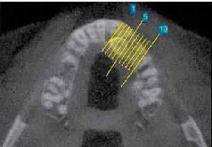
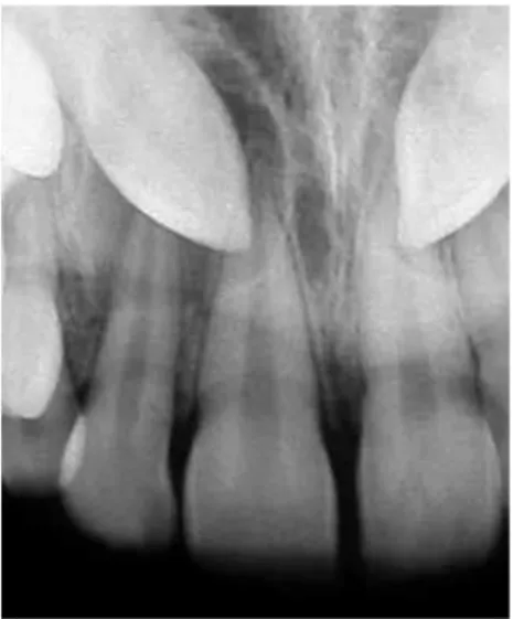
The location of the canine by means of images must be technically the best possible. To do so, occlusal ra-diographs may be used (Fig 1), standard frontal and lateral cephalometric radiographs, CAT scans (Figs 2 and 3), panoramic X-rays (Fig 4), although in most cases, the periapical films are sufficient for this pur-pose.3,8,11,12,15,16 The Clark technique performed with
the periapical films (Figs 5 and 6) indicates only if the tooth is buccally or palatally displaced.3,4,10,12 More
re-cently rapid prototyping can be used, a technique that involves the use of various technologies that apply data from computer-aided design files (CAD; abbreviation for “computer aided design”) to produce physical mod-els by a process of material addition. There are various rapid prototyping technologies and, among them, the most accurate and of better quality are laser sintering (SLS), stereolithography (SLA) and PolyJet.6,7
In these radiographic examinations, the outline of the lamina dura of the root process of non-erupted tooth is searched; suggestive images of apical disrup-tion; of the relationship of the root apex in the osse-ous space limited by the cortical bone of the maxillary sinus; the canine crown relationship with the roots of neighboring teeth; tooth positioning in the arch and presence of pathological processes.11
Prototyp-ing, on its turn, aims besides diagnosing an included maxillary canine impaction, create a specific acces-sory for tooth tractioning and assisting the surgeon
Figure 1 - Occlusal radiograph of impacted canine due to cleft palate.
Figure 2 - CT scan for localization of the impacted canine.
Figure 3 - CT scan for localization of the impacted canine.
Autogenous transplantation
Surgical procedure which extracts the retained canine and immediately transplants it to an artificial alveolus in the alveolar ridge. There is frequent risk of pulp necrosis, root resorption and loss of trans-planted tooth.8
Extraction of retained canine
Should be considered when it is ankylosed and prevents the realization of a transplant or a traction-ing; or with external and/or internal root resorp-tion; or with severe root disrupresorp-tion; or with a severe impaction for example, with the canine located be-tween the roots of central and lateral incisors; or if the occlusion is acceptable, with the first premolar occupying the place of a canine; or, with pathological changes such as cysts or infections, and if the patient refuses to undergo orthodontic treatment.
In some of these cases, patient rehabilitation is required for treatment complementation after extraction. Orthodontic rehabilitation can be per-formed promoting space closure by the movement of neighboring teeth since the premolar is on the same axial position of the canine, the prosthetic orthodon-tic treatment recovering space for the installation of dental prosthesis, or simply if there is enough space, the placement of a fixed prosthesis.8,10,11
Figure 5 - Localization of periapical radiograph us-ing the technique of Clark.
Figure 6 - Periapical radiograph positioning using the Clark technique with displacement for canine buccal or palatine position evaluation.
in intraoperative tooth exposure and individualized accessory bonding.6,7
Canine location is of primary importance for the determination of access to the tooth and appropriate surgical procedure, as well as the direction of orth-odontic force application.11
Treatment alternatives
After individual assessment of malocclusion and with the various current mean of diagnosis available, it is possible to prevent or even avoid the impactions and/or ectopic eruption of canines, being this a rel-evant fact in clinical planning.
In this way, conducing an ectopic tooth to its prop-er position in the dental arch is regarded as ideal for health, function and patient’s esthetic and the clinician can consider various options for treatment, including:
Extraction of deciduous canine
Is indicated when the permanent canine has the potential of eruption, or is with half of its root formed and being not so much horizontal.8,10
Orthodontic tractioning
Currently, with the possibility of early diagnosis and the development of orthodontic-surgical tech-niques, the more conservative treatments are more desirable, such as the orthodontic surgical trac-tions, followed by leveling and alignment of teeth in the arch until enough space is obtained to accom-modate the canine. This treatment represents the preferred procedure in the permanent dentition and consists of the surgical access to the retained canine for fixation of the orthodontic accessory, by which a force is applied to perform its traction until the correct positioning in the dental arch.1,3,5,8,9,10,16,17
Orthodontic-surgical approach of buccally and palatally impacted canine
Surgical access for tooth exposure depends on the definition, by radiographic examination, of the posi-tion in which the tooth is located in the arch.9 There
are several methods for surgical exposure of impact-ed canines and tractioning in order to lead them to occlusion. The methods most commonly used are:
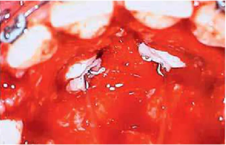
1) Surgical exposure, allowing the natural erup-tion (apically reposierup-tioned flap); and 2) surgical expo-sure and placement of an orthodontic accessory (Fig 7), for further tractioning (gingival flap repositioned in its original position). Orthodontic forces are sub-sequently applied to move the impacted teeth.1,11
The first option is used when the canine presents with a favorable axial inclination, not needing to be uprighted during its eruption. The canine erup-tion is monitored by means of X-rays, using some reference points as the neighbouring teeth or the orthodontic archwire. The disadvantage of this pro-cedure is about the time, because the eruption is slow and spontaneous, prolonging treatment time. The second option consists of surgical exposure of the tooth and the placement of an auxiliary device. Therefore, direct bonding buttons and orthodontic brackets for traction of impacted teeth have become the preferred techniques because, in addition to the easiness, it requires minor tissue removal and surgi-cal extension to access the tooth crown. However, as a disadvantage, there is the risk of accessory debond-ing because of the difficulty in drydebond-ing the operatdebond-ing field with consequent deficient bonding, needing an-other surgery for new fixation in case of the non-use of hydrophilic primers.1,2,3,5,11
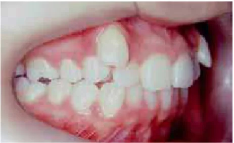
The buccal impaction is less frequent and usually caused by insufficiency in arch length. As a result, the canine is in a higher position in the alveolar bone and erupts ectopically in the alveolar mucosa (Figs 8, 9 and 10). Teeth buccally impacted in a favorable verti-cal position must be treated by simple surgiverti-cal expo-sure and natural eruption of the tooth and in some cases, with orthodontic tractioning.2,3,9,10,11,13,17
Force system
The buccally impacted canine should be to ex-truded in order to obtain arch alignment and the force system necessary to get this type of movement is a simple application of extrusive force on the ca-nine. This force system can be easily achieved by using a cantilever so that the posterior teeth experi-ence an intrusive force and a anterior tipping mo-ment, while canines erupt.1,17
The main objective in the treatment of buccal impacted teeth consists in creating and preserving a inserted functional band gum. This inserted gum can be achieved by means of an retail repositioned by apical or laterally, or when it is necessary, for a in-serted graft gum free, thus avoiding future gingival recessions and marginal alveolar bone loss.11
is accompanied by a system balance with an intru-sive force and anterior tipping moment on the pos-terior teeth. Buccal canine tipping is achieved by applying a simple force in brackets. When moving the canines buccally the molar tips lingually and undergoes a mesial rotation toward the palate. It is recommended to use cantilevers on the molar tube connected to the canines to promote the eruption of palatally impacted canines.1
During buccal or palatal impacted canine trac-tion, one must be careful with the direction of the applied force, because this should not promote traction towards the roots of neighbouring teeth, to avoid causing trauma and external root resorp-tions. It is recommended, initially, to obtain space in the dental arches before the traction and also the use of force of low intensity (no more than 60 g) and the employment of archwires of sufficient rigidity (0.018 x 0.025-in) to avoid deflections which may undermine the movements.1,2,3,5,9
Traction methods
Several therapeutic approaches have been em-ployed and presented in the orthodontic literature to obtain the appropriate positioning of impacted ca-nines in proper alignment and levelling of dental arch-es. Among them, we highlight orthodontic loops built with reduced size wire (0.6 mm) which can be bonded to the palatine arches or buccally with Adam’s clasps in removable appliances, also on second-order bends, introduced in leveling arches, as well as elastic wires with shape memory and elastomeric chains.2,3,5,17
a) Use of superelastic wires — obtaining space for the placement of the impacted canine can be reached by the use of nickel titanium superelastic loop (Fig 11). This loop can be kept in this position
as a stabilization method.
After initial leveling and alignment, the upper arch was stabilized by means of rectangular archwire 0.019 x 0.025-in and lingually interconnecting the right and left side molars by a transpalatal bar. Then canine surgical exposure and accessory bonding is performed. The main stainless steel arch is removed and the NiTi superelastic archwire is positioned (Fig 12), occurring deflection until it properly fits into the slot of the bracket on the canine.1
b) Use of removable appliances — after the expo-sure of the canine the working model for the remov-able appliance construction is obtained, which must include Adam’s and drop type clasps for retention in premolars, in addition to a buccal support structure with a hook for the use of 3/16 diameter elastic from the hook to the canine accessory.
The disadvantage of this system is the employ-ment of intermittent force and, therefore, patient cooperation is necessary in how to use the appliance and change elastics to keep the traction force.1,2,3
Another system that can be used in buccal ca-nine traction is the incorporation of loops welded to the horizontal bar of the Adam’s clasp. The force intended for canine traction will be due to this loop activation and must be applied on the accessory bonded to the tooth.1
c) Use of cantilevers — systems that provide the proper control for canine movement, associated with a smaller commitment of anchorage units. It is built with titanium-molybdenum wire (TMA) 0.017 x 0.025-in. Loop activation must focus on activation points contained in the loop shape and observing the pre-activation bends. The big advantage found in this method consists in the possibility to work with a de-fined force system.1
Figure 9 - Frontal view of ectopic upper canines.
Figure 8 - Right side view of ectopic upper ca-nine.
d) Use of elastomeric chains — system that pro-vides control over the magnitude and direction of the force applied to the tooth to be tractioned.2,3,4
Orth-odontic traction to bring the impacted tooth to the arch should start as soon as possible after surgery.3
DISCUSSION
The upper canine despite being the tooth of most frequent eruption anomalies after third molars, was considered by most authors as one of the most impor-tant teeth of the dental arch, and a correct diagnosis and clinical approaches are necessary in cases where it is impacted. Their retention covers a small portion of the population ranging, according to the authors, from 0.9 to 2.5%1,2,3,6,8,11,12,16 and it occurs two to three
times more in females than in males (although the source is unknown, there is an assumption that this occurs due to a lower transverse dimension of upper arch in female gender), it generally occurs unilaterally (75 to 95% of cases) palatally (60 to 80%)8,12,17 and in
8% of cases are impacted bilaterally.16,17
In general, most authors advocate that for a good treat-ment prognosis the conduct for canine location must be accomplished through clinical and radiographic evalu-ation (teleradiographies, periapical, occlusal computed tomographies and, more recently, rapid prototyping) and treatment alternatives analyzed according to the particu-larities of each case after careful evaluation of the ortho-dontist and professionals of distincted areas.2,3,4,8,15
The orthodontic-surgical conduct is one of the most used options in cases where there is buccal or palatal impaction.2,3,9,10,17 Most employed methods
are surgical exposure allowing natural eruption,10
surgical exposure for bonding of an orthodontic ac-cessory and further traction.1,3,11
Various methods are suggested for the traction and alignment of impacted canines, among them the orthodontic removable or fixed appliances, the use of anchorage in the same arch or opposite arch.2,3,5,15,17
The use of the lower arch as anchorage makes it dif-ficult to control the direction and magnitude of the force applied. Another form of treatment would in-volve the use of a removable device at an early stage and subsequently install of the fixed orthodontic ap-pliance for case finishing.
The use of a removable device presents some ad-vantages as transferring anchorage to the palate1 or
lower arch15 a more vertical component when
sup-ported in the lower arch, but despite these advantag-es the orthodontist must bear in mind that this type of device depends on the cooperation of the patient and so the completion of the case can be compro-mised.1,2,3,15 The fixed orthodontic appliance provides
greater control and effectiveness of the force applied, and in most cases there is a need to correct some oth-er type of associated malocclusion and to open and keep the space for the tooth to be tractioned, using specific accessories as loops.
With regard to the force system for traction of buccally or palatally impacted canines, one must be careful with the direction of the applied force, because this should not direct traction to the roots of neighbouring teeth, not to cause trauma and external root resorptions. It is recommended, ini-tially, to gain space in dental arches before traction and also the use of force of low intensity (no more than 60 g) and the employment of sufficiently rigid archwires (0.018 x 0.025-in) not to deflect which may undermine movement control.1,2,3,5,9
CONCLUSIONS
1) The permanent upper canine presents a high prevalence of impaction, and this anomaly occurs more frequently in the palate and in the female gender.
2) The correct diagnosis of the position of an impacted tooth, in addition to the essential clinical examination, must be followed of a meticulous radiographic examination. 3) The adequate time for surgical and
orthodon-tic intervention is when the tooth presents more than half of its root formation.
4) Surgical exposure of the dental crown should be conservative and the flap must be com-pletely repositioned allowing tooth eruption. 5) The support for the traction force is prefera-bly anchoraged over retangular orthodontic wire, in order to occur minimal deflection and the least amount of undesirable effects.
1. Almeida RR, Fuziy A, Almeida MR, Almeida-Pedrin RR, Henriques JFC, Insabralde CMB. Abordagem da impactação e/ou erupção ectópica dos caninos permanentes: considerações gerais, diagnóstico e terapêutica. Rev Dental Press Ortod Ortop Facial. 2001;6(1):93-116.
2. Bishara SE. Impacted maxillary canines: a review. Am J Orthod Dentofacial Orthop. 1992;101(2):159-71.
3. Capellette M, Capellette M Jr, Fernandes LCM, Oliveira AP, Yamamoto LH, Shido FT, et al. Caninos permanentes retidos por palatino: diagnóstico e terapêutica- uma sugestão técnica de tratamento. Rev Dental Press Ortod Ortop Facial. 2008;13(1):60-73.
4. Cernochova P, Kanovska K, Krupa P. Morfology and position of the root apex in impacted maxillary canines. Scripta Med (BRNO). 2003;76(1):9-20. 5. Crescini A, Clauser C, Giorgetti R, Cortellini P, Pini Prato GP. Tunnel traction of
infraosseous impacted maxillary canines. A three-year periodontal follow-up. Am J Orthod Dentofacial Orthop. 1994;105(1):61-72.
6. Faber J, Berto PM. Rapid prototyping as a tool for diagnosis and treatment planning for maxillary canine impaction. Am J Orthod Dentofacial Orthop. 2006;129(4):583-9. 7. Faber J. Jorge Faber responde: O que é a prototipagem e como ela pode ser
utilizada para avaliação de dentes inclusos? Rev Dental Press Ortod Ortop Facial. 2006;4(6):9-15.
8. Henriques JFC, Machado DT, Hayasaki SM, Janson GRP. Uma das alternativas de tratamento da maloclusão com os caninos superiores retidos e os inferiores em infravestibuloversão: apresentação de um caso clínico. Rev Dental Press Ortod Ortop Facial. 2002;7(8):61-9.
9. Kokich VG. Surgical and orthodontic management of impacted maxillary canines.
REFERENCES
Am J Orthod Dentofacial Orthop. 2004;126(3):278-83.
10. Leite HR, Oliveira GS, Oliveira GS, Brito HHA. Labially displaced ectopically erupting maxillary permanent canine: interceptive treatment and long-term results. Am J Orthod Dentofacial Orthop. 2005;128(2):241-51.
11. Martins DR, Kawakami RY, Henriques JFC, Janson GRP. Impactação dentária: condutas clínicas - apresentação de casos clínicos. Rev Dental Press Ortod Ortop Facial. 1998;3(1):12-22.
12. Martins PP, Gurgel JA, Sant’Ana E, Ferreira Júnior O, Henriques JFC. Avaliação radiográfica da localização de caninos superiores não irrompidos. Rev Dental Press Orthod Ortop Facial. 2005;10(4):106-14.
13. Peng CL, Su YY, Lee SY. Unilateral horizontally impacted maxillary canine and first premolar treated with a double archwire technique. Angle Orthod. 2006;76(3);502-9.
14. Rossato C, Romero E. Canino superior impactado: considerações gerais e apresentação de caso clínico. Unopar Cient Ciênc Biol Saúde. 2001;3(1):21-9. 15. Sinha PK, Nanda RS. O controle de caninos superiores impactados por meio de
ancoragem mandibular. Rev Dental Press Ortod Ortop Facial. 2000;5(1):68-71. 16. Stewart JA, Giover KE, Williamson PC, Lam EWN, Major PW. Factors that relate
to treatment duration for patients with palatally impacted maxillary canines. Am J Orthod Dentofacial Orthop. 2001;119(3):216-25.