Regulation of
sodB
in
H. pylori
Beth M. Carpenter1, Hanan Gancz1, Reyda P. Gonzalez-Nieves1, Abby L. West2, Jeannette M. Whitmire1, Sarah L. J. Michel2, D. Scott Merrell1*
1Department of Microbiology and Immunology, Uniformed Services University of the Health Sciences, Bethesda, Maryland, United States of America,2Department of Pharmaceutical Sciences, School of Pharmacy, University of Maryland, Baltimore, Maryland, United States of America
Abstract
Helicobacter pyloriis a significant human pathogen that has adapted to survive the many stresses found within the gastric environment. Superoxide Dismutase (SodB) is an important factor that helpsH. pyloricombat oxidative stress.sodBwas previously shown to be repressed by the Ferric Uptake Regulator (Fur) in the absence of iron (apo-Fur regulation) [1]. Herein, we show thataporegulation is not fully conserved among all strains of H. pylori.apo-Fur dependent changes in sodB
expression are not observed under iron deplete conditions inH. pyloristrains G27, HPAG1, or J99. However, Fur regulation ofpfrandamiEoccurs as expected. Comparative analysis of the Fur coding sequence between G27 and 26695 revealed a single amino acid difference, which was not responsible for the alteredsodBregulation. Comparison of thesodBpromoters from G27 and 26695 also revealed a single nucleotide difference within the predicted Fur binding site. Alteration of this nucleotide in G27 to that of 26695 restoredapo-Fur dependentsodBregulation, indicating that a single base difference is at least partially responsible for the difference insodBregulation observed among theseH. pyloristrains. Fur binding studies revealed that alteration of this single nucleotide in G27 increased the affinity of Fur for thesodBpromoter. Additionally, the single base change in G27 enabled thesodBpromoter to bind toapo-Fur with affinities similar to the 26695sodBpromoter. Taken together these data indicate that this nucleotide residue is important for direct apo-Fur binding to the sodB
promoter.
Citation:Carpenter BM, Gancz H, Gonzalez-Nieves RP, West AL, Whitmire JM, et al. (2009) A Single Nucleotide Change Affects Fur-Dependent Regulation ofsodB inH. pylori. PLoS ONE 4(4): e5369. doi:10.1371/journal.pone.0005369
Editor:Niyaz Ahmed, University of Hyderabad, India
ReceivedAugust 25, 2008;AcceptedMarch 27, 2009;PublishedApril 28, 2009
This is an open-access article distributed under the terms of the Creative Commons Public Domain declaration which stipulates that, once placed in the public domain, this work may be freely reproduced, distributed, transmitted, modified, built upon, or otherwise used by anyone for any lawful purpose.
Funding:Research in the laboratory of D. Scott Merrell is made possible by grants R073LA from USUHS and AI065529 from the NIAID. Contents of this manuscript are the sole responsibility of the authors and do not necessarily represent the official views of the NIH or the DOD. The funders has no role in study design, data collection and analysis, decision to publish, or preparation of the manuscript.
Competing Interests:The authors have declared that no competing interests exist.
* E-mail: [email protected]
Introduction
Helicobacter pyloriis an important human pathogen that infects over 50% of the world’s population [2]. While infection is predominantly asymptomatic, this bacterium is associated with development of gastritis, peptic ulcer disease, mucosa-associated lymphoid tissue lymphoma, and gastric adenocarcinoma. Infection often occurs early in childhood and persists throughout a person’s lifetime unless they are treated with specific antibiotics [3]. Given its propensity for chronic colonization and the substantial number of infected individuals, H. pylori imposes a significant disease burden worldwide.
This microaerophilic, Gram negative bacterium is interesting in that it colonizes and survives within the gastric mucosa of the human stomach.H. pyloriis well suited to life within this niche and has many factors that enable it to thrive there [2,4]. One such factor, the Ferricuptake regulator (Fur), functions as a transcrip-tional regulator that is involved in maintaining iron homeostasis [5]. Iron is essential for bacterial survival and is a co-factor in a variety of proteins; however, iron is redox active and can promote oxidative damage making it imperative that intracellular iron levels are tightly controlled. One particularly deleterious reaction that free iron can promote is reaction with reactive oxygen species (ROS) to form highly reactive hydroxyl radicals via Fenton
chemistry. Hydroxyl radicals cause DNA and cellular damage that eventually lead to cell death. Thus, cells must strive to maintain a balance between insufficient and excess iron. Fur is involved in preserving this fine balance inH. pylori, and consequently, it is not surprising thatfurhas been shown to be critical for colonization in both gerbil and murine models of infection [6,7].
Fur is conserved in a wide variety of bacterial species and functions similarly in all of them by repressing gene expression under conditions of sufficient cellular iron. When Fur is bound to its iron (Fe2+) co-factor, it binds to specific regions in iron-regulated promoters called Fur Boxes and blocks the binding of RNA polymerase. Genes regulated in this manner are often associated with iron acquisition and are repressed under iron replete conditions to prevent the harmful effects of iron overload. WhileH. pyloriFur has been found to repress a set of genes in its iron-bound state, it has also uniquely been found to repress an additional set of genes in the absence of the iron cofactor, i.e. when Fur is in itsapoform.apo-Fur regulation involves repression of an iron storage gene and occurs under iron limited conditions [8].
pylorithat is not directly linked to iron metabolism, but is involved with the oxidative stress response, issuperoxide dismutase (sodB) [1]. SodB was first identified inH. pyloriin 1993 and was shown to be iron co-factored like theEscherichia coliFeSod with 53.5% identity between the two proteins [9]. However, unlikeE. coliFeSod, which is localized within the cytosol of the bacterium, H. pyloriSodB is associated with the cell surface [9]. SodB is the only identified Sod in
H. pyloriand has been shown to be critical for survivalin vivo[10]. Also,sodB deficient mutants are more sensitive to O2 as well as
exhibit a higher rate of spontaneous mutation [10,11]. Interestingly,
H. pylori sodBmutants have been shown to harbor more free iron within their cells than WT bacteria [11].
Globally, Sods are responsible for combating oxidative stress (both internal and external) by converting superoxide radicals into hydrogen peroxide and oxygen. Superoxide radicals are formed as a by-product of metabolism and, if left unchecked, can react with ferric iron (Fe3+) to form hydrogen peroxide, which in turn feeds
the Fenton Reaction [12] and is detrimental to the cell. Sods prevent the interaction of iron and superoxide radicals as well as block the formation of hydroxyl radicals from hydrogen peroxide [12]. In this way, the role of Fur as the primary regulator of iron uptake and the role of SodB as the primary defense against superoxide radicals in H. pyloriare linked. In keeping with this,
sodB has been shown to be regulated by apo-Fur such that it is repressed under circumstances where iron is severely limited [1]. This regulation appears to be direct since Electrophoretic Mobility Shift Assays showed that Fur specifically binds to the sodB
promoter in the absence of iron [1]. Herein we describe a series of experiments that define a single polymorphic nucleotide within the
H. pylori sodB promoter that is important for apo-Fur dependent regulation. Moreover, we show that alterations in this single base result in strain specific responses to iron limitation.
Materials and Methods
Bacterial strains and growth
Strains and plasmids used in this study are listed in Table 1, and primer sequences are listed in Table 2. Strains ofH. pylori were maintained as frozen stocks at280uC in brain heart infusion broth (BD) supplemented with 10% fetal bovine serum (Gibco) and 20% glycerol (EMD Chemicals, Inc.). Bacterial strains were grown on horse blood agar (HBA) plates which contained 4% Columbia agar base (Neogen Corporation), 5% defibrinated horse blood (HemoStat Laboratories, Dixon, CA), 0.2% b-cyclodextrin (Sigma), 10mg/ml vancomycin (Amresco), 5mg/ml cefsulodin (Sigma), 2.5 U/ml polymyxin B (Sigma), 5mg/ml trimethoprim (Sigma), and 8mg/ml amphotericin B (Amresco). Liquid cultures of H. pyloriwere grown in brucella broth (Neogen Corporation) supplemented with 10% fetal bovine serum and 10mg/ml vancomycin at 37uC with shaking at 100 rpm. As noted in Table 1, where appropriate, cultures and plates were supplement-ed with 8mg/ml chloramphenicol (Cm) (EMD Chemicals, Inc.) and/or 25mg/ml kanamycin (Kan) (Gibco). In addition, where detailed in the Materials and Methods, some HBA plates contained 5% sucrose (Suc) (Sigma). Both liquid and plate cultures were grown under microaerophilic conditions (5% O2, 10% CO2,
and 85% N2) generated with an Anoxomat gas evacuation and
replacement system (Spiral Biotech) in gas evacuation jars.
H. pyloristrains used in this study are all derivatives of G27 [13] and 26695 [14,15], with the exception of WTH. pylori J99 [16] and HPAG1 [17]. Afur(HP1027) mutant of G27, DSM300, was utilized in this work and contains a deletion insertion of the fur
coding sequence with thecatgene fromCampylobacter coliconferring Cm resistance as previously described [18]. This DHP1027::cat
construct was also naturally transformed into 26695 to create an analogous fur mutation in this strain background and is called DSM357. Exponential phase cultures were grown for 20 hrs, and stationary phase cultures were grown for 44 hrs.
Creation of thesodBpromoter fusion plasmid
A transcriptional fusion of thesodB(HP0389) promoter to the promoterless gfpmut3 on the transcriptional reporter plasmid, pTM117, was constructed as previously described [18]. Briefly, the
sodBpromoter of WT G27 was PCR amplified using sodB-F1 and sodB-R1 primers, which incorporate SacII and BamHI restriction sites, respectively. The resulting PCR fragment was subcloned into pGEM-T Easy (Promega) and digested with SacII (New England
Table 1.Plasmids and strains used in this study.
Plasmid or strain Description Reference
Plasmids
pTM117 Modified pHP666 to includeE. coli
origin andropgene,aphA-3cassette (Kanr), multiple cloning site, and a
promoterlessgfpmut3gene
[18]
pDSM236 pTM117sodBpromoter::gfpmut3fusion This study
pDSM368 pTM117pfrpromoter::gfpmut3fusion [18]
pKSF-II pEK::kan-sacB [19,20]
pDSM386 pGEM-T Easy::Dfur This study
pDSM387 pGEM-T Easy::Dfur::kan-sacB This study
pDSM469 pGEM-T Easy::DsodB This study
pDSM475 pGEM-T Easy::DsodB::kan-sacB This study
pDSM481 pGEM-T Easy::sodBC-5A This study
pDSM429 pGEM-T Easy::26695fur This study
pDSM430 pET21A::26695fur This study
pKD4 kantemplate plasmid [22]
pKD46 Red recombinase expression plasmid [22]
H. pyloristrains
G27 WTH. pylori [13]
DSM300 G27Dfur::cat, Cmr [18]
26695 WTH. pylori [14,15]
DSM357 26695Dfur::cat, Cmr This study
DSM238 G27 (pDSM236), Kanr This study
DSM308 DSM300 (pDSM236), KanrCmr This study
DSM369 G27 (pDSM368), Kanr [18]
DSM370 DSM300 (pDSM368), KanrCmr [18]
DSM391 G27Dfur::kan-sacB, KanrSucs This study
DSM403 G27,fur26695, SucrKans This study
DSM480 G27DsodB::kan-sacB, KanrSucs This study
DSM491 G27sodBC-5A, SucrKans This study
J99 WTH. pylori [16]
HPAG1 WTH. pylori [17]
E. colistrains
DSM328 K12 (pKD46), Ampr, Temps [22]
DSM355 K12Dfur, Kanr This study
DSM326 BL21 DE3 Rosetta/pLysS, Cmr This study
DSM365 BL21 DE3 Rosetta/pLysSDfur, Kanr, Cmr This study
DSM431 BL21Dfur(pDSM430) Ampr, Cmr, Kanr This study
Biolabs) and BamHI (Invitrogen). The resulting promoter fragment was then ligated into the appropriately digested pTM117 vector to create pDSM236. The fusion was confirmed by PCR amplification with sodB-F1 and gfp-1 [18] primers and by sequencing with the aphA3-2 primer [18]. pDSM236 was naturally transformed into WT G27 and DSM300, and transfor-mants were selected on HBA plates containing 25mg/ml Kan and
25mg/ml Kan plus 8mg/ml Cm, respectively. The WT strain bearing pDSM236 was designated DSM238, and DSM300 bearing pDSM236 was designated DSM308.
GFP reporter assays
The ability of the sodB transcriptional fusion to drive the expression of GFP was assessed using flow cytometry as described Table 2.Primers used in this study.
Primerb Sequence (59-39)a Reference
sodBpromoter primers
sodB-F1 (SacII) CCGCGGCGCCATTGACCAATTTCAG This study
sodB-R1 (BamHI) GGATCCGCAACTCTCGTAATGTAAAC This study
Screening and Sequencing primers
gfp-1 AAGTCGTGCTGCTTCATGTG [18]
aphA3-2 CGGTGATATTCTCATTTTAGCC [18]
sacBSCN-F2 CGAATCGAATTCAGGAAC This study
HpKanSacSCN-R GGGAAGTTCTATGCTTATGG This study
HpsodBSCN-R GCTCGCTTCTTTAAACTCAACC This study
Cloning primers
FurCF (XbaI) TCTAGAAAGGCTCACTCTACCCTATT [18]
HpUKanSacR (XhoI, SmaI) CTCTTGGCATTCTTTACACCACACCCCGGGAGGCTCGAGGCTGATATCTTCCTTATCCG This study
HpDKanSacF (XhoI, SmaI) CGGATAAGGAAGATATCAGCCTCGAGCCTCCCGGGGTGTGGTGTAAAGAATGCCAAGAG This study
HpDKanSacR CGCAGCGATAAAGGCGTGGTG This study
FurCR (SalI) GTCGACAAGACTTTCACCTGGAAACGC [18]
USod-F GCTTTATCGCCCACTTTCAAG This study
USod-R CCACAATAGCCGTAACGCTTACCCGGGAGGCTCGAGCATGTTTTCTCCTTGTGATTAG This study
DSod-F CTAATCACAAGGAGAAAACATGCTCGAGCCTCCCGGGTAAGCGTTACGGCTATTGTGG This study
DSod-R GGCATGGAATTGTCAATCC This study
SodBMt-R GATAGCCTTATTGTAATC This study
SodBMt-F GATTACAATAAGGCTATC This study
HP_Fur_expression F2 (NdeI) CATATGAAAAGATTAGAAACTTTGGAATCCATTTT This study
HP_Fur_expression R2 (Xho1) CTCGAGTTATTAACATTCACTCTCTTGG This study
Red_EC_Fur_F GAGCTGTAACTCTCGCTTTTCTTATTTCCCTTGCATGTGTAGGCTGGAGCTGCTTC This study
Red_EC_Fur_R TCATGTCTACGCCGTATTAATAGATAATGCCAATCACCATATGAATATCCTCCTTAGTTC This study
RPA primers
amiE-RPA-F GGTTTGCCTGGGTTGGAT [7]
amiE-RPA-R GATTTTGCGGTATTTTG [7]
pfr-RPA-F GCGGCTGAAGAATACGAG [18]
pfr-RPA-R CTGATCAGCCAAATACAA [18]
sodB-RPA-F AAGCCCTGTAGCGTTTGATT This study
sodB-RPA-R CCCAATTCCAACCAGAGCCA This study
fur RPA F GAGCGCTTGAGGATGTCTATC [18]
fur RPA R GTGATCATGGTGTTCTTTAGC [18]
EMSA primers
G27 sodB EMSA-F CTACAAAATTTGCATAACG This study
26695 sodB EMSA-F CCACAAAATTTGCATAAAG This study
sodB EMSA-R GCAACTCTCGTAATGTAAAC This study
rpoB EMSA-F CCAAAGAGGGTAAAGAGAGCG This study
rpoB EMSA-R CCTCTCCATCGCTTCTCTAAC This study
aRestriction endonuclease sites are underlined, and linker bases are in bold type. bImportant restriction sites are included in parentheses.
previously [18]. Briefly, DSM238 was grown overnight in liquid culture with and without the iron chelator, 2,29-dipyridyl (dpp) (Sigma) at a final concentration of 60mM, and DSM308 was grown overnight in the absence of chelator. As a comparison, the previously characterized strains, DSM369 and DSM370, which bear pfr (nonheme iron-containing ferritin) transcriptional fusion plasmids in WT andDfur G27, respectively, were grown in the same manner [18]. Following overnight growth, 1.5 ml of each culture were pelleted and resuspended in 1 ml of sterile 16
phosphate-buffered saline. Bacterial clumps and culture debris were subsequently removed by passing the resuspended culture through a 1.2-mm Acrodisc PSF syringe filter (Pall). Flow cytometry analysis was performed using a Beckman Coulter Epics XL-MCL flow cytometer with a laser setting of 750 V for thepfr
fusion construct and 900 V for the sodB fusion construct. 100,000 events were collected for each assay. WinList 3D, version 6.0 (Verity Software House) was used to analyze the flow cytometry data.
Creation of the ‘‘Fur swap’’ Strain
To exchange thefurcoding sequence, we first created a G27 strain containing the counter-selectablekan-sacBcassette previously described by Copass, et al [19]. This cassette contains the sacB
gene from Bacillus subtilis, which confers Suc sensitivity and is expressed under the control of theflaApromoter ofH. pylori, and the aphA3 gene from Campylobacter coli, which confers Kan resistance. A 340 bp region upstream of the G27 fur coding sequence was PCR amplified using primers FurCF1 [18] and HpUKanSacR, and a 339 bp region downstream of thefurcoding sequence was PCR amplified using primers HpDKanSacF and HpDKanSacR. HpUKanSacR and HpDKanSacF were designed to incorporate XhoI and SmaI restriction endonuclease sites. Each of these products were purified and mixed in a Splicing by Overlap Extension (SOE) PCR reaction using the FurCF1 and HpDkanSacR primers. The resulting 679 bp product was subcloned into pGEM-T Easy creating pDSM386. The kan-sacB
cassette was liberated from pKSF-II [19,20] by sequential double digestion with XhoI (New England Biolabs) and SmaI (New England Biolabs), and this fragment was ligated to the appropri-ately digested pDSM386 to create pDSM387. This plasmid was naturally transformed into WT G27, and transformants were selected on HBA plates containing Kan. Double crossover homologous recombination of pDSM387 with the WT chromo-some results in the complete deletion of thefur(HP1027) coding sequence and replacement with the upstream fur-kan-sacB -downstream fur product. The resulting transformants were patched on 5% Suc HBA plates to ensure Suc sensitivity, and proper integration into the chromosome was confirmed by PCR with sacBSCN-F2 and HpKanSacSCN-R primers, which lie within the sacB gene and downstream of fur, respectively. One such transformant was named DSM391.
To create the ‘‘Fur swap’’ strain, a 923 bp product of theH. pylori26695 genome was amplified using the FurCF and FurCR primers. This product, which includes thefurcoding sequence and a portion of the upstream and downstream regions, was purified and naturally transformed into DSM391. Transformants were selected on 5% Suc HBA plates and patched onto Kan HBA plates to ensure Kan sensitivity. Double crossover homologous recombination resulted in the replacement of thekan-sacBcassette with thefurcoding sequence of 26695, and this strain was named DSM403. Proper integration was confirmed by PCR with the FurCF and FurCR primers and by sequencing with the FurCR primer. DSM403 expresses 26695furfrom the nativefurlocus in a G27 strain background.
Creation of a ‘‘25 bp swap’’ mutation in thesodBpromoter The sodB promoter from G27 was sequenced using primers USod-F and DSod-R and compared to the known sequence of the
sodBpromoter from 26695 [14]. This comparison revealed a single base pair (bp) difference within the predicted Fur Box [1] at the25 position relative to the start of transcription. The ‘‘25 bp swap’’ mutation within thesodBpromoter of G27 was created using SOE PCR and the kan-sacB cassette from pKSF-II. A 297 bp region upstream and a 329 bp region downstream of sodB were PCR amplified from G27 using primer pairs USod-F and USod-R and DSod-F and DSod-R, respectively. USod-R and DSod-F contain XhoI and SmaI restriction endonuclease sites to allow for the directional cloning of thekan-sacB fragment. The upstream and downstream products were purified and mixed in a SOE PCR reaction with the USod-F and DSod-R primers. The resulting 626 bp SOE PCR product was subcloned into pGEM-T Easy to create pDSM469. pDSM469 and pKSF-II were each sequentially double digested with XhoI and SmaI, and the resulting fragments were ligated to create pDSM475. pDSM475 was naturally transformed into WT G27, and transformants were selected on Kan and then patched to verify sucrose sensitivity. Double crossover homologous recombination of pDSM475 into the G27 chromo-some results in the deletion of thesodBgene and replacement with thekan-sacBcassette. The resulting Kan resistant, sucrose sensitive strain, DSM480, was confirmed by PCR with sacBSCN-F2 and HpsodBSCN-R primers, the latter of which lies downstream ofsodB. The25 bp in the G27sodBpromoter was mutated from a C to an A using SOE PCR. First, primers USod-F and SodBMt-R were used to PCR amplify upstream of thesodBpromoter through to the25 bp and incorporate the C-5A mutation. Second, primers DSod-R and SodBMt-F were used to PCR amplify from the25 bp through to downstream of the sodB gene and to incorporate the C-5A mutation. These products were purified and combined in SOE PCR reaction using the USod-F and DSod-R primers. The resulting SOE PCR product was sublconed into pGEM-T Easy. The subcloned sodB
25 bp promoter mutation construct was designated pDSM481 and was confirmed by sequencing with the USod-F and DSod-R primers. pDSM481 was naturally transformed into DSM480 to integrate thesodB25 bp promoter mutation into the chromosome in place of thekan-sacBcassette. Transformants were selected as detailed above for the creation of DSM403. The resulting Suc resistant, Kan sensitive strain was named DSM491. Proper recombination was confirmed by PCR with the USod-F and DSod-R primers (yielding a 1,262 bp fragment) and by sequencing with both of those primers. DSM491 expressessodBwith the C-5A mutation from its native locus within the G27 chromosome.
RNase protection assays (RPAs)
iron-limited growth culture was allowed to grow overnight and was harvested the following morning for the stationary phase, iron-limited growth RNA samples. RNA was extracted as described previously [21]. RNase Protection Assays (RPAs) were performed as previously described [18] with 1.5mg of RNA usingsodB, pfr,
amiE, and/orfurriboprobes that were generated using the primer pairs listed in Table 2. In brief, riboprobes were generated with 50mCi [32P]UTP (Perkin-Elmer) and a Maxiscript kit (Applied Biosystems). The RPA III kit (Applied Biosystems) was used for the RPA reactions that were resolved on 5% acrylamide-16
Tris-borate-EDTA-8M urea denaturing gels. The gels were exposed to phosphor screens, and the phosphor screens were scanned using a FLA-5100 multifunctional scanner (Fujifilm). Analyses and quantitation of the RPAs were performed using the Multi-Gauge software (version 3.0, Fujifilm). In all cases, three to four biological repeats of each experiment were performed.
H. pylori Fur Expression and Purification
H. pylori 26695 Fur coding sequence was amplified using primers HP_Fur_expression F2 (NdeI) and HP_Fur_expression R2 (XhoI), and the PCR product was cloned into the pGEM-T easy vector (Promega) to create plasmid pDSM429. pDSM430 was created by proper digestion of pET21A (Novagen) and pDSM429 with NdeI and XhoI and ligation of the gel purified fragments. The Fur coding region in pDSM430 was sequenced to verify the construct. To prevent cross contamination ofH. pylori
recombinant Fur with E. coli endogenous Fur, an E. coli BL21 Rosetta Dfur strain was constructed using the Wanner method [22]. Briefly, the Kan resistance cassette was amplified from pKD4 [22] with primers Red_EC_Fur_F and Red_EC_Fur_R. This PCR product was introduced into arabinose inducedE. coliK-12 carrying the pKD46 plasmid [22] to create DSM355. DSM365 was created by transduction of DSM326 with P1L4 grown on DSM355. EndogenousE. coli Fur deletion was verified by PCR. pDSM430 was introduced into DSM365 to create DSM431, which was used for rFur induction. DSM431 was grown to mid log in Luria-Bertani (EMD Chemicals) medium and then induced with 0.5 mM IPTG (isopropyl-D-thiogalactopyranoside) (Sigma) at 30uC for 3 h. The cells were disrupted using French press (Amicon) and crude extracts were prepared from the IPTG-induced cells by centrifugation (5,000 rpm for 30 minutes). Protein purification was performed by fast-protein liquid chroma-tography; the cytoplasmic protein was first passed through a HiTrap SP column for ion-exchange-based purification with a salt gradient of 25 mM to 500 mM NaCl (obtained by using buffer A [50 mM sodium phosphate, 25 mM NaCl, pH 8.0] and buffer B [25 mM sodium phosphate, 500 mM NaCl, pH 8.0]). Peak fractions containing Fur protein from the ion-exchange procedure were collected and further purified based on size exclusion by using a Sephacryl-200 column (buffer C [50 mM sodium phosphate, 200 mM NaCl, pH 8.0]). rFur was partially concen-trated using an Amicon Ultra Centrifugal Filter Device (Millipore) to remove a portion of buffer C. Then an equal volume of EMSA binding buffer (BB) was added to the partially concentrated rFur with an additional 50% glycerol. rFur was further concentrated before being quantitated and stored at 220uC. The final concentration of the rFur stock was 2 mg/mL.
Electrophoretic Mobility Shift Assays (EMSAs)
A 120 bp region of thesodBpromoter (encompassing the Fur-box) [1] was PCR amplified using the following template and primer pairs: WT G27 and DSM491 (‘‘25 bp swap’’) with G27 sodB EMSA-F and sodB EMSA-R and WT 26695 with 26695 sodB EMSA-F and sodB EMSA-R. To serve as a negative control
in the EMSA studies, a 142 bp region of therpoB promoter was amplified from WT G27 using the rpoB EMSA-F and rpoB EMSA-R primer pair. Each PCR product was acrylamide gel purified and resuspended in 16Tris-EDTA (TE) buffer. 150 ng of each promoter region was end labeled with [32P] ATP (Perkin Elmer) using T4 polynucleotide kinase (New England Biolabs) as previously described [7]. The unincorporated nucleotide was removed using the MinElute Reaction Clean-up kit (Qiagen), and labeled promoter fragments were eluted twice with 10mL EB, and 50mL ofapo-BB was added to the eluted product.
EMSAs were performed under apo (iron-free) conditions as previously described for WT 26695 sodB [1]. Briefly, 1 ng of labeled sodB or rpoB promoter was mixed with 5mL of the following dilutions of the Fur stock: 1:1,875, 1:3,125, 1:15,625, and 1:78,125 and combined with 10mL of 26 apo-BB (24% glycerol, 40 mM Tris, pH 8.0, 150 mM KCl, 2 mM DTT, 600mg/mL bovine serum albumin, 200mM EDTA, and 0.1 mg/ mL sheared salmon sperm DNA). In addition, a no protein control reaction and a 100 ng cold (unlabeled) DNA competition reaction were performed. The cold competition reaction was performed with the highest concentration of Fur (1:1,875). All reactions were allowed to incubate at 37uC for 30 min. After the incubation, the reactions were separated on a 5% polyacrylamide gel (5% 19:1 acrylamide, 16Tris Glycine EDTA (TGE) buffer, 2.5% glycerol)
for 3 hours at 70 V in 16TGE buffer. The gels were then exposed to phosphor screens and scanned on a Storm 860 scanner (GE Healthcare). Analysis was performed using ImageQuant version 5.2 software (Molecular Dynamics).
Competition EMSA Studies
Competition studies were performed in a manner analogous to the EMSAs. Each labeledsodBpromoter fragment was combined with the 1:1,875 dilution of rFur,apo-BB, and either 5 ng, 10 ng, or 25 ng of cold (unlabeled)sodBpromoter from each of the three respective strains. A no competitor control was included for each labeledsodBpromoter fragment. In this manner, each labeledsodB
fragment (WT G27, ‘‘25 bp swap,’’ and WT 26695) competed for binding to Fur with its own unlabeledsodBfragment as well as to that of the other two strains. The incubations, electrophoresis, and analysis were performed as described for the EMSAs. Binding competition occurs as follows: PDP32zD<PDzDP32, where P = Fur, DP32= labeled DNA, and D = cold competitor. Thus, if
the competitor promoter fragment (D) can bind to Fur (P) with a higher affinity than the labeled promoter (DP32), then an increase
in the amount of unbound, labeled promoter (DP32) would be seen.
The percent of unbound, labeledsodBpromoter was quantitated for each competition EMSA using densitometry as a means of comparing the relative affinity of each promoter fragment for Fur.
Statistical Analysis
Two-tailed Student’s t-tests were performed using Microsoft Office Excel 2003.
Nucleotide sequence accession number
The nucleotide sequence of thesodBpromoter is available from GenBank under accession number EU888136. The G27 fur
sequence was previously reported [18] and is available as GenBank accession number EF537051.
Results
apo-Fur Regulation in H. pylori
In order to studyapo-Fur dependent regulation inH. pylori, the
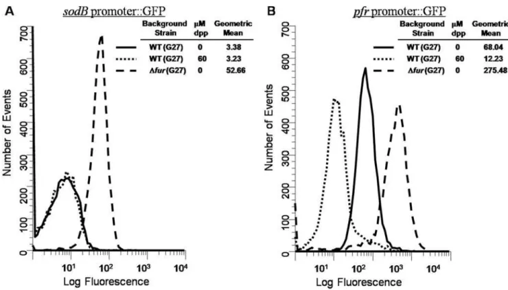
promoterlessgpfmut3gene in pTM117. Currently, these promoters represent the only known targets ofapo-Fur [1,8]. Given thisapo -regulation and since promoter activity can be measured by changes in fluorescence with our system, we expected to see a decrease in GFP fluorescence under iron limited conditions for both promoter fusions. However, as shown in Fig. 1A, the addition of iron chelator resulted in no change in the level of sodB
expression. This is in contrast topfr, where iron depletion resulted in strong repression ofpfrexpression (Fig. 1B). BothsodBandpfr
were upregulated in afurmutant (Fig. 1A and 1B) suggesting that both genes are repressed by Fur. However, the lack of responsiveness to iron chelation suggested thatsodB apo-regulation is not as expected in G27.
Sinceapo-Fur has been shown to have a lower affinity for the
sodBpromoter than thepfrpromoter, and since thegfpmut3allele encodes a long-lived GFP variant [23], we reasoned that we might not be able to detect small changes in GFP expression under the control of the sodB promoter under iron limited conditions. Therefore, we performed RPAs to further investigate the discrepancy between our results and results previously reported forsodBregulation in strain 26695 [1]. Additionally, we considered the fact that strain specific differences might be responsible for the discrepancy. Therefore, RPAs using a sodB riboprobe were performed on RNA isolated from WT andDfurderivatives from both G27 and 26695. pfrand amiE(aliphatic amidase) riboprobes were also used as control apo-Fur and iron-bound Fur regulated target genes, respectively. Fig. 2A shows results for all three riboprobes using RNA isolated from exponential phase cultures. Again, we observed that for G27 the level ofsodBexpression did
not change under iron-limited growth conditions (G) or under a harsher iron-depletion shock condition (S) that was added to ensure robust chelation as compared to normal (N) iron replete conditions.
Examination of sodB expression in 26695 revealed a smaller protected fragment than originally expected. However, sequence analysis revealed that the smaller fragment is due to a small region of mismatch between thesodBmRNA sequence in 26695 and the G27 template DNA used to generate the riboprobe. This mismatch causes a bubble of single stranded RNA to form and thus is subjected to RNase cleavage in the region of mismatch (data not shown). For WT 26695, a 2-fold decrease in sodB
expression was achieved under both limited growth and iron-depletion shock conditions, which agrees with the previous report [1]. This change is Fur-dependent as there is no change insodB
expression under either iron depletion condition in the absence of
fur.
Since it has been shown that growth phase strongly affects gene expression inH. pylori[21], we performed similar experiments on RNA harvested from stationary phase cultures. As shown in Fig. 2B, we obtained identical results with the exception that the fold decrease seen insodBexpression was less pronounced in 26695 in this growth phase. Again, there was no decrease in sodB
expression in G27, indicating that growth phase is not responsible for the differences in our results. Moreover, the difference insodB
regulation between the two strains is not the result of a generalized difference inapo-Fur regulation between G27 and 26695 since the appropriate decrease inpfr expression [8] was observed in both strains under iron-limited growth and iron-depletion shock
Figure 1. Flow Cytometry analysis ofsodBandpfrGFP reporters.Strains bearingsodB::gfpmut3orpfr::gfpmut3promoter fusions were grown overnight in either iron replete or iron depleted media. Changes in fluorescence were analyzed as described in the Materials and Methods. Results for thesodBpromoter fusions are displayed in Panel 1A, and results for thepfrpromoter fusions are displayed in Panel 1B. For both A and B, solid lines indicate the plasmid in WTH. pyloriG27 grown in iron replete conditions, dotted lines indicate the plasmid in WT bacteria grown in iron deplete conditions, and dashed lines indicate the plasmid inDfurbacteria grown in iron replete conditions. Fluorescence is measured in relative units, and the data are representative of multiple independent flow analyses.
conditions (Fig 2A and 2B). Furthermore, iron-bound Fur regulation of amiE was as expected [24] for both G27 and 26695; amiE expression was increased under both iron limited conditions (Fig. 2A and 2B). Taken in total, these data indicate thatapo-Fur regulation ofsodBis altered in G27 as compared to 26695.
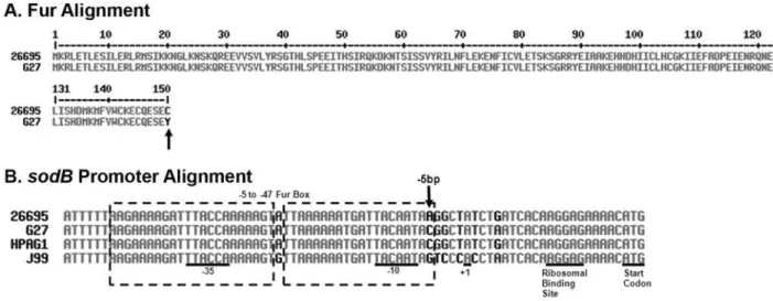
Analysis of the role an amino acid (AA) difference in Fur plays insodBregulation
Given the difference insodBregulation between the two strains, we reasoned that either a difference in Fur or a difference insodB
between the two strains was likely to be responsible for the change. We therefore aligned the predicted Fur amino acid sequence from G27 and 26695 to determine if there were any obvious differences between the two strains that might account for the differences in
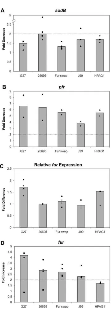
sodBregulation. As shown in Fig. 3A, the last AA was found to differ between the strains. In G27 AA 150 is a Tyr while in 26695 it is a Cys. To determine if this AA difference had any role in Fur-dependent regulation of sodB, a ‘‘Fur swap’’ strain was created, which completely replaced the G27furcoding sequence with the coding sequence from 26695. RPAs were then conducted on RNA harvested from WT G27, WT 26695, and the ‘‘Fur swap’’ strain. Results are shown in Fig. 4. In order to show the reproducibility of the data, RPA data is represented in a graphical format. In this manner the fold change for each strain and biological repeat is displayed as a point on the graph. Additionally, the median fold change is depicted as a bar to allow for easy comparison between the strains. Because the decrease insodBexpression in 26695 is most pronounced in exponential phase, only results of RPAs performed using exponential phase RNA are shown. Expressing Figure 2. Direct Comparison ofsodBRegulation inH. pyloriStrains G27 and 26695.WT andDfurstrains of G27 and 26695 were grown to exponential (A) and stationary (B) phase in iron replete and iron-limited (growth) media (60mM dpp). After growth overnight, one-half of the exponential phase, iron replete culture was removed for RNA isolation. 200mM dpp (final concentration) was added to create an iron-depletion shock condition to the remaining half of the iron replete cultures, and those cultures were grown for an additional hour prior to RNA isolation. The same procedure was applied the following day to the iron replete, stationary phase culture. After overnight growth, one-half of the iron-limited growth culture was removed for RNA isolation in exponential phase while the remaining half was allowed to grow into stationary phase, and RNA was isolated the following day. RNase Protection Assays (RPAs) were performed on RNA isolated from these strains usingsodB,pfr, andamiEriboprobes. Data for Exponential phase cultures are shown in Panel A, and data for Stationary phase cultures are shown in Panel B. Fold-changes are indicated below each pair and were calculated by comparing either the relative amount of protected riboprobe in the iron-depletion shock environment (S) or the relative amount of protected riboprobe in the iron limited growth environment (G) to the iron replete lane (N). These data are representative of multiple independent experiments.
26695 Fur in G27 (the ‘‘Fur swap’’ strain) did not restoreapo-Fur
sodBregulation in G27 under either limited growth or iron-depletion shock conditions (Fig. 4A and data not shown). However, apo-Fur regulation of pfr was as expected in all three strains (Fig. 4B and data not shown) [8]. Because the trends of the growth data for both thesodBandpfrRPA data were similar to the shock, the growth data has not been shown.
While the AA difference in Fur was apparently not responsible for the difference insodBregulation, we wondered if the levels of
furexpression were similar between the different strains. To test this, RPAs were performed on RNA isolated from all three strains using a furriboprobe. The basal level of fur expression in each strain was then compared to that of WT 26695 as shown in Fig. 4C. While the level offur expression in the G27 strain was slightly higher than in 26695, no substantial differences in fur
expression were found between the strains.
As Fur has been shown to be autoregulatory, repressing its own expression in the presence of iron [25,26], we also compared Fur autoregulation between G27, 26695, and the ‘‘Fur swap’’ strain.
furRPAs were performed on RNA isolated from each strain, and an increase in furexpression was seen for G27, 26695, and the ‘‘Fur swap’’ strain under iron-depletion shock conditions while little to no increase was seen under iron-limited growth conditions (Fig. 4D and data not shown). This data shows that Fur autoregulation is consistent in each strain and further supports the notion that the AA difference in Fur is not responsible for the difference insodBregulation between G27 and 26695.
RPA determination of the role the25 bp of the sodB
promoter plays insodBregulation
Since the difference insodBregulation between G27 and 26695 appeared not to be related to the difference in the Fur coding sequence, we next considered that there might be differences in thesodBpromoter between the strains that could account for the discrepancy in regulation. Therefore, we sequenced the sodB
promoter from G27 and compared it to the knownsodBpromoter sequence from 26695 [14]. As shown in Fig. 3B, a single base change was evident in the Fur Box. Previous DNA Footprint analysis showed that Fur protects a region that extends from
25 bp to 247 bp within thesodBpromoter [1]. At the 25 bp, G27 encodes a C while 26695 encodes an A. To determine if this nucleotide difference was important forsodBregulation, a ‘‘25 bp swap’’ strain was engineered such that the G27 promoter would encode an A at the25 bp position. RPAs were then conducted on RNA isolated from the ‘‘25 bp swap’’ strain along with WT G27 and WT 26695, and results are shown in Fig. 5. While sodB
expression remained unchanged in G27 under iron depletion shock conditions, a two-fold decrease in sodB expression was observed in the ‘‘25 bp swap’’ strain (Fig. 5A). The difference in fold decrease between G27 and the ‘‘25 bp swap’’ was statistically significant with a p-value of 0.006, as was the difference between G27 and 26695 with a p-value of 0.0001. While the fold decrease insodBexpression in the ‘‘25 bp swap’’ strain under iron-limited growth conditions did not reach 2-fold, it was consistently higher than its G27 counterpart (data not shown).apo-Fur regulation ofpfr
in each of these strains was similar and as expected [8] (Fig. 5B and data not shown). These data suggest that a single nucleotide difference within thesodBpromoter is at least partially responsible for the difference in regulation of this gene between G27 and 26695.
Comparison ofsodBregulation in various strains of H. pylori
Given the differences insodBregulation in G27 and 26695, we wondered if other H. pylori strains exhibited apo-Fur regulation similar to G27 or 26695. Therefore, we also examined J99 and HPAG1. Analysis of thesodB promoter sequences of these two additional strains showed that at the25 bp HPAG1 encodes a C similar to G27, and J99 encodes a G that is different from all other strains (Fig. 3B). Given that the A at the25 bp seems to be crucial for apo-Fur regulation of sodB, we predicted that these strains would show Fur regulation ofsodBsimilar to what was seen with G27. To test this, RPAs were performed on RNA isolated from J99 and HPAG1. As shown in Fig. 4, neither J99 nor HPAG1 displays the expected decrease insodBexpression [1]; both behave similarly to G27 (Fig. 4A). However,pfrexpression (Fig. 4B), basal levels offurexpression (Fig. 4C), andfurautoregulation (Fig. 4D) are preserved in J99 and HPAG1. Taken together, these data
Figure 3. Alignments of Fur and of thesodBpromoters.Panel A contains the alignment of the predicted Fur amino acid sequences of G27 and 26695. As indicated by an arrow, amino acid 150 is different between the two strains. Panel B contains thesodBpromoter alignment from G27, 26695, J99, and HPAG1 with essential promoter elements indicated. The predicted Fur Box ranges from bases25 to247 and is indicated by the dashed box [1]. The25 bp difference between the strains is indicated with an arrow in Panel B. Alignments for both panels were constructed using MultAlin software [37].
suggest that natural polymorphisms found at the25 bp of thesodB
promoter in differentH. pyloristrains affect the regulation ofsodB
byapo-Fur.
In vitrobinding of Fur to differentsodBpromoters
Given that the 25 bp in the sodB promoter appears to play some role in theapo-Fur regulation ofsodB, we next investigated the direct interaction ofapo-Fur with the varioussodBpromoters. To assay the binding of apo-Fur, we performed Electrophoretic Mobility Shift Assays (EMSAs) and competition studies for each
sodBpromoter (WT G27, ‘‘25 bp swap,’’ and WT 26695) using purified Fur underaporeaction conditions [1]. As shown in Fig. 6, Fur binds to and retards the mobility of each of the three sodB
promoters, but not the control rpoB promoter. Moreover, the addition of homologous unlabeledsodBpromoter DNA was able to compete for Fur binding with eachsodBpromoter thus confirming specific interaction between Fur and thesodBpromoters (Fig. 6).
Becauseapo-Fur was able to bind to and shift each of the three
sodBpromoter fragments and because our expression data showed that the25 bp was important for regulation, we reasoned that the various promoter fragments should show differences in their affinity for Fur. To test this, each labeledsodBpromoter fragment was competed with varying concentrations of its own (homologous) unlabeled promoter fragment as well as with each of the other unlabeled promoter fragments. The success of the competition was then measured by quantitating the percent of unbound probe resulting from each competition reaction such that
PDP32zD<PDzDP32, where P = Fur, DP32= labeled DNA,
and D = cold competitor. As shown in Fig. 7, the various promoter fragments showed differences in affinity such that 26695$25 bp.G27. In all cases, the 26695 and25 bp promoter were better able to compete for Fur binding as the largest percentages of unbound labeled promoter fragment are observed with these two promoters in comparison to the WT G27 sodB
promoter. Taken together with the expression data, these data indicate that the25 bp is important for Fur interaction at thesodB
promoter.
Discussion
Given how pleomorphic H. pylori is, it is not surprising that genes may be regulated differently in different strains. Indeed, there have been several instances of this reported in the literature in recent years involving acid-response and CrdRS [27], vacA
regulation [28], virulence gene regulationin vivo[29], andcagAand
vacA expression in response to salt [30]. In addition, a single nucleotide polymorphism upstream of the Fur-box was found to alter Fur regulation ofIrgAin two different strains ofE. coli[31] indicating that there may be more to Fur regulation in other organisms than just binding at the recognition sequence. This study adds to that body of knowledge and is the first to explore the differences in Fur regulation among different strains ofH. pylori. Figure 4. Strain specific differences insodBregulation.Various
H. pyloristrains were grown to exponential phase as described in the
Materials and Methods, and RNA was isolated from iron replete and iron-depleted shock conditions. RPAs were performed usingsodB,pfr, andfur riboprobes and results are displayed in Panels A, B, and D, respectively. Basal levels of fur expression relative to the level of expression in 26695 are depicted in Panel C. Fold decrease in expression forsodB andpfr, fold increase forfur, and relative levels of basalfur
apo-Fur regulation remains a unique form of Fur regulation found only inH. pylori. Additionally, our understanding of this type of regulation is currently limited as only two apo-Fur repressed genes, sodB [1] and pfr [8], have been characterized. Here we present evidence thatH. pylorishows strain specific differences in
sodB apo-regulation that are partially controlled by a natural polymorphism found at the 25 bp of the sodB promoter. Alteration of this single nucleotide in the G27 promoter to resemble the residue found in 26695 resulted in alteration of G27
sodBregulation that mimicked regulation seen in 26695. Based on this observation, we accurately predicted that two other commonly used strains ofH. pylori, J99 and HPAG1, would show alteredsodB
regulation since they each encode a different nucleotide at the25 position within thesodBpromoter.
The importance of the 25 bp within the sodB promoter is further supported by our EMSA competition data. At low concentrations of competitor DNA, the ‘‘25 bp swap’’ promoter
is able to bind toapo-Fur with an affinity similar to WT 26695 while WT G27 exhibits weaker binding. At higher concentrations of competitor, the affinity of the ‘‘25 bp swap’’ promoter forapo -Fur is still greater than WT G27 but slightly less than WT 26695. Thus, it appears that strain specific regulation of sodBis due to differences in the affinity of Fur for the various promoters and that natural polymorphisms at the25 bp are largely responsible for this differential regulation.
The significance of thesodBpolymorphism inH. pylorifitness, especiallyin vivo, is currently unclear. However, the affinity ofapo -Fur for thesodBpromoter in 26695 was reported to be relatively weak (Kd= 260 nM) [1], and based upon our competition data it is
likely even weaker in G27. As Ernst, et al. suggested, a weak affinity betweenapo-Fur and thesodBpromoter makes physiolog-ical sense, as SodB is the only defense H. pylori has against superoxide radical damage [1,10]. Therefore, it would be ill-advised to repress sodBunder conditions where any iron is still Figure 5. Role of the25 bp insodBregulation.WT G27, WT 26695, and the ‘‘25 bp swap’’ strain were grown as described in the Materials and Methods, and RNA was isolated under iron replete and iron-depletion shock conditions. RPAs were performed on RNA isolated from 4 biologically independent experiments usingsodBandpfrriboprobes. Data fromsodBRPAs are presented in Panel A, and data frompfrRPAs are presented in Panel B. Each square, diamond, triangle, and circle represent the average fold decrease calculated from three technical repeats with each independent set of RNA for each strain and growth condition combination. Median fold decrease is represented as a bar for each combination, and the dotted-dashed line represents the 2-fold significance cut-off.*p-value of 0.0001.#
available, since iron catalyzed oxidative damage could still be possible [1]. In keeping with this, some strains of H. pylorimay have evolved to either inactivateapo-Fur regulation ofsodB, or to weaken repression by decreasing the Fur/sodB binding affinity. Also of note, as shown in Fig. 2, in the absence of Fur, iron chelation results in slight increases in sodB (and pfr) perhaps suggesting the presence of additional regulatory proteins that ensure proper expression of this critical factor.
Furthermore, it is interesting to speculate that strains, which possess sequences similar to 26695, might actually show decreased
in vivofitness due to decreased expression ofsodBin the iron limited environment of the stomach. Analysis of the sodB promoter sequence in the efficient gerbil colonizing strain B128 (isolate 7.13) [32] revealed that B128, similar to G27, encodes a C at the25 bp (data not shown). Therefore, studies could potentially be designed with this strain that would allow for the determination of whether directapo-Fur regulation ofsodBprovides a competitive advantage toH. pylori in vivo.
Currently, little is understood about the sequences recognized by
H. pyloriFur that dictate binding of the protein at target promoters. This is true of both iron-bound andapoforms of Fur. InE. coli, Fur binding has been shown to involve recognition of a well-conserved consensus sequence called a Fur Box. This Fur Box consists of two
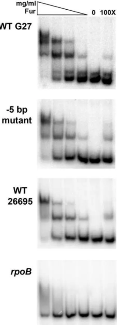
Figure 6. Fur binding to the sodB promoters. EMSAs were performed by incubating various concentrations of purified Fur with radiolabeled fragments of the WT G27, ‘‘25 bp swap,’’ and WT 26695
sodB promoters as well as the negative control promoter, rpoB, as detailed in the Materials and Methods. In the first four lanes, the Fur concentrations are indicated by the triangle from highest to lowest and range from 1.07mg/mL to 0.026mg/mL. A no protein control for each promoter is found in the fifth lanes. The last lane shows the 1006cold
(unlabeled) competition control for each promoter fragment, which were each performed with the highest concentration of Fur (1.07mg/ mL). Fur exhibits specific interaction with each of thesodBpromoters, and no interaction with therpoBpromoter except for very little non-specific binding at the highest Fur concentration. These data are representative of multiple independent EMSA experiments.
doi:10.1371/journal.pone.0005369.g006
Figure 7. Competitive Binding Studies. To assess the relative affinity of Fur for each of the sodB promoter fragments (WT G27, ‘‘25 bp swap,’’ and WT 26695), Fur was incubated with each radiolabeled promoter and 56, 106, or 256 the amount of
homologous or heterologous unlabeledsodBpromoter fragments as described in the Materials and Methods. For each labeled promoter, lane one contains a no competition control. Lanes two to four, five to seven, and eight to ten contain the competition EMSAs with unlabeled WT G27, ‘‘25 bp swap,’’ and WT 26695sodBfragments, respectively. The percent of labeled promoter that is outcompeted and remains unbound in each lane is given below each image. These data are representative of multiple independent experiments.
9 bp inverted repeat sequences separated by a single A nucleotide to create a 19 bp palindromic sequence as follows: GATAATGA-TAATCATTATC [33]. This sequence can also be interpreted as a series of three hexameric repeats of NATA/TAT [34]. However, in
H. pylori thisE. coli Fur Box is not conserved, and consensus is currently ill-defined. For iron-bound Fur regulation, the binding sequence occurs in A/T-rich regions in the target promoter oftentimes with repeats of AAT [8,24,25,35,36]. There is no defined consensus sequence forapo-Fur binding given that the two promoters of the known apo-Fur regulated genes, pfr and sodB, share only minimal homology [1,8]. In an organism that has about 60% A/T residues in its genome, a Fur Box consensus sequence that is comprised of mainly these two nucleotides does not seem to be an ideal approach for Fur regulation. Rather, inH. pyloriit is perhaps more plausible that both iron-bound andapo-Fur recognize unique DNA structures that are required for proper regulation of their target genes. The work presented here is the first to define a residue that is important forapo-Fur binding to thesodBtarget promoter. Future work from our group will focus on elucidating binding residues
important for both iron-bound andapo-Fur regulation with the hope that continued exploration of Fur regulation will provide greater understanding into the complexity of gene regulation in this important human pathogen.
Acknowledgments
We thank R. Peek for providing B128 isolate 7.13, C. Olsen for assistance with data presentation, R. Maier for providing the pET21A vector, A. vanVliet and J. Stoof for assistance with the EMSA protocol, E. Maynard and T. Dunn for assistance with protein purification, and members of the Merrell lab for help with cell harvesting.
Author Contributions
Conceived and designed the experiments: BMC HG DSM. Performed the experiments: BMC HG RPGN DSM. Analyzed the data: BMC JMW SLJM DSM. Contributed reagents/materials/analysis tools: ALW JMW SLJM. Wrote the paper: BMC SLJM DSM.
References
1. Ernst FD, Homuth G, Stoof J, Mader U, Waidner B, et al. (2005) Iron-responsive regulation of the Helicobacter pylori iron-cofactored superoxide dismutase SodB is mediated by Fur. J Bacteriol 187: 3687–3692.
2. Dunn BE, Cohen H, Blaser MJ (1997)Helicobacter pylori. Clin Microbiol Rev 10: 720–741.
3. Blaser MJ (1990) Helicobacter pylori and the pathogenesis of gastroduodenal inflammation. J Infect Dis 161: 626–633.
4. van Amsterdam K, van Vliet AH, Kusters JG, van der Ende A (2006) Of microbe and man: determinants ofHelicobacter pylori-related diseases. FEMS Microbiol Rev 30: 131–156.
5. Bereswill S, Lichte F, Vey T, Fassbinder F, Kist M (1998) Cloning and characterization of thefurgene fromHelicobacter pylori. FEMS Microbiol Lett 159: 193–200.
6. Bury-Mone S, Thiberge JM, Contreras M, Maitournam A, Labigne A, et al. (2004) Responsiveness to acidity via metal ion regulators mediates virulence in the gastric pathogenHelicobacter pylori. Mol Microbiol 53: 623–638.
7. Gancz H, Censini S, Merrell DS (2006) Iron and pH homeostasis intersect at the level of Fur regulation in the gastric pathogenHelicobacter pylori. Infect Immun 74: 602–614.
8. Delany I, Spohn G, Rappuoli R, Scarlato V (2001) The Fur repressor controls transcription of iron-activated and -repressed genes inHelicobacter pylori. Mol Microbiol 42: 1297–1309.
9. Spiegelhalder C, Gerstenecker B, Kersten A, Schiltz E, Kist M (1993) Purification of Helicobacter pylori superoxide dismutase and cloning and sequencing of the gene. Infect Immun 61: 5315–5325.
10. Seyler RW Jr, Olson JW, Maier RJ (2001)Superoxide dismutase-deficient mutants ofHelicobacter pyloriare hypersensitive to oxidative stress and defective in host colonization. Infect Immun 69: 4034–4040.
11. Wang G, Conover RC, Olczak AA, Alamuri P, Johnson MK, et al. (2005) Oxidative stress defense mechanisms to counter iron-promoted DNA damage in
Helicobacter pylori. Free Radic Res 39: 1183–1191.
12. Gutteridge JM, Maidt L, Poyer L (1990) Superoxide dismutase and Fenton chemistry. Reaction of ferric-EDTA complex and ferric-bipyridyl complex with hydrogen peroxide without the apparent formation of iron(II). Biochem J 269: 169–174.
13. Covacci A, Censini S, Bugnoli M, Petracca R, Burroni D, et al. (1993) Molecular characterization of the 128-kDa immunodominant antigen ofHelicobacter pylori
associated with cytotoxicity and duodenal ulcer. Proc Natl Acad Sci U S A 90: 5791–5795.
14. Tomb JF, White O, Kerlavage AR, Clayton RA, Sutton GG, et al. (1997) The complete genome sequence of the gastric pathogenHelicobacter pylori. Nature 388: 539–547.
15. Eaton KA, Morgan DR, Krakowka S (1989)Campylobacter pylorivirulence factors in gnotobiotic piglets. Infect Immun 57: 1119–1125.
16. Alm RA, Ling LS, Moir DT, King BL, Brown ED, et al. (1999) Genomic-sequence comparison of two unrelated isolates of the human gastric pathogen
Helicobacter pylori. Nature 397: 176–180.
17. Oh JD, Kling-Backhed H, Giannakis M, Xu J, Fulton RS, et al. (2006) The complete genome sequence of a chronic atrophic gastritisHelicobacter pyloristrain: evolution during disease progression. Proc Natl Acad Sci U S A 103: 9999–10004.
18. Carpenter BM, McDaniel TK, Whitmire JM, Gancz H, Guidotti S, et al. (2007) Expanding theHelicobacter pylorigenetic toolbox: modification of an endogenous plasmid for use as a transcriptional reporter and complementation vector. Appl Environ Microbiol 73: 7506–7514.
19. Copass M, Grandi G, Rappuoli R (1997) Introduction of unmarked mutations in theHelicobacter pylori vacAgene with a sucrose sensitivity marker. Infect Immun 65: 1949–1952.
20. Mehta N, Olson JW, Maier RJ (2003) Characterization ofHelicobacter pylorinickel metabolism accessory proteins needed for maturation of both urease and hydrogenase. J Bacteriol 185: 726–734.
21. Thompson LJ, Merrell DS, Neilan BA, Mitchell H, Lee A, et al. (2003) Gene expression profiling ofHelicobacter pylorireveals a growth-phase-dependent switch in virulence gene expression. Infect Immun 71: 2643–2655.
22. Datsenko KA, Wanner BL (2000) One-step inactivation of chromosomal genes inEscherichia coliK-12 using PCR products. Proc Natl Acad Sci U S A 97: 6640–6645.
23. Cormack BP, Valdivia RH, Falkow S (1996) FACS-optimized mutants of the green fluorescent protein (GFP). Gene 173: 33–38.
24. van Vliet AH, Stoof J, Poppelaars SW, Bereswill S, Homuth G, et al. (2003) Differential regulation of amidase- and formamidase-mediated ammonia production by theHelicobacter pylorifur repressor. J Biol Chem 278: 9052–9057. 25. Delany I, Spohn G, Pacheco AB, Ieva R, Alaimo C, et al. (2002) Autoregulation ofHelicobacter pyloriFur revealed by functional analysis of the iron-binding site. Mol Microbiol 46: 1107–1122.
26. Delany I, Spohn G, Rappuoli R, Scarlato V (2003) An anti-repression Fur operator upstream of the promoter is required for iron-mediated transcriptional autoregulation inHelicobacter pylori. Mol Microbiol 50: 1329–1338.
27. Pflock M, Muller S, Beier D (2007) The CrdRS (HP1365-HP1364) two-component system is not involved in pH-responsive gene regulation in the
Helicobacter pyloriStrains 26695 and G27. Curr Microbiol 54: 320–324. 28. Ayala G, Chihu L, Perales G, Fierros-Zarate G, Hansen LM, et al. (2004)
Quantitation ofH. pyloricytotoxin mRNA by real-time RT-PCR shows a wide expression range that does not correlate with promoter sequences. Microb Pathog 37: 163–167.
29. Gieseler S, Konig B, Konig W, Backert S (2005) Strain-specific expression profiles of virulence genes inHelicobacter pyloriduring infection of gastric epithelial cells and granulocytes. Microbes Infect 7: 437–447.
30. Gancz H, Jones KR, Merrell DS (2008) Sodium chloride affectsHelicobacter pylori
growth and gene expression. J Bacteriol 190: 4100–4105.
31. Rashid RA, Tarr PI, Moseley SL (2006) Expression of theEscherichia coliIrgA homolog adhesin is regulated by the ferric uptake regulation protein. Microb Pathog 41: 207–217.
32. Franco AT, Israel DA, Washington MK, Krishna U, Fox JG, et al. (2005) Activation of beta-catenin by carcinogenicHelicobacter pylori. Proc Natl Acad Sci U S A 102: 10646–10651.
33. de Lorenzo V, Wee S, Herrero M, Neilands JB (1987) Operator sequences of the aerobactin operon of plasmid ColV-K30 binding the ferric uptake regulation (fur) repressor. J Bacteriol 169: 2624–2630.
34. Escolar L, Perez-Martin J, de Lorenzo V (1998) Binding of the Fur (ferric uptake regulator) repressor ofEscherichia colito arrays of the GATAAT sequence. J Mol Biol 283: 537–547.
35. Delany I, Ieva R, Soragni A, Hilleringmann M, Rappuoli R, et al. (2005) In vitro analysis of protein-operator interactions of the NikR and Fur metal-responsive regulators of coregulated genes inHelicobacter pylori. J Bacteriol 187: 7703–7715. 36. Delany I, Pacheco AB, Spohn G, Rappuoli R, Scarlato V (2001) Iron-dependent transcription of the frpB gene of Helicobacter pylori is controlled by the Fur repressor protein. J Bacteriol 183: 4932–4937.