Article
Printed in Brazil - ©2017 Sociedade Brasileira de Química0103 - 5053 $6.00+0.00*e-mail: [email protected]
Antiproliferative Activity of Dibenzoylmethanes from Root Bark of
Muellera filipes
(
Benth) M.J. Silva & A.M.G. Azevedo
Érica L. Santos,a Juliana Jo,a Francisco A. Marques,a Ana Maria G. A. Tozzi,b
Ana Lúcia T. G. Ruizc and Beatriz Helena L. N. S. Maia*,a
aDepartamento de Química, Universidade Federal do Paraná, Centro Politécnico, 81531-990
Curitiba-PR, Brazil
bDepartamento de Biologia Vegetal, Instituto de Biologia, Universidade Estadual de Campinas,
13083-970 Campinas-SP, Brazil
cDivisão de Farmacologia e Toxicologia, Centro Pluridisciplinar de Pesquisas Químicas Biológicas
e Agrícolas (CPQBA), Universidade Estadual de Campinas, 13148-218 Paulínia-SP, Brazil
Three new dibenzoylmethane derivatives, (E )-3,4-methylenedioxy-2’-methoxy-2’’,2’’-dimethylpyrano-(5’’,6’’:3’,4’)-7-methoxychalcone, 2’-hydroxy-5’(3’’’’,3’’’’-dimethylallyl)-8-(α,α-dimethylallyl)-furano-(4’’,5’’:3’4’)-dibenzoylmethane and 2’-O -(3’’,3’’-dimethylallyl)-8-(α,α-dimethylallyl)-furano-(4’’,5’’:3’4’)-dibenzoylmethane, were isolated from a dichloromethane extract from the root bark of Muellera filipes (Benth.) M.J. Silva & A.M.G. Azevedo, along with two dibenzoylmethane derivatives, 2’-methoxy-8-(α,α -dimethylallyl)-furano-(4’’,5’’:3’,4’)-dibenzoylmethane and 2’-hydroxy-8-(α,α -dimethylallyl)-2”,2”-dimethylpyrano-(5”,6”:3’,4’)-dibenzoylmethane; one flavanone,isolonchocarpin and one flavone, pongaglabrone, which had been previously isolated from other sources. The molecular structures of the new dibenzoylmethane derivatives were determined by analysis of their spectral data (1H and 13C nuclear magnetic resonance (NMR), 2D-NMR, NOE (nuclear Overhauser effect) and HRMS (high resolution mass spectrometry). The known compounds were identified using 1D experiments and comparison with spectral data from the literature. 2’-O -(3’’,3’’-Dimethylallyl)-8-(α,α-dimethylallyl)-furano-(4’’,5’’:3’4’)-dibenzoylmethane displayed strong activity against the human cancer cell line OVCAR-3 (ovarian), and 2’-methoxy-8-(α,α -dimethylallyl)-furano-(4’’,5’’:3’,4’)-dibenzoylmethane showed high selectivity for cell lines NCI-H460 (lung) and UACC-62 (melanoma).
Keywords:Muellera filipes, Fabaceae, flavonoids, dibenzoylmethanes, antiproliferative activity
Introduction
In continuation of our studies on flavonoids from the species of Lonchocarpus (Fabaceae) occurring in Brazil, we examined Lonchocarpus filipes Benth., which, based on molecular data and morphological analysis, was reclassified as Muellera filipes (Benth.) M.J. Silva & A.M.G. Azevedo, together with Muellera montana A.M.G. Azevedo (previously Lonchocarpus montanus A.M.G. Azevedo).1,2
The genus Muellera L.f. has been treated as a synonym of Lonchocarpus Kunth (Fabaceae) and formerly included two species, M. moniliformis and M. fluvialis. Based on the new classification, this genus comprises 26 species, mainly in South America.2,3
Phytochemical data obtained for Lonchocarpus and the allied genera Muellera and Dahlstedtia allowed the characterization of many secondary metabolites, mainly flavonoid structural types.4,5 Dibenzoylmethane
is a β-diketone compound belonging to a small group of flavonoids that are rarely found in nature, but has been isolated from L. costaricensis, L. latifolius,D. floribunda (previously L. subglaucescens), D. muehlbergiana (previously L. muehlbergianus), D. glaziovii, M. fluvialis (previously L. fluvialis), M. montana and M. filipes.1,4,6-13
skin damage from UV radiation in sunlight.14,15 Significant
antitumor, antimalarial and trypanocidal activities have recently been reported for a number of dibenzoylmethanes and synthetic derivatives.16
We report here the isolation and characterization of three new dibenzoylmethane derivatives (1-3) together with four known compounds (4-7), from the root bark of M. filipes. The antiproliferative activity of the dibenzoylmethane derivatives was evaluated.
Experimental
General experimental procedures
1D and 2D nuclear magnetic resonance (NMR) data were recorded at 293 K in CDCl3 on a Brüker Avance DRX
400 operating at 9.4 T, observing 1H at 400.1 MHz and 13C at 100.6 MHz. Chemical shifts (d) are given in ppm
relative to TMS (d 0.00) as the internal standard. High resolution mass spectra (HRMS) were recorded on a Bruker Daltonics MicroQTOF mass spectrometer equipped with an electrospray ionization (ESI) source and GC-MS (gas chromatography-mass spectrometry) data were acquired in a Shimadzu GC-17A chromatograph interfaced with a MS-QP5050A mass spectrometer. Optical rotations were measured in CHCl3 solutions at room temperature on a
Jasco polarimeter model P-2000. The UV-Vis spectra were obtained in CHCl3 on a UV-2401PC (Shimadzu)
spectrophotometer system. Column chromatography (CC) separations were on silica gel 60 (70-230 mesh, Merck). Thin layer chromatography (TLC) was pre-coated silica gel plates (60 F254 Merck, 0.25 mm, aluminum) and
pre-coated silica gel plates (60 PF254 Merck, 1 mm, glass) were
used for preparative TLC. Compounds were detected by UV (λ = 254 and 366 nm) irradiation and with solution of anisaldehyde, followed by heating.
Plant material
The roots of M. filipes were collected in the Ecological Park, State University of Campinas (UNICAMP, Campinas-SP, Brazil) in September 2009. The plant was identified by Dr A. M. G. A. Tozzi from the Biology Institute of UNICAMP. A voucher specimen (A.M.G.A. Tozzi 143605) is deposited at the herbarium of UNICAMP.
Extraction and isolation
Dried and pulverized roots barks (137.1 g) of M. filipes were successively extracted with petroleum ether (50-60 °C), dichloromethane and methanol for 60 h in a Soxhlet
apparatus. Removal of the solvents from the extracts under reduced pressure gave the petroleum ether extract as viscous yellow oil (3.29 g), while the dichloromethane (5.27 g) yield amorphous brown solid and the methanol extract (22.45 g) gave a brown gum. Part of the dichloromethane extract (4.0 g) was fractionated by silica gel CC (0.063-0.200 mm, 50.0 × 4.0 cm) eluted first with petroleum ether. The eluent polarity was gradually increased by addition of ethyl acetate and then methanol to furnish 201 fractions (30 mL each) which were reduced to 32 groups after TLC analysis. Most of the compounds were found in four groups ranging from fractions 32 to 91. A sample of each was further fractionated by successive preparative TLC (silica gel) respectively run with petroleum ether:ethyl acetate (25:75), petroleum ether:dichloromethane (20:80) and petroleum ether:dichloromethane (10:90). Flavonoids were visualized under UV light (λ= 254 and 366 nm) and recovered from TLC plates by extraction with dichloromethane to furnish dibenzoylmethanes derivatives 1 (8.5 mg), 2 (10.0 mg),
3 (53.4 mg), 4 (58.2 mg), 5 (8.3 mg), 6 (4.5 mg) and
7 (3.6 mg).
Filipone A (1)
Viscous yellowish oil; silica gel 60 F254, petroleum
ether/ethyl acetate (20:80). λmax / nm 267.9, 319.0; 1H NMR
(400 MHz, CDCl3) d 7.40 (d, 1H, J 8.4 Hz, H-6’), 7.00 (dd,
1H, J 8.0, 1.6 Hz, H-6), 6.92 (d, 1H, J 1.6 Hz, H-2), 6.72 (d, 1H, J 8.0 Hz, H-5), 6.57 (d, 1H, J 10.0 Hz, H-4’’), 6.54 (d, 1H, J 8.4 Hz, H-5’), 6.24 (s, 1H, H-8), 5.94 (s, 2H, H-2’’’), 5.66 (d, 1H, J 10.0 Hz, H-3’’), 3.85 (s, 3H, R-OCH3), 3.80
(s, 3H, Ar-OCH3), 1.42 (s, 6H, H-2’’); 13C NMR (100 MHz,
CDCl3) d 190.0 (C-9), 170.2 (C-7), 156.7 (C-4’), 155.4
(C-2’), 148.9 (C-4), 146.9 (C-3), 131.1 (C-6’), 130.3 (C-3’’), 129.0 (C-1), 127.2 (C-1’), 123.7 (C-6), 116.6 (C-4’’), 114.5 (C-3’), 112.4 (C-5’), 109.6 (C-5), 107.6 (C-2), 101.9 (C-8), 101.2 (C-2’’’), 77.6 (C-2’’), 63.3 (Ar-OCH3), 56.4 (R-OCH3),
27.9 (2’’-CH3); HRMS (pESI) calcd. for C23H22O6 [M + Na]+:
417.1314; found: 417.1319.
Filipone B (2)
Viscous yellowish oil; [α]25
D −9.04 (c 0.36, CHCl3). λmax / nm 238.6, 348.9; 1H NMR (400 MHz, CDCl
3) d13.16
(s, 1H, OH), 7.95 (m, 2H, H-2 and H-6), 7.58 (d, 1H, 2.0 Hz, H-2’’), 7.56 (sl, 1H, H-6’), 7.53 (dt, 1H, J 7.2, 2.0 Hz, H-4), 7.42 (m, 2H, H-3 and H-5), 6.97 (d, 1H, J 2.0 Hz, H-3’’), 6.20 (dd, 1H, J 17.6, 10.8 Hz, H-2’’’), 5.54 (s, 1H, H-8), 5.33 (tq, 1H, J 7.2, 1.3 Hz, H-2’’’’), 5.03 (dd, 1H, J 17.6, 0.8 Hz, H-3a’’’), 4.98 (dd, 1H, J 10.8, 0.8 Hz, H-3b’’’), 3.50 (d, 2H, J 7.6 Hz, H-1’’’’), 1.79 (sl, 3H, 5’’’’-CH3),
1.73 (sl, 3H, 4’’’’-CH3), 1.33 (sl, 3H, 1’’’-CH3), 1.31 (sl,
194.0 (C-7), 158.5 (C-2’), 158.5 (C-4’), 144.4 (C-2’’), 137.9 (C-1), 134.1 (3’’’’), 133.2 (C-4), 145.8 (C-2’’’), 128.7 (C-6), 128.7 (C-2), 128.4 (C-3), 128.4 (C-5), 125.6 (C-6’), 120.9 (2’’’’), 117.5 (C-5’), 117.5 (C-3’), 114.6 (C-1’), 112.0 (C-3a’’’), 112.0 (C-3b’’’), 105.3 (C-3’’), 62.1 (C-8), 41.5 (C-1’’’), 27.3 (C-1’’’’), 26.3 (1’’’-CH3), 26.0 (1’’’-CH3),
25.7 (5’’’’-CH3), 17.8 (4’’’’-CH3); HRMS (pESI) calcd. for
C27H28O4 [M + Na]+: 439.1886; found: 439.1887.
Filipone C (3)
Amorphousyellowish powder; [α]25
D −4.82 (c 0.43,
CHCl3). λmax / nm 240.6, 387.0; 1H NMR (400 MHz, CDCl3)
d 8.01 (m, 2H, H-2 and H-6), 7.58 (d, 1H, J 2.2 Hz, H-2’’), 7.50 (tt, 1H, J 7.3, 1.2 Hz, H-4), 7.40 (m, 2H, H-3 and H-5), 7.40 (m, 1H, H-6’), 7.18 (dd, 1H, J 8.6, 0.9 Hz, H-5’), 6.91 (dd, 1H, J 2.2, 0.9 Hz, H-3’’), 6.09 (dd, 1H, J 17.4, 10.7 Hz, H-2’’’), 5.87 (s, 1H, H-8), 5.31 (tq, 1H, J 7.0, 1.3 Hz, H-2’’’’), 4.91 (dd, 1H, J 17.4, 1.1 Hz, H-3a’’’), 4.83 (dd, 1H, J 10.7, 1.1 Hz, H-3b’’’), 4.74 (m, 1H, H-1a’’’’), 4.65 (m, 1H, H-1b’’’’), 1.64 (s, 3H, 4’’’’-CH3), 1.49 (s, 3H,
4’’’’-CH3), 1.19 (s, 6H, 1’’’’-CH3); 13C NMR (100 MHz,
CDCl3) d 197.9 (C-9), 195.8 (C-7), 158.4 (C-4’), 151.3
(C-2’), 145.8 (C-2’’’), 144.7 (C-2’’), 139.3 (C-3’’’’), 138.7 (C-1), 132.5 (C-4), 128.8 (C-6), 128.8 (C-2), 128.3 (C-1’), 128.2 (C-5), 128.2 (C-3), 126.7 (C-6’), 119.1 (C-2’’’’), 118.7 (C-3’), 111.4 (C-3’’’), 106.8 (C-5’), 105.5 (C-3’’), 70.4 (C-1’’’’), 66.5 (C-8), 41.5 (C-1’’’), 26.1 (1’’’-CH3),
25.6 (5’’’’-CH3), 17.8 (4’’’’-CH3); HRMS (pESI) calcd. for
C27H28O4 [M + Na]+: 439.1886; found: 439.1915.
2’-Methoxy-furano-(2’’,3’’,4’,3’)-dibenzoylmethane (4)
Viscous yellowish oil; [α]20
D +28.31 (c 0.84, CH2Cl2).
Spectroscopic data matched well with literature.6
2’-Hydroxy-8-(α,α -dimethylallyl)-2”,2”-dimethylpyrano-(5”,6”:3’,4’)-dibenzoylmethane (5)
Yellow needles; [α]25
D −8.68 (c 0.80, MeOH).
Spectroscopic data identical to the literature.13
Isolonchocarpin (6)
Yellow needles. 1H and 13C NMR data were coincident
with the literature data.17
3’,4’-Methylenedioxy-furano-(2”,3”:7,8)-flavone (7)
Colorless crystals. Spectroscopic data were compared with the literature data.5
Antiproliferative activity
The pure compounds were evaluated for in vitro cytotoxic activities against human tumor cell lines MCF-7
(breast), NCI-H460 (lung), UACC-62 (melanoma), PC-3 (prostate), HT-29 (colon), K562 (leukaemia), OVCAR-3 (ovarian) and NCI-ADR/RES (ovarian expressing phenotype multiple drugs resistance) were provided by National Cancer Institute (NCI). Stock cultures were grown in medium containing 5 mL RPMI 1640 (GIBCO BRL) supplemented with 5% fetal bovine serum. Gentamicine (50 µg mL-1) was added to experimental cultures. Cells
in 96 well plates (100 µL cells well-1) were exposed to
sample concentrations in DMSO/RPMI (0.25, 2.5, 25 and 250 µg mL-1) at 37 °C, 5% of CO
2 in air for 48 h.
Doxorubicin was used as positive control. After 48 h of treatment, cells were fixed with 50% trichloroacetic acid and cell proliferation determined by spectrophotometric quantification (540 nm) of cellular protein content using sulforhodamine B assay and total growth inhibition (TGI) was extracted from concentration-response curves. The concentration-response curve was determined through non-linear regression analysis using Origin 8.0 (OriginLab Corporation) software.18,19
Results and Discussion
Previous studies identified one flavone and three dibenzoylmethane derivatives from L. filipes (M. filipes).1 In
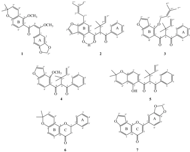
the present study, the dichloromethane extract of M. filipes root bark was fractionated and purified by a combination of chromatographic analyses (CC, TLC and preparative TLC). We describe the isolation and identification of one dibenzoylmethane derivative (1), four dibenzoylmethane derivatives with α,α-dimethylallyl (prenyl) substituent attached to the central carbon (C-8) (2-5), one flavanone (6), and one flavone (7) (Figure 1).
Compound 1 was obtained as a viscous yellowish oil. The 1H NMR spectrum showed the characteristic signals
for a 2,2-dimethylchromene moiety at d 1.42 (s, 6H, 2’’-CH3), 5.66 (d, 1H, J 10.0 Hz, H-3’’) and 6.57 (dd, 1H,
J 10.0, 0.5 Hz, H-4’’). This was confirmed in the 13C NMR
spectrum by the signals at d 27.9 (2’’-CH3), 77.6 (C-2’’),
130.3 (C-3’’) and 116.6 (C-4’’). The 2,2-dimethylchromene group located at ring B in the angular position, i.e., linked in C-3’/C-4’, was confirmed in the 1H NMR spectrum by the
signals of two ortho coupled hydrogens atd 6.54 (dd,1H, J 8.4, 0.8 Hz, H-5’) and 7.40 (d, 1H, J 8.4 Hz, H-6’). The methylenedioxy group was shown in the 1H NMR spectrum
(C-9) and 170.2 (C-7) in the 13C NMR were in accordance
with a non-enolizable dibenzoylmethane skeleton.20,21
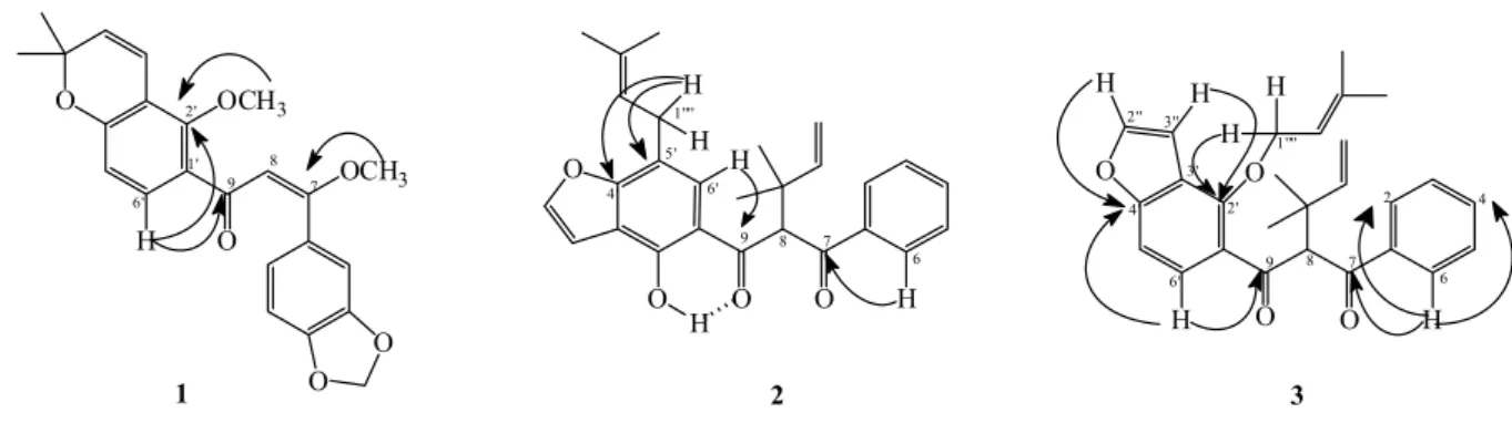
In the heteronuclear multiple bond correlation (HMBC) experiment (Figure 2), the correlations of the hydrogen signals at d 3.85 (7-OCH3) with the carbon at d170.2
(C-7), and of d 3.80 (2’-OCH3) with d 155.4 (C-2’) were
in agreement with the presence of a β-methoxyl group (7-OCH3) and a methoxyl group bonded to the benzene
ring (2’-OCH3), respectively. The location of a β-methoxyl
group at C-7 was further confirmed by correlations of the hydrogen signal at d 6.24 (H-8) with the carbon at d 170.2 (C-7). The stereochemistry E of double bond (C-7/C-8) was proposed based on the study carried out with the isomers of β-methoxychalcones.22 The correlations of the hydrogen
signal at d 7.40 (H-6’) with 190.0 (C-9) and 155.4 (C-2’) showed that the carbonyl and methoxyl groups are bonded to the tetrasubstituted ring B of β-methoxychalcone.
On the basis of this spectroscopic evidence, the structure of the new compound 1 (Figure 1) was established as (E)-3,4-methylenedioxy-2’methoxy-2’’,2’’-dimethylpyrano- (5’’,6’’:3’,4’)-7 methoxychalcone, named filipone A.
Compound 2 was also isolated as a viscous yellowish oil. In the 1H NMR spectrum, signals for a monosubstituted
aromatic ring [d 7.95 (m, 2H, H-2, H-6), 7.53 (dt, 1H, J 7.2, 2.0 Hz, H-4) and 7.42 (m, 2H, H-3, H-5)]; one
α,α-dimethylallyl group [d 6.20 (dd, 1H, J 17.6, 10.8 Hz, H-2’’’), 4.98 (d, 1H, J 10.8, 0.8 Hz, H-3b’’’), 5.03 (dd, 1H, J 17.6, 0.8 Hz, H-3a’’’), 1.31 (s, 3H) and 1.33 (s, 3H)]; one uncoupled methynic hydrogen [d 5.54 (s, 1H, H-8)]; one furan ring [d 6.97 (d, 1H, J 2.0 Hz, H-3’’) and 7.58 (d, 1H, J 2.0 Hz, H-2’’)]; and one hydrogen bonded by hydroxy group [d 13.16 (s, 1H, 2’-OH)] were observed. The isoprenyl group was confirmed in the 1H NMR spectrum
by the signals at d 3.50 (d, 2H, J 7.6 Hz, H-1’’’’), 5.33 (ts, 1H, J 7.2, 1.6 Hz, H-2’’’’), 1.73 (s, 3H, 4’’’’-CH3), and
1.79 (s, 3H, 5’’’’-CH3). The signals of two carbonyl groups
(d199.8 and 194.0) in the 13C NMR were in accordance
with a non-enolizable dibenzoylmethane skeleton.6 The 13C NMR spectrum and DEPT 135° indicated the presence
of 10 quaternary carbons, 11 CH, 2 CH2 and 4 CH3. In
the HMBC experiment (Figure 2), the correlations of the hydrogen signals at d 7.95 (H-2 and H-6) with the carbon at d 194.0, and d 7.56 (H-6’) with the carbon at d 199.8 confirmed the position of the carbonyl groups in C-7 and C-9, respectively. The hydrogen signal at d 3.50 (H-1’’’’) showed correlations with the carbons at d 117.5 (C-5’),
125.6 (C-6’) and 158.7 (C-4’) suggesting the presence of the isoprenyl group in the B ring benzoyl moiety. These data suggest two structural possibilities concerning the alternative isoprenyl group, located in C-3’ or C-5’ of the B ring. In the NOE (nuclear Overhauser effect) difference experiment (Figure 3), irradiation of the isoprenyl group hydrogens at H-1’’’’ (d 3.50) enhanced the signals at H-6’ (d 7.56) and H-5’’’’ (d 1.74), while irradiation of one aromatic hydrogen at H-6’ (d 7.56) further enhanced the same isoprenyl group signal at H-1’’’’ (d 3.50) and H-8 (d 5.54). Irradiation of the furan ring hydrogen at H-3’’ (d 6.97) enhanced the signal at H-2’’ (d 7.58). These findings are in agreement with structure 2 (Figure 3).
The MS spectrum showed the base peak at m/z 105 and a peak at m/z 229, which can be interpreted respectively as the C-7/C-8 and C-9/C-8 bond cleavage of a dibenzoylmethane, confirming the location of the α,α-dimethylallyl group on C-8. The presence of α,α-dimethylallyl was further confirmed by a peak at m/z 279 (Scheme 1). Therefore, the new compound 2 was determined as 2’-hydroxy-5’-(3’’’’,3’’’’-dimethylallyl)-8-(α,α ’-dimethylallyl)-furano-[4’’,5’’:3’,4’]-dibenzoylmethane, named filipone B.
Compound 3 was isolated as a yellow amorphous solid. Its 1H and 13C NMR spectra showed close similarities to
compound 2, in the presence of one α,α-dimethylallyl group, one furan ring and two carbonyl groups, shown in the
13C NMR spectrum by the signals at d 195.8 (C-7) and 197.9
(C-9). However, compound 3 has an O-prenyl group on the
B ring, shown in the 1H NMR spectrum by the signals at
d 4.74 (m, 1H, H-1’’’’), 4.65 (m, 1H, H-1’’’’), 5.31 (tq, 1H, J 7.0, 1.3, H-2’’’’), 1.49 (s, 3H, 4’’’’-CH3) and 1.64 (s, 3H,
5’’’’-CH3). This was confirmed in the 13C NMR spectrum
by the signals at d 70.4 (C-1’’’’), 119.1 (C-2’’’’), 139.3 (C-3’’’’), 17.9 (C-4’’’’) and 25.6 (C-5’’’’), respectively. In the HMBC experiment (Figure 2), the correlations of the hydrogen signals at d 7.58 (H-2’’) with the carbon at
d 158.4 (C-4’), d6.91 (H-3’’) with the carbon at d 118.7 (C-3’), 4.74 (H-1a’’’’), and 4.65 (H-1b’’’’) with the carbon at d 151.3 (C-2’), and of the signal at d 7.40 (H-6’) with the carbons at d 151.3 (C-2’), 158.4 (C-4’), and 197.9 (C-9) were in agreement with the tetrasubstituted ring B, while the signal at d 1.19 (1’’’-CH3) with the carbon at d 66.5
(C-8) confirmed the α,α-dimethylallyl group connected at C-8. The correlation of the hydrogen signal at d8.01 (H-6) with carbons at d 128.8 (C-2), 132.5 (C-4), and 195.8 (C-7) confirmed the presence of a monosubstituted ring A. These data suggest two possible isomers for structure 3. In the NOE difference experiment (Figure 3), irradiation of the O-prenyl group hydrogens at d 4.74 (H-1’’’’a) and 4.65 (H-1’’’’b) further enhanced the signal of furan ring hydrogen at d 6.91 (H-3’’) and methinic hydrogen of an
α,α-dimethylallyl group at d 1.19 (1’’’-CH3), suggesting
that the furan ring was connected at an angle to the B ring, with the O-prenyl group adjacent to the furan ring and located at carbon C-2’. In the MS spectrum, the fragmentation pathway is analogous to that of 2 (Scheme 1).
Figure 2. Key correlations of compounds 1-3, determined from the HMBC experiment.
On the basis of the above evidence, the structure of the new compound 3 was established as 2’-O- (3’’’’,3’’’’-dimethylallyl)-8-(α,α -dimethylallyl)-furano-(4’’,5’’:3’,4’)-dibenzoylmethane, named filipone C.
The known compounds (4-7) were characterized by comparison of the respective spectral data with those found in the literature.5,6,13,17
Dibenzoylmethanes can coexist with their equivalent with an α,α-dimethylallyl (prenyl) group affixed to the central carbon (C-8). Dibenzoylmethane derivatives (2,
3, 4 and 5) with asubstituent on C-8 are extremely rare in nature. They have been found only in the three closely related genera, Lonchocarpus, Dahlstedtia and Muellera, mainly in species from Brazil such as D. muehlbergiana,10
D. glaziovii,11M. montana,13 and M. filipes,1 except for
L. latifolius which occurs in Venezuela, Bolivia and Mexico,8 and M. fluvialis which occurs in Argentina,
Paraguay and Bolivia.12 The abundance of these compounds
in M. filipes reinforces its allocation together with Muellera montana in the new classification of the genera Lonchocarpus, Dahlstedtia and Muellera.2 The structures of
the most abundant dibenzoylmethanes (3 and 4) furnished by M. filipes are biosynthetically related.13,23,24 The presence
of compounds considered biomarkers in a small group of closely related genera can support inferences about biogenetic and evolutionary aspects, based on the metabolic pathways that govern the synthesis of these compounds.5,25
Dibenzoylmethane derivatives 3 and 4 were assayed for cytotoxicity, using eight human cancer cell lines: MCF-7 (breast), NCI-H460 (lung), UACC-62 (melanoma), PC-3
(prostate), HT-29 (colon), K-562 (leukemia), OVCAR-3 (ovarian) and NCI-ADR/RES (ovarian expressing a multiple drug resistant phenotype). The inhibitory effects of compounds 3 and 4 were assessed by varying the concentrations (0.60 to 600 µM) and the results were obtained after 48 h using the sulforhodamine B method. All assays were performed in triplicate, and the results were expressed as the concentration-dependent parameter TGI (concentration that produces total growth inhibition or cytostatic effect) (Table 1).
Compound 3 was selective for the OVCAR-3 (ovarian) cell line, but less selective than the positive control (doxorubicin), based on the TGI values (Table 1). On the other hand, compound 4 showed strong activity against the NCI-H460 (lung) cell line (TGI = 6.46 µM) and was selective for the K-562 (leukemia), OVCAR-3 (ovarian), NCI-ADR/RES (ovarian expressing a multiple drug resistant phenotype) and UACC-62 (melanoma) cell lines compared to the positive control (doxorubicin). This therapeutic activity against melanoma cells is particularly interesting for antimelanoma studies, because dibenzoylmethane derivatives can protect against UVA/UVB radiation, which plays a significant role in sunlight-induced skin damage.15,26 Compounds 1-2 and
5-7 were not tested for antiproliferative activity due to the small amount of each sample available.
Conclusions
Scrutiny of the structural variations may enable c h e m o t a x o n o m i c p r o p o s a l s t o b e d r aw n ; t h u s dibenzoylmethane derivatives with asubstituent on C-8 characterize some species of the genera Lonchocarpus, Dahlstedtia and Muellera of the family Fabaceae. The occurrence of these compounds in Muellera filipes reinforces its allocation together with Muellera montana in the new classification of these three closely related genera, based on the phylogenetic and morphological analyses. Compounds 3 and 4 showedhigh selectivity against human tumor cell lines, and compound 4 is a candidate for further study as a sunscreen against UVA/UVB radiation.
Table 1. Total growth inhibition (TGI) (µM) of dibenzoylmethane derivatives 3 and 4 against human cancer cells
Compound Cell lines
UACC-62 MCF7 NCI-ADR/RES NCI-H460 PC-3 OVCAR-3 HT-29 K562 VERO
3 > 600 > 600 > 600 > 600 > 600 52.35 > 600 > 600 > 600
4 91.67 > 600 85.43 6.46 > 600 125.96 > 600 32.80 > 600
Doxorubicina 0.33 1.84 > 45 1.42 2.30 8.11 > 45 0.55 > 45
aReference drug (positive control). Human cancer cell lines: UACC-62 (melanoma); MCF-7 (breast); NCI-ADR/RES (ovarian expressing multiple drug resistant phenotype); NCI-H460 (lung); PC-3 (prostate); OVCAR-3 (ovarian); HT-29 (colon); K-562 (leukemia); VERO (renal, normal cells, green monkey).
Supplementary Information
1D and 2D NMR spectra and HRMS data for compounds 1-3 are available free of charge at http://jbcs. sbq.org.br as PDF file.
Acknowledgments
The authors are grateful to Conselho Nacional de Desenvolvimento Científico e Tecnológico (CNPq) for a scholarship and financial support.
References
1. Santos, E. L.; Costa, E. V.; Marques, F. A.; Vaz, N. P.; Magalhães, E. G.; Tozzi, A. M. A.; Maia, B. H. L. N. S.; Quim. Nova2009, 9, 2255.
2. da Silva, M. J.; Queiroz, L. P.; Azevedo-Tozzi, A. M. G.; Lewis, G. P.; Sousa A. P.; Taxon 2012, 61, 93.
3. da Silva, M. J.; Azevedo-Tozzi, A. M. G.; Phytotaxa2011, 29, 41.
4. Cassidy, C. E.; Setzer, W. N.; Nat. Prod. J.2011, 1, 75. 5. Garcez, F. R.; Scramin, S.; do Nascimento, M. C.; Mors, W.
B.; Phytochemistry 1988, 27, 1079.
6. Magalhães,A. F.; Tozzi, A. M. G. A.; Magalhães, E. G.; Blanco, I. S.; Nogueira, M. A.; Phytochemistry1997, 46, 1029. 7. Waterman, P. G.; Mahmoud, E. N.; Phytochemistry1985, 24,
571.
8. Magalhães, A. F.; Tozzi, A. M. A.; Magalhães, E. G.; Nogueira, M. A.; Queiroz, S. C. N.; Phytochemistry2000, 55, 787. 9. Magalhães, A. F.; Azevedo-Tozzi, A. M. G.; Maia, B. H. L. N.
S.; Magalhães, E. G.; Phytochemistry1996, 42, 1459. 10. Magalhães, A. F.; Tozzi, A. M. G. A.; Magalhães, E. G.; Blanco,
I. S.; Soriano, M. P. C.; An.Acad. Bras. Cienc.2004, 76, 651. 11. Canzi,E. F.; Marques, F. A.; Teixeira, S. D.; Tozzi, A. M. G.
A.; Silva, M. J.; Duarte, R. M. T.; Ruiz, A. L. T. G.; Monteiro, P. A.; Carvalho, J. E.; Maia, B. H. L. N. S.; J. Braz. Chem. Soc.
2014, 6, 995.
13. Magalhães, A. F.; Tozzi, A. M. G. A.; Magalhães, E. G.; Sannomiya, M.; Soriano, M. P. C.; Perez, M.; An. Acad. Bras. Cienc.2007, 79, 351.
14. Nogueira, M. A.; Magalhães, E. G.; Magalhães, A. F.; Biloti, D. N.; Laverde, A.; Pessine, F. B.; Carvalho, J. E.; Kohn, L. K.; Antônio, M. A.; Marsaioli, A. J.; Il Farmaco2003, 58, 1163. 15. Atarashi, K.; Takano, M.; Kato, S.; Kuma, H.; Nakanishi, M.;
Nakanishi, M.; Tokura, T.; J. Photochem. Photobiol. B 2012,
113, 56.
16. Santos, D. A. P.; Braga, P. A. C.; Silva, M. F. G. F.; Fernandes, J. B.; Vieira, P. C.; Magalhães, A. F.; Magalhães, E. G.; Marsaioli, A. J.; Moraes, V. R. S.; Rattray, L.; Croft, S. L.; J. Pharm. Pharmacol. 2009, 61, 257.
17. Rao, E. V.; Raju, N. R.; Phytochemistry1979, 18, 1581. 18. Shoemaker, R. H.; Nat. Rev. Cancer2006, 6, 813.
19. Spindola, H. M.; Carvalho, J. E.; Ruiz, A. L. T. G.; Rodrigues, R. A. F.; Denny, C.; Sousa, I. M. O.; Tamashiro, J. Y.; Foglio, M. A.; J. Braz. Chem. Soc.2009, 20, 569.
20. Parmar, V. S.; Rathore, J. S.; Jain, R.; Malone, J. F.;
Phytochemistry1989, 28, 591.
21. Fukai, T.; Nishizaka, J.; Nomura, T.; Phytochemistry1994, 35, 515.
22. Kiuchi, F.; Chen, X.; Tsuda, Y.; Chem. Pharm. Bull. 1990, 38, 1862.
23. Gottlieb, O. R.; Micromolecular Evolution, Systematic and Ecology - An Essay into a Novel Botanical Discipline, 1st ed.; Springer-Verlag Berlin Heidelberg: Berlin, German, 1982. 24. Chamberlajin, T. R.; Collins, J. F.; Grundon, M. F.; Chem.
Commun.1969, 1269.
25. Wink, M.; Phytochemistry2003, 64, 3.
26. Simeoni, S.; Scalia, S.; Benson, H. A. E.; Int. J. Pharm.2004,
280, 163.