A bacteriological study of hospital beds
before and after disinfection with phenolic
disinfectant
1
Denise de Andrade,
2Emília L. S. Angerami,
2and Carlos Roberto Padovani
3In hospitals, one of the ways to control microbial contamination is by disinfecting the furni-ture used by patients. This study’s main objective was to evaluate the microbiological condi-tion of hospital mattresses before and after such disinfeccondi-tion, in order to identify bacteria that are epidemiologically important in nosocomial infection, such as Staphylococcus aureusand
Pseudomonas aeruginosa.RODAC plates with two different culture media were used to collect specimens. Patient beds were selected according to previously established criteria, and surface areas on the mattresses were chosen at random. From the total of 1 040 plate cultures from 52 mattresses, positive results were obtained from 500 of them (48.1%), 263 before dis-infection and 237 after disdis-infection. Considering the selectivity of the culture media, the posi-tivity rate was high. There were high prevalences of S. aureusboth before and after mattress disinfection. The study results suggest that the usual disinfection procedures, instead of di-minishing the number of microbes, merely displace them from one part of the mattress to an-other, and the number of microorganisms remains the same.
ABSTRACT
Within the hospital environment, inanimate objects can provide
oppor-tunities for contact with and trans-mission of microorganisms. Hospital disinfection can help in breaking the epidemiological chain of infection. However, many in the scientific med-ical community consider the inani-mate environment to be of less impor-tance in the incidence of hospital infections, and some professionals have confused “less important” with “irrelevant.” Nevertheless, the inani-mate environment plays a less impor-tant role only after basic environmen-tal pollution control measures are established, that is, when minimal cleanliness and disinfection standards are followed (1).
It has long been recognized that there is an urgent need to carry out
studies that can help improve the quality of care, as well as lower the rate of nosocomial infections and the costs of hospitalization. However, the fight against nosocomial infections is a complex problem that produces many disputes and disagreements.
The nursing profession has tried to direct its activities toward diminishing the possible risks that trigger hospital infections. One of the activities that nurses perform daily to keep the hos-pital environment biologically safe is the disinfection of patient “units.” The patient unit has been defined as the aggregate spaces and pieces of furni-ture for use by each patient. While its components vary from one hospital to another, the unit is basically made up
1 Adapted from: Angelo, D de AD. A
manuten-ção de um ambiente hospitalar biologicamente seguro: avaliação microbiológica dos leitos de um hospital geral antes e depois de sua limpeza termi-nal. [Maintaining a biologically safe hospital envi-ronment: microbiological evaluation of the hospi-tal beds in a general hospihospi-tal before and after terminal disinfection] [doctoral thesis]. Ribeirão Preto, São Paulo, Brasil: Escola de Enfermagem de Ribeirão Preto da Universidade de São Paulo; 1998.
2 Departamento de Enfermagem Geral e
Especia-lizada da Escola de Enfermagem de Ribeirão Preto da Universidade de São Paulo, Ribeirão Preto, São Paulo, Brasil. Send correspondence to: Denise de Andrade, Department of General and Specialized Nursing, Av. Bandeirantes 3900, Campus da USP, 14040-902, Ribeirão Preto, SP, Brasil; e-mail: dan-drade @glete.eerp.usp.br
3 Instituto de Biociências do Campus da
of a bed, mattress, chair, step stool, bell, and night table with items for per-sonal use (2–4).
The traditional nursing literature recognizes two types of unit disinfec-tion, regular and terminal. Regular disinfection refers to the disinfection performed daily on some parts of the unit and on personal objects after their use. In contrast to this, terminal disin-fection of a unit involves all of the unit elements and is done when the patient vacates his bed after he is discharged from the hospital, dies, is transferred, or completes isolation treatment. This type of disinfection is also recommended when a patient has occupied a bed for a long period of time.
In general, both types of disinfection are meant to remove dirt and to impede the spread of microorganisms that col-onize the horizontal surfaces of furni-ture. These microorganisms include
Staphylococcus aureus, Clostridium diffi-cile, Pseudomonas spp., Proteus spp., Ser-ratia marcescens, andCandidaspp. (5).
When disinfecting the patient unit, the use of chemical products with effi-cacious germicidal action has been rec-ommended to remove and destroy surface microorganisms. Due to the use of germicidal substances, the ter-minology related to “cleaning” the pa-tient unit has been changed to “disin-fecting,” as evidenced in the literature (6–8).
In Brazil, the disinfection products for hospital use must contain phenolic active ingredients, or organic or inor-ganic compounds that release active chlorine, or ammonia or alcohol qua-ternaries, or other ingredients comply-ing with current legislation (1, 7, 9).
The hospital environment offers ex-cellent conditions for the propagation of microorganisms, in spite of disinfec-tants, antibiotics, and chemotherapeu-tics. In addition, with their immune systems weakened by illness, surgery, or accidents, patients generally make good hosts for microorganisms.
Further evidence of the need to maintain a biologically safe hospital environment comes from the work by Angelo et al. (10). It showed that in 23 hospitals in the interior of the state of São Paulo, disinfection activities were
characterized by a lack of investment in technology, a variety of procedures, and inadequate attention to the scien-tific principles considered standard in the literature.
Despite the diversity of pathogens involved in hospital infections, a great number of medical studies center on
S. aureus and Pseudomonas aeruginosa,
which both play important roles in the incidence of hospital infections during all times of the year. These two mi-croorganisms appear as contagious agents harbored by health profession-als and by hospital and medical arti-cles, equipment, and other inanimate objects.
The possibilities of a person acquir-ing an infection depend on various factors. Specifically in relation to the infective dose of S. aureus, Marples (11) showed that an inoculation of more than 106 colony-forming units
(CFUs) can produce pus in healthy skin and that the same is possible with a much lower dose (102 CFUs) in
trau-matized or occluded skin. In the body,
S. aureus is harbored mainly in the an-terior region of the nasal cavities; how-ever, it can also colonize skin lesions (surgical, burns, eczemas, decubitus ulcers, and others), the perineum, and the rectum.
Hands have always been considered an important body area from which to obtain samples of S. aureus and other microorganisms. In hospitals, hands are recognized as one of the chief car-riers of bacteria from an infected pa-tient to another papa-tient or to a care-giver (12–14).
It is generally considered improba-ble that the microorganisms present in patient secretions, blood, or other exu-dates would contaminate the sur-rounding air, as the microorganisms are restricted to the surfaces where they are deposited. We believe that, of all hospital surfaces, mattresses have the greatest possibility of holding or-ganic matter and microorganisms. Nevertheless, a study by Mendonça (15) indicated that the patient contam-inates his immediate environment. In a bacteriological analysis the same phage type was found on patient bed linens and on other patient-unit
com-ponents at various distances. The nearer to the patient, the greater was the concentration of microorganisms.
Given that positive relationship of distance and concentration of microor-ganisms, we believe that the mattress is the element within the patient unit that can harbor the greatest concentra-tion of pathogenic agents. The mat-tress is the unit element with which the patient has the most contact, espe-cially taking into account that the bed linens are changed daily. In addition, the mattress serves as a depository for organic and inorganic impurities.
Following this line of thinking, we formulated a comparative study on the microbiological condition of hospital mattresses before and after terminal disinfection of the patient unit, with a view toward improving terminal-disinfection procedures. Because of the diversity of microorganisms that can be isolated from hospital mat-tresses, we decided to focus our atten-tion on S. aureusand P. aeruginosa. Our study had two specific objectives: 1) to measure contamination of mattresses by S. aureus and P. aeruginosa before and after terminal disinfection of the patient unit and 2) to evaluate the ef-fectiveness of the disinfection proce-dure in terms of changes in microbial density.
MATERIALS AND METHODS
The present study was carried out in 1998 in a general public teaching and research hospital in a city located in the interior of the state of São Paulo, Brazil. In light of our interest in evalu-ating the microbiological conditions of mattresses in that hospital before and after terminal disinfection of the pa-tient unit, we chose a random sample of hospital service areas. Nursing staff members do the disinfection in those areas, following procedures recom-mended in the literature (3, 4, 8). Ter-minal cleaning is structured according to basic principles of disinfection. In short, it is a manual procedure, with actions that are mechanical (friction) and chemical (disinfectant solution and phenol detergent). The phenol
used is a combination of two phenols (paratertiary butyl phenol and ortho-benzil parachlorophenol), which was applied following manufacturer’s rec-ommendations for its effective antimi-crobial action, including preparation of the solution, concentration, and length of exposure.
Of the 258 active beds in the service units investigated, we took samples from 52 mattresses, or approximately 20% of the total. We collected samples from each mattress before and after terminal disinfection of the patient unit. We waited approximately 60 minutes before collecting the after-disinfection samples since phenol has residual effects that are an important factor in reducing the microbial load.
The 52 mattresses sampled included two types of foam mattresses, 18 mat-tresses covered with cotton fabric and 34 covered with plastic.
Specimens from the mattresses were collected on RODAC™ (Replicate Or-ganism Detection and Counting) plates, which are recommended in many stud-ies for quantification of microbial sur-face contamination. Each plate has a ca-pacity of 15 to 20 mL, with 16 mL as the ideal volume (16). The RODAC plates were primed with one of two culture media, either Bacto Chapman Stone medium (“CH”) or Bacto cetrimide agar medium (“CT”) (17).
Bacto Chapman Stone medium is in-dicated for the isolation and quantifi-cation of Staphylococcus colonies. We used it to count the colonies of Staphy-lococcusand also to verify the presence of S. aureus.
Bacto cetrimide agar medium is used to isolate and culture P. aerugi-nosa. That culture medium typically inhibits the growth of Escherichia coli
and of S. aureus while producing an excellent growth of P. aeruginosa, along with a change of color in the medium to greenish blue or greenish yellow, due to the presence of two pigments, pyocyanin (green) and fluorescein (yellow).
Considering the study objectives, materials used for data collection, and the mattress dimensions, we found it necessary to establish a representative sampling approach. Given the total
size of the mattress and the dimen-sions of the RODAC plates, we di-vided each mattress into 136 quad-rants, from which samples were chosen by lot.
Specimens were collected from the side of the mattress with which the pa-tient had had the most contact. We placed the culture plates with their re-spective media in the places selected by lot and pressed the plates lightly against the mattress for one minute. For each of the 52 mattresses, we per-formed the procedure five times be-fore terminal disinfection and five times after terminal disinfection, with both of the culture media, for a total of 1 040 cultures.
In the laboratory, the plates were in-cubated for 48 hours at 37 °C. All the plates, with their respective culture media, were analyzed for odor, mor-phology of the colonies, color, size, height, borders, and number of colonies. In order to facilitate and stan-dardize the readings, we established three different levels of macroscopic growth: absent, countable, and un-countable. Plates that showed no macroscopic growth of colonies were categorized as “absent,” plates with macroscopic growth of up to 130 colonies were labeled as “countable,” and plates showing macroscopic growth of more than 130 colonies were categorized as “uncountable.”
The plates with cetrimide culture medium underwent general observa-tions and counts and were also checked regularly for changes of color and odor, since this culture medium turns greenish and acquires a sweetish smell in the presence of P. aeruginosa.
The plates with Chapman culture medium were submitted to counts of the total colonies and analysis of the colonies as to their size and appear-ance. All species of Staphylococcuswill develop in that culture medium, but only S. aureus produces fermentation of the mannitol that is in that medium, so the colonies have a different appear-ance. To ascertain that the colonies were S. aureus, we tested for coagulase, using the technique recommended by Blair (18) and Bayliss and Hall (19). The most generally accepted
explana-tion for the coagulase-test reacexplana-tion is that S. aureusis capable of producing coagulase, an “enzyme-like” protein that activates the prothrombin present in serum, transforming it into throm-bin, which in turn acts upon fibrinogen to produce fibrin clots.
For our statistical analysis we ap-plied the Goodman test of confidence intervals for multinomial proportions (20), using a computer program pro-duced by Curi and Moraes (21), to compare, for each of the two types of mattress coverings and two culture media, the proportions of changes in the number of colonies before and after terminal disinfection.
For our analysis, we considered the type of mattress and the culture medium and then generated three classification categories: “worsening” (the number of colonies increased), “holding” (the number of colonies stayed the same), and “improving” (the number of colonies decreased).
Results where the comparisons were statistically significant (P< 0.05) were annotated in capital letters next to the observed values. Two proportions with at least one letter in common indicate that there was no significant difference in regard to changes in the number of colonies after terminal disinfection.
RESULTS
Frequency of positive and negative cultures according to culture media before and after terminal
disinfection of mattresses
Out of the total of 1 040 plates used to collect specimens from the 52 mat-tresses, 500 of the plates (48.1%) were positive, that is, produced the growth of colonies (Table 1). Of these 500 posi-tive cultures, 263 were gathered before disinfection and 237 were gathered after disinfection. These results did not show a significant difference between positive values before and after termi-nal disinfection.
the mattresses, 241 of them (92.7%) had positive cultures. Of the 260 Chap-man plates used after terminal disin-fection, 210 (80.8%) had positive cul-tures. Thus, in contrast to the total
number of positive cultures obtained before and after disinfection, with the Chapman plates there was a signifi-cant reduction (P < 0.0001) in positiv-ity from before to after disinfection of the mattresses.
Out of the total of the 451 Chapman plates that had positive cultures, 286 (63.4%) showed positive results for
S. aureus. Of those 286, 150 were from cultures done before disinfection and 136 were from cultures done after dis-infection, a difference that was not sta-tistically significant.
Taken together, the results with the Chapman culture medium give the impression of there only being a bet-ter distribution of the microbial load on the mattress after disinfection, in-stead of a decrease in the number of microbes.
Similarly, the differences found with the cetrimide culture medium
be-fore and after disinfection were not significant.
In addition, in the culture-positive plates containing cetrimide, we did not find growth of pigment-produc-ing P. aeruginosa either before or after disinfection of the mattresses. It is possible that the colonies that did ap-pear in the cetrimide culture medium were strains of Pseudomonas without pigment or were some other gram-negative bacteria that does not pro-duce changes in the color of this cul-ture medium.
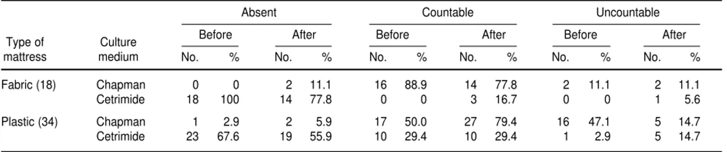
Number of mattresses with absent, countable, and uncountable microorganism colonies before and after terminal disinfection
The 52 beds were divided into three groupings according to the number of microorganism colonies found before and after terminal disinfection: absent (no macroscopic growth of colonies), countable (macroscopic growth of up to 130 colonies), and uncountable
(macroscopic growth of more than 130 colonies). For the 18 fabric mattresses sampled with the Chapman culture medium, 16 (88.9%) had countable colonies before terminal disinfection and 14 (77.8%) had countable colonies after terminal disinfection (Table 2).
Also with the Chapman culture medium, there were similar percent-ages of the two types of mattresses with countable colonies after terminal disinfection.
The cetrimide agar culture medium produced results that conflicted with those obtained with the Chapman medium. For the 18 fabric mattresses, all 18 (100%) were in the “absent” cat-egory before terminal cleaning, and 14 (77.8%) were in that grouping after ter-minal cleaning. For the 34 plastic mat-tresses, 23 (67.6%) were in the “absent” category before terminal cleaning, and 19 (55.9%) were in that grouping after terminal cleaning.
The results of positivity with the cetrimide medium were considered low. However, there were some trou-bling variations before and after disin-fection. We did not find any “count-able” or “uncount“count-able” colony growth with any of the 18 fabric mattresses before disinfection. Nevertheless, af-ter disinfection, 3 of those mattresses (16.7%) had countable colonies and 1 of them (5.6%) had uncountable colonies of microorganisms. However, this change was not statistically signifi-cant. In the case of the 34 plastic mat-tresses, with disinfection the number of mattresses from which colonies were absent changed from 23 to 19, the number with countable colonies re-mained at 10, and the number with
182 Andrade et al. • Hospital beds before and after disinfection
TABLE 1. Number and percent of positive and negative cultures from 52 beds before and after terminal disinfection of mat-tresses, for Chapman (CH) and cetrimide (CT) culture media, Brazil, 1998
Before disinfection After disinfection
CH CT CH CT
Culture No. % No. % No. % No. %
Positive 241 92.7 22 8.5 210 80.8 27 10.4 Negative 19 7.3 238 91.5 50 19.2 233 89.6
Total 260 260 260 260
TABLE 2. Number and percent of mattresses with absent, countable, and uncountable microorganism colonies before and after terminal disinfection, Brazil, 1998
Absent Countable Uncountable
Before After Before After Before After
No. % No. % No. % No. % No. % No. %
Fabric (18) Chapman 0 0 2 11.1 16 88.9 14 77.8 2 11.1 2 11.1
Cetrimide 18 100 14 77.8 0 0 3 16.7 0 0 1 5.6
Plastic (34) Chapman 1 2.9 2 5.9 17 50.0 27 79.4 16 47.1 5 14.7
Cetrimide 23 67.6 19 55.9 10 29.4 10 29.4 1 2.9 5 14.7
Type of mattress
uncountable colonies increased from 1 to 5.
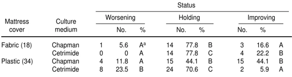
Frequency of changes in colony status after terminal disinfection of mattresses
We compared the number of colonies before and after disinfection of the 52 mattresses for the two culture media. We used those data to assess the number and frequency of changes in that status in terms of the three
cat-egories mentioned earlier: worsening, holding, and improving (Table 3).
Using Goodman’s test we obtained the following results by comparing values for the categories (P < 0.05):
Plastic CT: Holding > Worsening > Improving
Plastic CH: (Holding = Improving) > Worsening
Fabric CT: Holding > Improving > Worsening
Fabric CH: Holding > (Improving > Worsening)
It can thus be seen that, in general, a holding status was the most frequent result for both culture media and both types of mattress coverings. In other words, it appears that the disinfection procedures used did not change the microbial load on the mattresses.
CONCLUSIONS
Our study has brought up important considerations related to the disinfec-tion procedure for hospital mattresses and has led to reflections on the possi-bilities of contagion, especially if we re-ally want to ensure maximum effi-ciency in that disinfection procedure.
Recognizing the part that patients, the environment, and contacts play in the epidemiological chain leading to hospital infections, we health profes-sionals urgently need to break the links in that chain of contagion, using the means of prevention and control appropriate to each specific situation. We will thus take a giant step in re-ducing the spread of microorganisms. TABLE 3. Changes in the status (number and percent) of microorganism colonies with
ter-minal disinfection of the mattresses, Brazil, 1998
Status
Worsening Holding Improving
No. % No. % No. %
Fabric (18) Chapman 1 5.6 Aa 14 77.8 B 3 16.6 A
Cetrimide 0 0 A 14 77.8 C 4 22.2 B
Plastic (34) Chapman 4 11.8 A 15 44.1 B 15 44.1 B
Cetrimide 8 23.5 B 24 70.6 C 2 5.9 A
aTwo proportions with at least one letter in common indicate there was no significant difference in regard to changes in the
number of colonies after terminal disinfection.
Culture medium Mattress
cover
1. Scarpitta CRM. Limpeza e desinfecção de arti-gos hospitalares — limpeza e desinfecção de áreas hospitalares. In: Rodrigues EAC et al. Infecções hospitalares — prevenção e con-trole. São Paulo: Sarvier; 1997. p. 411–421. 2. Fuerst EV, Wolff L, Weitzel MH.
Fundamen-tos de enfermagem — o humanitarismo e as ciências na enfermagem. 5th ed. São Paulo: In-teramericana; 1977.
3. Kawamoto EE, Fortes JI. Fundamentos de en-fermagem. São Paulo: Editora Pedagógica e Universitária/EPU; 1986.
4. Mussi NM et al. Técnicas fundamentais de en-fermagem. São Paulo: Atheneu; 1995. 5. Pannuti CS. A importância do meio ambiente
hospitalar. In: Rodrigues EAC et al. Infecções hospitalares — prevenção e controle. São Paulo: Sarvier; 1997. pp. 449–454.
6. Brasil, Ministério da Saúde. Processamento de artigos e superfícies em estabelecimentos de saúde. 2nd ed. Brasília: Coordenação de Con-trole de Infecções Hospitalares, Ministério da Saúde; 1994.
7. Brasil, Ministério da Saúde. Portaria n. 2616. Diário Oficial da União, 1998 12 Maio. 8. Ferraz AEP et al. Introdução à enfermagem.
Ribeirão Preto: Centro Interescolar do Hospi-tal das Clínicas da Faculdade de Medicina de Ribeirão Preto; 1993.
9. Pedrosa TMG, Macedo RM. Desinfecção e es-terilização química e líquida. In: Couto RC et
al. Infecção hospitalar — epidemiologia e con-trole. São Paulo: MEDSI; 1997. pp. 203–217. 10. Angelo D de AD, Angerami ELS, Oliveira
Santos BM, Bispo A. Avaliação da limpeza de unidade terminal do paciente em hospitais do interior do estado de São Paulo. In: Anais do Congresso Brasileiro de Controle de In-fecção Hospitalar. Rio de Janeiro, Brasil; 1996. p. 111.
11. Marples RR. Local infections — experimental aspects. Journal of the Society of Cosmetic Chemists 1976;27:449–457.
12. Larson L, McGinley K, Grove G, Lalbot G. Physiologic and microbiologic changes in skin related to frequent hand washing. Infect Con-trol 1986;7(2):59–63.
13. Oliveira Santos BM, Aguilar OM, Takakura MS. Colonização simultânea de Staphylococcus aureusna cavidade nasal e mãos de portadores sãos de um hospital escola. Rev Microbiol 1990;21(4):309–314.
14. Corrêa I. Avaliação do procedimento da lavagem das mãos no plano assistencial à criança portadora de diarréia aguda bacte-riana [doctoral thesis]. Campinas, São Paulo, Brasil: Universidade de Campinas; 1995. 15. Mendonça CP. Estudos sobre Staphylococcus
aureus (portadores e infecções hospitalares) num hospital geral de Araraquara, S.P., 1964– 1975 [“livre-docência” thesis]. Araraquara, São Paulo, Brasil: Faculdade de Farmácia e
Odontologia de Araraquara, Universidade Estadual Paulista; 1976.
16. Hall LB, Harnett MJ. Measurement of the bac-terial contamination on surfaces in hospitals. Public Health Rep 1964;79:1021–1024. 17. Difco Laboratories. Difco manual: dehydrated
culture media and reagents for microbiology. 10th ed. Detroit, Michigan: Difco Laborato-ries; 1984.
18. Blair JE. Laboratory diagnosis of staphylococ-cal infections. Bull World Health Organ 1958; 18;(3):291–307.
19. Bayliss BG, Hall ER. Plasma coagulation by organism other than Staphylococcus aureus. J Bacteriol 1965; 89(11):101–105.
20. Goodman LA. On simultaneous confidence intervals for multinomial proportions. Tech-nometrics 1965; 7(2): 247–254.
21. Curi PR, Moraes RV. Associação, homogenei-dade e contrastes entre proporções em tabelas contendo distribuições multinomiais. Ciência e Cultura 1981;33(5):712–722.
Manuscript received on 1 December 1998. Revised ver-sion accepted for publication on 27 August 1999.
En los hospitales, una de las formas de controlar la contaminación microbiana es la desinfección de los muebles y utensilios utilizados por los pacientes. El principal ob-jetivo del presente estudio consistió en evaluar las condiciones microbiológicas de los colchones de hospital antes y después de la desinfección, a fin de identificar bacterias epidemiológicamente importantes en las infecciones nosocomiales, tales como Staphy-lococcus aureusy Pseudomonas aeruginosa.Para recolectar las muestras se utilizaron pla-cas RODAC con dos medios de cultivo diferentes. Las camas de los pacientes fueron seleccionadas según criterios establecidos anteriormente y las áreas de la superficie de los colchones fueron elegidas aleatoriamente. De un total de 1 040 placas de cultivo procedentes de 52 colchones, 500 (48,1%) proporcionaron resultados positivos: 263 antes de la desinfección y 237 después de ella. Teniendo en cuenta la selectividad de los medios de cultivo, la tasa de positividad fue elevada. La prevalencia de S. aureus
fue alta, tanto antes como después de la desinfección de los colchones. Los resultados de este estudio indican que los procedimientos de desinfección habituales, en vez de reducir el número de microbios, lo único que hacen es desplazarlos de una parte del colchón a otra, pero el número de microorganismos se mantiene.
RESUMEN
Estudio bacteriológico
de las camas de hospital
antes y después
de la desinfección con fenol
184 Andrade et al. • Hospital beds before and after disinfection
Congreso Mundial de Bioética
Fecha: 20 a 24 de junio de 2000
Lugar: Gijón, Asturias, España
Este Congreso Mundial de Bioética marcará un hito en las reflexiones sobre los aspec-tos bioéticos de los adelanaspec-tos científicos y técnicos que se vislumbran para el siglo XXI. Pres-tigiosos profesionales de todo el mundo se ocuparán, durante las jornadas del Congreso, de examinar a fondo la aplicación de los conocimientos científicos y tecnológicos. Se debatirán diferentes conjuntos temáticos relacionados con la bioética en una serie de mesas redondas, ponencias y comunicaciones.
El ambicioso programa de este encuentro internacional ha sido elaborado por un comité científico de reconocido prestigio y convocado por la Sociedad Internacional de Bioética (SIBI), fundada en 1987. El Principado de Asturias, donde se celebrará el encuentro, ha sido punto de referencia en la protección y el desarrollo de los derechos humanos y de las libertades funda-mentales. Fue en él donde tuvo lugar en 1997 la Convención sobre los Derechos Humanos y la Biomedicina.
Información: Congreso Mundial de Bioética Sociedad Internacional de Bioética (SIBI)
Calle Maternidad 2, 3o 33207 Gijón, España Teléfono: 985-17-60-06
Fax: 985-17-55-07