A Method for Recording Urethral Pressure
Profiles in Female Rats
Shengfei Xu1, Xiaohui Li1, Lei Xu1, Biao Chen1, Huibing Tan2, Guanghui Du1*
1Department of Urology, Tongji Hospital, Tongji Medical College, Huazhong University of Science and Technology, Wuhan, Hubei Province, P. R. China,2Department of Anatomy, Liaoning Medical College, Jinzhou, Liaoning Province, P. R. China
Abstract
Aims
Urethral pressure profile (UPP) and leak-point pressure (LPP) measurements as well as external urethral sphincter (EUS) electromyography (EMG) and videourodynamic analyses are the primary methods for evaluating urethral function in humans. However, UPP record-ing in female rats, a widely used animal model, is challengrecord-ing due to their small body sizes. This study reports a novel method for recording UPP in female rats.
Materials and Methods
Seventeen anesthetized female rats were studied. LPP data for 14 rats were included. The other 3 rats were excluded because of death or abnormal urogenital organs. UPP curves were recorded using a modified water-perfusion catheter system, with the lateral hole facing the 3-, 6-, 9-, and 12-o’clock positions in a randomized sequence. LPP, functional urethral length (FUL) and maximum urethral closure pressure (MUCP) were analyzed.
Results
The mean LPP was 64.39±20.29 cm H2O. The mean FUL and MUCP values at the 3-, 6-,
9-, and 12-o’clock positions were 12.90±1.20, 16.70±1.95, 13.90±2.42, and 11.60±0.97
mm, respectively, and 38.70±11.85, 33.90±11.82, 37.40±11.95, and 71.90±23.01 cm
H2O, respectively. The FUL at the 6-o’clock position and MUCP at the 12-o’clock position were significantly greater than those at the other 3 positions. The FUL and MUCP of repeated UPP recordings were not significantly different than those of the first recordings.
Conclusions
UPP recording using a modified method based on a water-perfusion catheter system is feasi-ble and replicafeasi-ble in female rats. It produces UPP curves that sensitively and appreciably reflect detailed pressure changes at different points within the urethra and thus provides oppor-tunity to evaluate urethral structures, especially the urethral sphincter, in detail. These results may enhance the utility of female rat models in research of urinary sphincter mechanisms. OPEN ACCESS
Citation:Xu S, Li X, Xu L, Chen B, Tan H, Du G (2015) A Method for Recording Urethral Pressure Profiles in Female Rats. PLoS ONE 10(10): e0140851. doi:10.1371/journal.pone.0140851
Editor:Michael Bader, Max-Delbrück Center for Molecular Medicine (MDC), GERMANY
Received:June 1, 2015
Accepted:October 1, 2015
Published:October 26, 2015
Copyright:© 2015 Xu et al. This is an open access article distributed under the terms of theCreative Commons Attribution License, which permits unrestricted use, distribution, and reproduction in any medium, provided the original author and source are credited.
Data Availability Statement:All relevant data are within the paper and its Supporting Information files.
Funding:This study was supported by National Natural Science Foundation of China (Nos: 30772290). GHD obtained funding. The funder had no role in study design, data collection and analysis, decision to publish, or preparation of the manuscript.
Introduction
Due to their ready availability, female rats are widely used for investigations of lower urinary tract functions and the pathophysiology of certain common clinical entities, such as urinary incontinence, urinary sphincter injury, and bladder outlet obstruction, as well as for the devel-opment of new therapeutic modalities [1–6]. Urethral pressure profile (UPP) and leak-point pressure (LPP) assessments as well as external urethral sphincter (EUS) electromyography (EMG), and videourodynamic analyses are the primary methods for evaluating urethral func-tion in humans [7–12]. However, while LPP measurement via suprapubic catheterization and EUS-EMG with surgically implanted electrodes are well-established and generally accepted methods for use in rats, recording UPP in this animal is challenging due to its small body size [1–6,10–11]. To the best of our knowledge, only one previous study has record UPP curves in female rats, and it was conducted with a 1.4 Fr. Mikro-Tip catheter pressure transducer by Walters and colleagues in 2006 [13]. The latest research progress was made by a German group, who used a novel micro-tip catheter to record UPP in female minipigs [14].
UPP recording is a classic technique used in basic science and clinical practice for determin-ing urinary sphincter dysfunction as a source of genuine stress incontinence or urethral obstruction [7,15–19]. Brown and Wickham were the first to describe and report a method for recording a constant UPP curve using a water-perfusion catheter system [20]. This technique provides measurements of pressure at consecutive points along the entire length of the urethra, and the sizes of these points are equal to those of the side holes in the catheter. This method has undergone many modifications and thorough investigations to standardize the water-per-fusion rate, withdrawal speed, catheter diameter, and number and sizes of the catheter side holes [21]. These standards can be applied in humans and larger animal models; however, as stated above, rat UPP recording presents considerable technical difficulties because of the ani-mal’s small body size. Small urethral diameters do not allow for the introduction of transure-thral catheters with large diameters. Therefore, the fluid-filled balloon catheter and multiple-channel catheter that are typically used in human urodynamic studies are not suitable for rat UPP recording. The rat bladder volume is also small. The direct use of Brown and Wickham’s technique generally results in overextension of the bladder or an overflow of bladder urine, which interferes with the accurate measurement of urethral pressure. Therefore, UPP record-ing methods that are commonly used in humans are not directly suitable for use in rats.
We modified the technique described by Brown and Wickham in our female rat UPP recordings to allow for the recording of UPP curves that mimics those for humans. We used a 3 Fr. single-channel catheter with one side hole for the measurement of urethral pressure, which allowed for easy transurethral catheterization and good pressure transmission. The suprapubic bladder catheter remained open to allow fluid from the water-perfusion catheter to flow freely from the bladder during the UPP recordings and to overcome the limitation of the small bladder volume. UPP curves that sensitively and appreciably reflected detailed pressure changes at different points within the urethra were successfully recorded in female rats using these modifications.
Materials and Methods
Ethics Statement
performed under sodium pentobarbital anesthesia, and all efforts were made to minimize ani-mal suffering.
Animals and Experimental Design
Seventeen female virgin Sprague-Dawley rats (8–12 weeks, 250–300 g) were used in this study. All animals were maintained in standard housing cages with free access to food and water and normal day/night cycling before the study. Sodium pentobarbital anesthesia (30 mg/kg intra-peritoneally) was administered at study initiation. Each anesthetized animal was placed into the prone position on a foam plastic board using an elastic band and nails. A urodynamic device with a water-perfusion catheter system (Laborie, Toronto, Canada) was used for the whole-animal urodynamic study (Fig 1). Normal saline (Huayun shuanghe, Wuhan, China) was used for all intravesical and water-perfusion catheter infusions.
Suprapubic Bladder Catheterization and LPP Determination
LPP was first determined using a suprapubic bladder tube in all of the female rats. A 15-cm-length 5 Fr. catheter (made from a ureteral catheter) was inserted into the bladder dome through an abdominal incision, and a purse-string suture was used to close the bladder dome incision tightly. The position of this catheter was adjusted so that only 0.5 cm was maintained in the bladder. The rest of the catheter was tunneled subcutaneously and secured with a hitch suture to the rectus fascia and lower abdominal skin to prevent slippage. The dimensions of this catheter and the transurethral water-perfusion catheter used for UPP recording were cho-sen based on the small body sizes of the female rats. Equal pressure transmission between the two catheters was demonstratedin vitroby elevating both ends of the transurethral and bladder catheters to a certain height after setting the pressure to 0 andin vivoby pressing the bladder
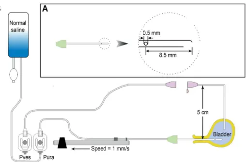
Fig 1. Schematic drawing of the setup for urethral pressure profile recording.(A) A lateral hole water-perfusion catheter, used for transurethral catheterization and UPP recording in female rats. (B) The general arrangement of the urodynamic device and the animal. Note that the suprapubic bladder catheter for the measuring of bladder pressure is disconnected and that both ends of this catheter are fixed at 5 cm above the 0 level (see text for details).Pves= vesical pressure transducer;Pure=urethral pressure transducer.
body when both ends of the catheters were positioned in the bladder. The bladder catheter was connected to a pressure transducer within a urodynamic device via a tube with a 3-way connec-tor. The bladder pressure value was digitized and recorded using urodynamic device software. Pressure activity was monitored on a computer screen, and the data were saved in the com-puter. The pressure transducer and pressure transmission tube system were examined to ensure that all air bubbles were eliminated, and the instrument was calibrated. Subsequently, 0.2 to 0.4 mL of room-temperature normal saline was infused slowly into the bladder. Stabiliza-tion of the initial bladder pressure at 15 ± 5 cm H2O was preferred prior to performing the LPP
tests. The bladder body was pressed using a cotton stick, and urine leakage at the external mea-tus of the urethra was observed simultaneously, as previously described [22,23]. The bladder pressure at the time that leakage appeared at the meatus was recorded as the LPP. This process was repeated 3 to 4 times, and the average value was used for statistical analyses.
Transurethral Catheterization and UPP Recording
UPP recordings were performed after LPP determinations. Rats have a relatively small bladder volume, and suprapubic bladder catheterization would further decrease this volume. Thus, saline infusion via a transurethral catheter during UPP recording would extend the bladder and increase bladder pressure, which could affect the UPP parameters. Therefore, we disconnected the suprapubic bladder and pressure transducer tubes to allow the fluid in the bladder to flow out freely. This procedure resulted in stabilization of the bladder volume and pressure during the UPP recordings and prevented the possible interference of variations in bladder volume and pressure on the UPP. The transurethral catheter opening and disconnected bladder catheter opening were set to the atmospheric pressure at the pubic symphysis level (i.e., set to 0), and cali-bration was performed. The end of the disconnected bladder pressure transducer tube and the outside opening of the suprapubic bladder catheter were fixed at 5 cm above the pubic symphysis to maintain the bladder pressure at 5 cm H2O throughout the entire UPP recording process.
A 5-cm 3 Fr. catheter made from an epidural anesthesia tube was prepared prior to the experiment (Fig 1B). These catheters generally have 3 fanning side holes near the end. We sealed 2 of the side holes with glue so that only one hole remained open. This catheter was con-nected to a pressure transducer via an adaptor and mounted to a mechanical withdrawal appa-ratus of the urodynamic device. The catheter was passed into the bladder via the urethra. Saline was infused via this transurethral catheter at 0.5 mL/min during UPP recording, and the cathe-ter was simultaneously withdrawn at 1 mm/s by the mechanical withdrawal apparatus. Bladder pressure and urethral pressure values were digitized and recorded using urodynamic device software. The pressure readings were monitored on the computer screen, and the data were saved in the computer. The UPP recordings were performed with the lateral water-perfusion hole facing the 3-, 6-, 9-, and 12-o’clock positions in a randomized sequence in each rat, and four UPP curves were obtained at these 4 positions. These recordings were repeated in a ran-domized sequence to assess data reproducibility if the rat was performing well.
The rats were sacrificed at the end of this series of tests and autopsied to identify abnormali-ties in the urogenital system. Data from rats with grossly pathological organs were excluded from statistical analyses.
The general arrangement of the urodynamic device and the animal are presented inFig 1
andS1 Fig.
Statistical Analyses
Kolmogorov-Smirnov (KS) test. Comparisons of the four positions of maximum urethral closure pressure (MUCP) among the data sets were performed using the Kruskal-Wallis test, followed by pair-wise multiple comparisons. Differences between the primary and repeated measurements of the MUCP and FUL were tested using paired-samples t tests (yielding two-tailedpvalues). The consistency of the UPP measurements between the pri-mary and repeated recordings was assessed by calculating intra-class correlation coeffi-cients (ICCs). In all cases, aP<0.05 was considered statistically significant.
Results
Seventeen adult female rats were prepared for the LPP test. Three of these rats exhibited unsta-ble respiration during the procedure and ultimately died, and one of them exhibited a vaginal cervix mass at autopsy. The data from these 3 rats were excluded from analyses. The mean LPP of the remaining 14 rats was 64.39 ± 20.29 cm H2O. These 14 rats were used in the next step
for UPP recording.
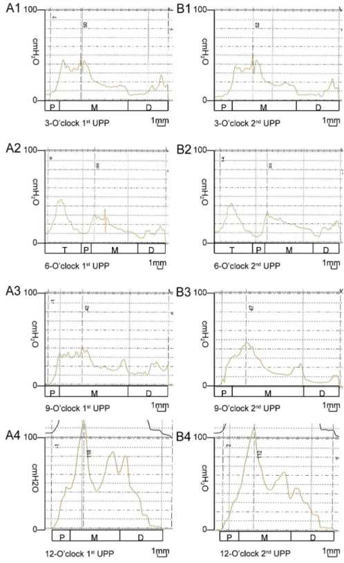
UPP recording was not completed in 4 of the 14 rats because of difficulties associated with transurethral catheterization. Therefore, UPP values from 10 rats were analyzed.Fig 2shows an example of the UPP curves recorded in 1 female rat, including the primary and repeated recordings. The FUL values at the 3-, 6-, 9-, and 12-o’clock positions were 12.90 ± 1.20, 16.70 ± 1.95, 13.90 ± 2.42, and 11.60 ± 0.97 mm, respectively, and the MUCP values at these positions were 38.70 ± 11.85, 33.90 ± 11.82, 37.40 ± 11.95, and 71.90 ± 23.01 cm H2O,
respec-tively. The highest MUCP value was observed at the 12-o’clock position compared with the other 3 positions (12- vs. 3-,P= 0.001; 12- vs. 6-, P<0.001; 12- vs. 9-, P<0.001), in addition to the lowest FUL value (12- vs. 6-, P<0.001; 12- vs. 3-, P = 0.084; 12- vs. 9-, P = 0.019). The lowest MUCP value was observed at the 6-o’clock position (6- vs. 12-,P<0.001; 6- vs. 3-,
P= 0.473; 6- vs. 9-,P= 0.737), in addition to the highest FUL value (6- vs. 12-,P<0.001; 6- vs. 3-,P= 0.005; 6- vs. 9-,P= 0.027). Similar MUCP (P= 0.702) and FUL (P= 0.540) values were detected at the 3- and 9-o’clock positions (Fig 3A and 3B). UPP recordings at all 4 positions were repeated in 9 rats.Table 1shows the MUCP and FUL values obtained from the primary and repeated UPP recordings. There were no significant differences between these values, dem-onstrating the high reproducibility of the present method of UPP recording (Pvalues are listed inTable 1). The ICC values for the MUCP and FUL are listed inTable 2. These values generally received high reliability scores. The ICC values for the MUCP between the primary and repeat recordings ranged from 0.838 to 0.960 with excellent reproducibility, and those for the FUL ranged from 0.672 to 0.844 with good reproducibility, further demonstrating the high repro-ducibility of this method of UPP recording.
Discussion
We present a modified method for the recording of UPP in female rats. This method was devel-oped on the basis of a water perfusion catheter system that was first described by Brown and Wickham. Our modifications utilize the most widely available urodynamic device and catheters.
Fig 2. Example UPP recorded using a modified water-perfusion catheter system in 1 female rat.The left column (A1, A2, A3, and A4) shows the UPP curves, recorded primarily with the side holes oriented at the 3-, 6-, 9-, and 12-o’clock positions, respectively. The right column (B1, B2, B3, and B4) shows the repeated UPP curves, recorded with the side holes also oriented at the 3-, 6-, 9-, and 12-o’clock positions, respectively. Note the consistency between the primary and repeated UPP curves for the same position. The curves at different positions display remarkable variations in the patterns and pressure values. T: bladder trigone, P: proximal, M: mid, D: distal.
which cannot accommodate the volume of water that typically flows in during a single UPP recording. An open suprapubic bladder catheter was used to resolve this problem by allowing the bladder water to flow out freely. The bladder pressure was fixed at approximately 5 cm H2O when we fixed the external end of the suprapubic bladder catheter and the end of the tube
connected to the bladder pressure transducer at 5 cm above the pubic symphysis. This method is simple and provides reliable and reproducible results. In this study, the entire length of the urethral wall, from the bladder neck to the external meatus, was evaluated. Unique UPP curves were recorded at 4 positions for each female rat. These preliminary results demonstrated that the UPP curves recorded using this method sensitively and appreciably reflected the different pressures exerted by the urethral wall. These female rats exhibited a rela-tively consistent UPP pattern, which was highly reproducible, and each urethra exhibited a characteristic pressure profile. The whole UPP curve can be divided into 3 segments. The first segment revealed a definite progressive increase in pressures, which represented the values at recording sites in the proximal urethral segment. The pressures in the second segment peaked and maintained a high level for a certain length, which represented the values at recording sites in the mid-urethral segment. The pressures in the third segment progressively dropped and ended at approximately the 0 level, which represented the distal urethral segment. The
Fig 3. Comparisons of MUCP and FUL values at 4 different positions.(A) The MUCP values at 4 different positions. The bars represent the mean±SD of the measurements. The highest MUCP value was observed
at the 12-o’clock position compared with the other 3 positions (12- vs. 3-,P= 0.001; 12- vs. 6-,P<0.001;
12-vs. 9-,P<0.001). The MUCP values at the 3- and 9-o’clock positions are insignificantly different (3- vs. 9-,
P= 0.702). (B) The FUL values at 4 different positions. The bars represent the mean±SD. The longest FUL was observed at the 6-o’clock position compared with the other 3 positions (6- vs. 12-,P<0.001; 6- vs. 3-, P= 0.005; 6- vs. 9-,P= 0.027). The FUL values at the 3- and 9-o’clock positions were similar (P= 0.540).
doi:10.1371/journal.pone.0140851.g003
Table 1. Comparisons of UPP parameters between the primary and repeatedrecordings (N = 9).
Paired differences of MUCP Paired differences of FUL
Positions Mean±SD 95% CI p Mean±SD 95% CI p
3-o’clock primary-repeated 2.56±4.30 -0.75,5.86 0.113 -0.44±0.73 -1.00,0.11 0.104
6-o’clock primary-repeated 2.78±4.55 -0.72,6.27 0.104 -0.22±1.64 -1.48,1.04 0.695
9-o’clock primary-repeated -0.11±5.97 -4.70,4.48 0.957 0.22±1.71 -1.10,1.54 0.708
12-o’clock primary-repeated 4.22±6.18 -0.53,8.97 0.075 -0.44±0.88 -1.12,0.23 0.169
The data are reported as the mean±SD.
Pvalues were calculated by the paired samples t-test. MUCP: maximum urethral closure pressure
FUL: functional urethral length; 95% CI: 95% confidence interval SD: standard deviation.
anatomical localizations of points of high and low pressures were easily performed using cathe-ter calibration. Therefore, this method provides a useful tool for the investigation of the anat-omy and physiology of urethra in this small animal species.
The UPP curves in female rats bear many similarities to those of women. Similar patterns of UPP curves and pressure levels of MUCP were reported previously in human females [16,17]. The most intriguing result is that the positional differences demonstrated UPP recordings in women were also demonstrated in female rat UPP recordings [16,17]. Only a few papers in the literature discuss the directional differences of UPP in human females, and two contrary opin-ions exist [16,17]. Some groups consider these differences to be artifacts. A hypothesis has been proposed that the passage of a flexible but straight catheter through a curved urethra results in application of additional forces on the transducer caused by catheter bending [15,28]. Other groups consider these differences to be physiological phenomena. An active perineal contrac-tion may explain the anisotropic rotacontrac-tional pressure variacontrac-tions in the urethra [14,29]. Our observations revealed new facts about these positional differences. The patterns of UPPs and their parameters between the left and right halves (i.e., 3 o’clock and 9 o’clock, respectively) were symmetrical. These observations suggest that positional differences in UPP should not be oversimplified as artifacts. These specific features of pressure distribution in the female rat ure-thra and their relationships with ureure-thral anatomical structures and physiological status will be addressed in future studies.
In human urodynamic studies, LPP and UPP measurements as well as EUS-EMG and videourodynamic analyses are commonly performed to examine urethral function or patho-physiology. LPP and UPP measurements are quantitative, while EUS-EMG and videourody-namic analyses are semi-quantitative. In clinical practice/basic science, various combinations of these methods are needed, depending on the diagnosis/scientific purpose. Most of these methods, with the exception of UPP measurement, have been widely used on small-sized animals, such as rats. LPP measurements and EMG have been successfully performed in anes-thetized and fully awake rats [1,10,11]. Our methodology provides a simple technique for per-forming UPP recordings in anesthetized rats. With further modifications, there is possibility of incorporating UPP in fully awake rats urodynamic study, which will develop an assessment protocol in rat models in close analogy to the urodynamic assessment used clinically in humans. The development of this protocol will further enhance the valve of rat model in the study of lower urinary tract dysfunction.
Table 2. Consistency between the primary and repeated UPP recordings (N = 9).
MUCP FUL
Positions ICC 95% CI ICC 95% CI
3-o’clock 0.926 0.708 0.983 0.844 0.455 0.963
6-o’clock 0.916 0.675 0.980 0.672 0.070 0.915
9-o’clock 0.838 0.437 0.961 0.738 0.198 0.934
12-o’clock 0.960 0.835 0.991 0.720 0.162 0.929
The data are reported as the mean±SD. MUCP: maximum urethral closure pressure FUL: functional urethral length
ICC: intra-class correlation coefficient; 95% CI: 95% confidence interval.
Study Limitations
The present study has several limitations as a primary methodological investigation. First, the suprapubic bladder catheter was open to the air to overcome the limitation of the small bladder volumes of female rats, which obviously does not represent the physiological situation. The physiological pressure level was approached by lifting the suprapubic bladder catheter opening to a height of 5 cm H2O in our study. However, the stress UPP was still not able to be
deter-mined. The stress UPP in female rats may be determined by lifting the suprapubic bladder catheter opening to a different height range (e.g., 30 cm H2O or more) or connecting an elastic
bag to enlarge the bladder volume. Further investigations should be conducted to standardize UPP and stress UPP recordings.
Second, we used sodium pentobarbital for anesthesia because this anesthetic is the most widely used in our laboratory, and it provides satisfactory anesthesia to allow for completion of UPP recordings. However, different anesthetics may have different effects on urodynamic parameters, such as cystometry and LPP [30–32]. The present study focused on the modifica-tion of the UPP recording technique, and we did not investigate whether different anesthetics differentially affected UPP recording. Future studies comparing different anesthetics on UPP recording outcomes should be performed to address this issue.
Finally, a full understanding of how each unique UPP curve is formed and what factors affect the UPP parameters is essential to a better understanding of the physiological relevance and clini-cal implications of these recordings. The present study did not address this complex issue. Eluci-dation of the corresponding relationships between UPP curve patterns and the anatomical and histological structures in the female rat urethra will be discussed in subsequent reports.
Conclusions
UPP recording using a modified method based on a water-perfusion catheter system is feasible and reproducible in female rats. This study is the first report of UPP recordings in female rats that are similar to those that have been reported in women. These results will likely enhance the utility of the female rat model forin vivoinvestigations of urethral function and of the mechanisms of urinary continence in humans.
Supporting Information
S1 Fig. The use of a modified water-perfusion catheter system with a urodynamic device for the recording of UPP in female rats.(A) The general arrangement of the urodynamic device and animal. The pressure transducer and rat pubic symphysis were set at same level. The tube end of the pressure transducer and the side hole of the water-perfusion catheter were always set to 0 at the pubic symphysis. (B) The water-perfusion catheter with 1 side hole for transure-thral catheterization and recording the uretransure-thral pressure. (C) The water-perfusion catheter was inserted into bladder transurethrally and later mounted to the mechanical withdrawer. (D) The suprapubic bladder catheter and the pressure transducer tube were disconnected, and both ends were opened to air. The ends were fixed 5 cm above the 0 level. Therefore, the water perfusing into the bladder during UPP recordings flowed out freely to avoid overextension of the bladder. The bladder pressure was fixed at 5 cm H2O (see text for details).
(TIF)
Acknowledgments
Zheng and Dr. Ping Yin (Public Health College, Huazhong University of Science and Technol-ogy, Wuhan, China) for their excellent statistical support and Mr. Yu Wang (Liaoning Medecal College, Jinzhou, China) for his work of draftingFig 1.
Author Contributions
Conceived and designed the experiments: SFX XHL BC HBT GHD. Performed the experi-ments: SFX XHL LX BC GHD. Analyzed the data: SFX XHL GHD HBT. Contributed reagents/ materials/analysis tools: SFX XHL LX BC HBT GHD. Wrote the paper: SFX GHD.
References
1. Oyama T, Kawai Y, Oka M. Tramadol enhances urethral continence function throughμ-opioid receptors
in rats. Neurourol Urodyn. 2013; 32: 98–103. doi:10.1002/nau.22274PMID:22674657
2. Chang HY, Havton LA. Anatomical tracer injections into the lower urinary tract may compromise cysto-metry and external urethral sphincter electromyography in female rats. Neuroscience. 2010; 166: 212–
219. doi:10.1016/j.neuroscience.2009.11.037PMID:20004710
3. Gasbarro G, Lin DL, Vurbic D, Quisno A, Kinley B, Daneshgari F, et al. Voiding function in obese and type 2 diabetic female rats. Am J Physiol Renal Physiol. 2010; 298: F72–F77. doi:10.1152/ajprenal. 00309.2009PMID:19889955
4. Rodríguez LV, Chen S, Jack GS, de Almeida F, Lee KW, Zhang R. New objective measures to quantify stress urinary incontinence in a novel durable animal model of intrinsic sphincter deficiency. Am J Phy-siol Regul Integr Comp PhyPhy-siol. 2005; 288: R1332–R1338. PMID:15650117
5. Peng CW, Chen JJ, Chang HY, de Groat WC, Cheng CL. External urethral sphincter activity in a rat model of pudendal nerve injury. Neurourol Urodyn. 2006; 25: 388–396. PMID:16637068
6. Corcos J, Loutochin O, Campeau L, Eliopoulos N, Bouchentouf M, Blok B, et al. Bone marrow mesen-chymal stromal cell therapy for external urethral sphincter restoration in a rat model of stress urinary incontinence. Neurourol Urodyn. 2011; 30: 447–455. doi:10.1002/nau.20998PMID:21412824
7. Zheng J, Xu K, Sun Y, Sun C, Ding Q, Fang Z. Evaluation of urodynamic findings before and after mid-urethral tape sling operation for female stress urinary incontinence. J Minim Invasive Gynecol. 2013; 20: 482–486. doi:10.1016/j.jmig.2013.02.002PMID:23567094
8. Sirls LT, Richter HE, Litman HJ, Kenton K, Lemack GE, Lukacz ES, et al. The effect of urodynamic test-ing on clinical diagnosis, treatment plan and outcomes in women undergotest-ing stress urinary inconti-nence surgery. J Urol. 2013; 189: 204–209. doi:10.1016/j.juro.2012.09.050PMID:22982425
9. Zimmern P, Litman H, Nager C, Sirls L, Kraus SR, Kenton K, et al. Pre-operative urodynamics in women with stress urinary incontinence increases physician confidence, but does not improve out-comes. Neurourol Urodyn. 2014; 33: 302–306. doi:10.1002/nau.22398PMID:23553613
10. LaPallo BK, Wolpaw JR, Chen XY, Carp JS. Contribution of the external urethral sphincter to urinary void size in unanesthetized unrestrained rats. Neurourol Urodyn. 2015 May 20. doi:10.1002/nau. 22789[Epub ahead of print] PMID:25995074
11. Schneider MP, Huqhes FM Jr, Enqmann AK, Purves JT, Kasper H, Tedaldi M, et al. A novel urody-namic model for lower urinary tract assessment in awake rats. BJU Int. 2015; Suppl 6: 8–15. doi:10. 1111/bju.13039PMID:25597776
12. Suzuki Bellucci CH, Wöllner J, Greqorini F, Birnböck D, Kozomara M, Mehnert U, et al. External urethral sphincter pressure measurement: an accurate method for the diagnosis of detrusor external sphincter dyssynergia? PLoS One. 2012; 7: e37996. doi:10.1371/journal.pone.0037996PMID:22701539
13. Walters RD, McMurray G, Brading AF. Comparison of the urethral properties of the female guinea pig and rat. Neurourol Urodyn. 2006; 25:62–69. PMID:16224796
14. Klünder M, Amend B, Vaeqler M, Kelp A, Feuer R, Sievert KD, et al. High definition urethral pressure profilometry: Evaluating a novel microtip catheter. Neurourol Urodyn. 2015; doi:10.1002/nau.22835 [Epub ahead of print]. PMID:26207994
15. Valentini FA, Robain G, Marti BG. Is a sequence of tests during urethral pressure profilometry corre-lated with symptoms assessment in women? Int Braz J Urol. 2012; 38: 809–817. PMID:23302401
16. Vereecken RL, Cornelissen JM. Rotational differences in urethral pressure in incontinence women. Urol Int. 1985; 40: 201–205. PMID:4049577
18. Khullar V, Cardozo L. The urethra (UPP, MUPP, Instability, LPP). Eur Urol. 1998; 34: 20–22. PMID: 9705549
19. Almeida FG, Bruschini H, Srougi M. Correlation between urethral sphincter activity and Valsalva leak point pressure at different bladder distentions: revisiting the urethral pressure profile. J Urol. 2005; 174: 1312–1315. PMID:16145410
20. Brown M, Wickham JE. The urethral pressure profile. Br J Urol. 1969; 41: 211–217. PMID:5814020
21. Ghoneim MD, Rottembourg JL, Fretin J, Susset JG. Urethral pressure profile. Standardization of tech-nique and study of reproducibility. Urology. 1975; 5: 632–637. PMID:1168962
22. Jiang HH, Pan HQ, Gustilo-Ashby MA, Gill B, Glaab J, Zaszczurynski P, et al. Dual simulated childbirth injuries result in slowed recovery of pudendal nerve and urethral function. Neurourol Urodyn. 2009; 28: 229–235. doi:10.1002/nau.20632PMID:18973146
23. Song QX, Baloq BM, Kerns J, Lin DL, Sun Y, Damaser MS, et al. Long-term effects of simulated child-birth injury on function and innervation of the urethra. Neurourol Urodyn. 2015; 34: 381–386. doi:10. 1002/nau.22561PMID:24501018
24. Tanagho EA, Meyers FH, Smith DR. Urethral resistance: its components and implications. I. Smooth muscle components. Invest Urol. 1969; 7: 136–149. PMID:4309609
25. Donker PJ, Ivanovici F, Noach EL. Analyses of the urethral pressure profile by means of electromyogra-phy and the administration of drugs. Br J Urol. 1972; 44: 180–193. PMID:4339396
26. Enhorning G. Simultaneous recording of intervesical and intraurethral pressure. Acta Chir Scand Suppl. 1961; 276: 1–68. PMID:13696922
27. Hanzal E, Berger E, Koelbl H. Reliability of the urethral closure pressure profile during stress in the diagnosis of genuine stress incontinence. Br J Urol. 1991; 68: 369–371. PMID:1933156
28. Griffiths D. The pressure within a collapsed tube, with special reference tourethral pressure. Phys Med Biol. 1985; 30: 951–963. PMID:4048278
29. Rossier AB, Fam BA. 5-microtransducer catheter in evaluation of neurogenic bladder function. Urology. 1986; 27: 371–378. PMID:3962062
30. Cannon TW, Damaser MS. Effects of anesthesia on cystometry and leak point pressure of the female rat. Life Sci. 2001; 69: 1193–1202. PMID:11508351
31. Wuethrich PY, Kessler TM, Burkhard FC. The effects of thoracic epidurally administered drugs on ure-thral sphincter function in women: a pooled analysis. Pain Med. 2013; 14: 1248–1253. doi:10.1111/ pme.12128PMID:23614971