Polymorphisms with Disease Susceptibility and
Cardiovascular Risk in Spanish Rheumatoid Arthritis
Patients
Mercedes Garcı´a-Bermu´dez1, Carlos Gonza´lez-Juanatey2, Raquel Lo´pez-Mejı´as3, Marı´a Teruel1,
Alfonso Corrales3, Jose´ A. Miranda-Filloy4, Santos Castan˜eda5, Alejandro Balsa6, Benjamı´n Ferna´ndez-Gutierrez7, Isidoro Gonza´lez-A´ lvaro5, Carmen Go´mez-Vaquero8, Ricardo Blanco3, Javier Llorca9, Javier Martı´n1., Miguel A. Gonza´lez-Gay3
*.
1Instituto de Parasitologı´a y Biomedicina Lo´pez-Neyra, IPBLN-C.S.I.C., Granada, Spain,2Cardiology Division, Hospital Xeral-Calde, Lugo, Spain, 3Department of Rheumatology, Hospital Universitario Marque´s de Valdecilla, IFIMAV, Santander, Spain,4Department of Rheumatology, Hospital Xeral-Calde, Lugo, Spain,5Department of Rheumatology, Hospital Universitario La Princesa, IIS-Princesa, Madrid, Spain,6Rheumatology Unit, Hospital Universitario La Paz, Madrid, Spain,7Department of Rheumatology, Hospital Clinico San Carlos, Madrid, Spain,8Department of Rheumatology, Hospital Universitario de Bellvitge, IDIBELL, Barcelona, Spain,9Department of Epidemiology and Computational Biology, School of Medicine, University of Cantabria, and CIBER Epidemiologı´a y Salud Pu´blica (CIBERESP), IFIMAV, Santander, Spain
Abstract
Objective: Rheumatoid arthritis (RA) is a chronic inflammatory disease associated with increased cardiovascular (CV) mortality. SinceCD40-CD154 binding has direct consequences on inflammation process initiation, we aimed to replicate previous findings related to disease susceptibility in Spanish RA population. Furthermore, as the major complication in RA disease patients is the development of CV events due to accelerated atherosclerosis, and elevated levels of CD40L/CD154 are present in patients with acute myocardial infarction, we assessed the potential association ofCD40andCD154/CD40L
gene variants with CV risk in Spanish RA patients.
Methods:One thousand five hundred and seventy-five patients fulfilling the 1987 ACR classification criteria for RA and 1600 matched controls were genotyped for theCD40rs1883832, rs4810485 and rs1535045 andCD154rs3092952 and rs3092920 gene polymorphisms, using predesigned TaqMan single nucleotide polymorphism genotyping assays. Afterwards, we investigated the influence of CD40-CD154gene variants in the development of CV events. Also, in a subgroup of 273 patients without history of CV events, we assessed the influence of these polymorphisms in the risk of subclinical atherosclerosis determined by carotid ultrasonography.
Results:Nominally significant differences in the allele frequencies for the rs1883832CD40gene polymorphism between RA patients and controls were found (p= 0.038). Although we did not observe a significant association ofCD40-CD154gene variants with the development of CV events, an ANCOVA model adjusted for sex, age at the time of the ultrasonography assessment, follow-up time, traditional CV risk factors and anti-cyclic citrullinated peptide antibodies disclosed a significant association (p= 0.0047) betweenCD40rs1535045 polymorphism and carotid intima media thickness, a surrogate marker of atherosclerosis.
Conclusion: Data from our pilot study indicate a potential association of rs1883832 CD40 gene polymorphism with susceptibility to RA. Also, theCD40rs1535045 gene variant may influence development of subclinical atherosclerosis in RA patients.
Citation:Garcı´a-Bermu´dez M, Gonza´lez-Juanatey C, Lo´pez-Mejı´as R, Teruel M, Corrales A, et al. (2012) Study of Association ofCD40-CD154Gene Polymorphisms with Disease Susceptibility and Cardiovascular Risk in Spanish Rheumatoid Arthritis Patients. PLoS ONE 7(11): e49214. doi:10.1371/journal.pone.0049214
Editor:Gualtiero Colombo, Centro Cardiologico Monzino IRCCS, Italy
ReceivedApril 27, 2012;AcceptedOctober 5, 2012;PublishedNovember 15, 2012
Copyright:ß2012 Garcı´a-Bermu´dez et al. This is an open-access article distributed under the terms of the Creative Commons Attribution License, which permits unrestricted use, distribution, and reproduction in any medium, provided the original author and source are credited.
Funding:This work was supported by two grants from Fondo de Investigaciones Sanitarias PI06-0024 and PS09/00748 (Spanish National Health System; Ministry of Science and Innovation, Spain Government) and by RETICS Program, RD08/0075 (RIER) from Instituto de Salud Carlos III (ISCIII), National Health System (Spain) within the VI Programa Nacional de I+D+i 2008–2011 (FEDER). This work was supported in part by grants from the European IMI BTCure Program. MGB is a beneficiary of a grant from Fundacio´n Espan˜ola de Reumatologı´a. The funders had no role in study design, data collection and analysis, decision to publish, or preparation of the manuscript.
Competing Interests:The authors have declared that no competing interests exist. * E-mail: [email protected]
Introduction
Rheumatoid arthritis (RA), the prototype of inflammatory chronic disease, is characterized by a consistent increase of cardiovascular (CV) mortality and morbidity, mainly due to a process of accelerated atherosclerosis [1,2,3]. There is some evidence supporting that both immune dysregulation and a systemic inflammatory milieu predating and characterizing earlier phases of RA may be important pathogenic factors of vascular impairment in patients with this condition. Interesting findings arise from the observation that CD40–CD40 ligand (CD40L/ CD154) interaction, a crucial step in autoimmune disease pathogenesis, is thought to be involved in atherogenesis and plaque rupture in RA [4].
Once that CD40 interacts with its ligand (CD40L, aka CD154) on T cells, it acts as a transmembrane signal transducer leading to activation of intracellular kinases and transcription factors, giving rise to inflammatory responses [5]. CD40 signalling has been linked to pathogenic processes of chronic inflammatory and autoimmune diseases [6]. As a consequence of CD154/CD40L binding to CD40, release of cytokines and expression of adhesion molecules, as well as numerous inflammatory processes are triggered [7]. In addition, CD40/CD154 interactions may induce the expression of matrix metalloproteinases that degrade compo-nents of the atherosclerotic plaques and promote neovasculariza-tion, instability and plaque rupture leading to acute CV events or sudden death [8] and can play a prominent role in thrombotic events after plaque rupture [5]. This is relevant as the main cause of mortality and morbidity of RA is atherosclerosis, driving to CV events that are preceded by subclinical atherosclerosis.
CD40 is a 45–50 kDa membrane glycoprotein, member of the TNF-receptor superfamily, which acts as a receptor for CD154. CD40 has been reported to be constitutively and/or inducibly expressed on B cells, platelets, monocytes and macrophages, endothelial cells, smooth muscle cells, mast cells, fibroblasts, dendritic cells, neutrophils and T cells [9] and also in the synovial fluid of RA patients [10].
A Genome Wide Association Study and meta-analysis high-lighted the role of the major allele of the rs4810485 CD40 polymorphism on chromosome 20q13 in the susceptibility to RA [11,12]. Further replication studies in different populations [13,14,15] confirmed this association. Moreover, a recent report disclosed that the rs4810485 CD40 variant may affect CD40 mRNA and protein expression on B cells and monocytes [16], and is in high linkage disequilibrium (r2= 0.95) with rs1883832, which has been shown to influence the efficiency of CD40 protein translation by disrupting a Kozak sequence [17], a stretch of nucleotides that flanks the start codon important for the initiation of translation of a nascent mRNA molecule [18]. Indeed, the major allele of rs1883832 increases the translational efficacy of CD40 transcripts, resulting in a 15–32% increase in CD40 protein production [19]. Furthermore, major alleles of rs4810485 and rs1883832 CD40 genetic variants are associated with Grave’s disease (GD) [17]; and also minor alleles have been associated with multiple sclerosis [20,21], Crohn’s disease [20] and giant cell arteritis [22], although with relatively weak statistical evidence.
CD154, also known as CD40L or TNFSF5, is a 39 kDa transmembrane protein with particular importance in T-cell dependent humoral immune responses. CD154 is expressed on various cell types, including cell types present in atherosclerotic plaques such as endothelial cells, monocytes, macrophages and smooth muscle cells [23]. Increased levels of soluble CD154 (sCD154) have been found in patients with systemic lupus erythematosus (SLE), RA, and Sjo¨gren’s syndrome in association
with disease activity [24]. Moreover, serum levels of sCD154 are higher in patients with RA than in healthy individuals and they correlate with both IgM-RF and IgG-RF titers [25]. Located in chromosome Xq26,CD15423459A.G (rs3092952) variant has been reported to influence CD40L plasma levels, and those levels above median seem to reflect a prothrombotic state which can be managed with the use of antithrombotic treatment [26]. Also, rs3092920CD154polymorphism is in high linkage disequilibrium (r2= 0.97) with a microsatellite located in 39-UTR that has been associated in Spanish population with SLE [27], another autoimmune disease associated with accelerated atherosclerosis and increased risk of CV complications, and with RA susceptibility [28]. sCD40L is associated with measures of subclinical athero-sclerosis in the general population [8], and increased levels of sCD40L have been found in patients with acute myocardial infarction (MI) and unstable angina [7]. The role of theCD40Lin the susceptibility to autoimmune diseases has not been investigated as broadly as theCD40, mainly due to this gene being located on the 6 chromosome. The different prevalence of some of autoimmune diseases between both genders [29] can suggest that genes located on the6chromosome could be susceptibility factors in these diseases; however, few studies analyzing polymorphisms on this chromosome has been made. Mutations onCD40Lgene are associated with X-Linked Hyper-IgM syndrome; a family genetic disorder characterized by an increase of IgM level and a decrease of IgG and IgA [30]; other mutations and/or altered expression ofCD40L, commoncchain,FOXP3(forkhead box P3) among other genes encoded on the6chromosome, are known to be cause of immune disorders such as X-SCID and IPEX dramatically manifested in men, while females can compensate or reduce severity of the symptoms due to the second6chromosome. Taking together all these considerations, in the present study we aimed to confirm the role of CD40 polymorphisms in the susceptibility to RA in Spanish population. In a second step, we assessed whether polymorphisms in CD40-CD154 genes may influence the susceptibility to CV disease in RA. For this purpose, we analyzed the implication of these polymorphisms in the risk of CV events and presence of subclinical atherosclerosis in RA.
Results and Discussion
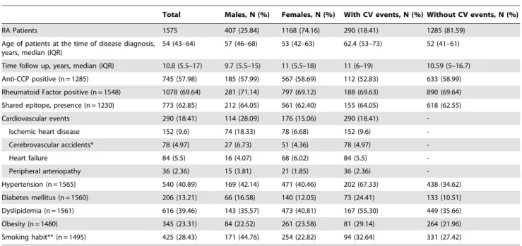
Information on the main demographical data, clinical charac-teristics of the RA patients (n = 1575) enrolled in the current study, CV risk factors and CV events of patients is shown inTable 1. Two hundred and ninety (18.41%, 114 men and 176 women) of these 1575 RA patients experienced clinically evident CV events: ischemic heart disease, heart failure, cerebrovascular accident or peripheral arteriopathy.
factors on the increased risk of cardiovascular disease of patients with rheumatoid arthritis, in our population we found no additive effect of smoking andCD40-CD154gene polymorphisms (data not shown).
Genotyping success rate was.95% overall. The estimated statistical power of the study to detect genetic modest effects like OR 1.17 in our cohort, was.75% for rs1883832, rs4810485 and rs1535045CD40polymorphisms; and 80% (in women population) and 46% (men) for rs3092952 and 63% (women) and 35% (men) for rs3092920 to detect an OR = 1.25 in the case of CD154 polymorphisms, assuming a prevalence of RA in Spanish population (0.5%) [38], under the condition ofaerror 0.05 and
berror 0.20.
The statistical power of this study to detect a difference between absence or presence of CV disease in RA patients with an estimated OR 1.25, a type I error rate of 0.05, type II error rate of 0.20, and 0.05% of population risk, was 61% for CD40 single nucleotide polymorphisms (SNPs) and between 48% (male) and 53% (female patients) for the rs3092952 polymorphism and between 34% (male) and 36% (female) for the rs3092920CD154 gene variant.
Minor Allele Frequencies of theCD40rs1883832, rs4810485 and rs1535045, and CD154 rs3092952 and rs3092920 Polymorphisms in RA Patients and Controls
Genotype frequencies were conformed to Hardy-Weinberg equilibrium (p.0.05) both in patients and controls for all genetic variants under study.
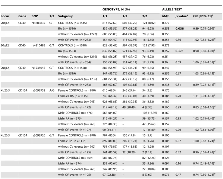
A nominally significant difference in the allele frequency of the CD40 rs1883832 gene polymorphism between RA patients and controls was seen (p= 0.038), supporting a role of this CD40
59UTR variant in the susceptibility to RA in Spanish population (Table 2). On the other hand, CD40 rs4810485 variant was marginally associated with disease susceptibility (p= 0.069). However, no association or trend was detected when allele or genotype frequencies of theCD40rs1535045 polymorphism in RA patients were compared with those observed in controls (Table 2). Allele frequencies observed for rs1883832 and rs4810485 CD40 genetic polymorphisms (27.7% and 27.2%, respectively) were slightly different from those reported in HapMap CEU population (22.5% and 23%, respectively). The rs1535045 allele frequency (24.7%) was similar to the reported in HapMap CEU population (25%).
Because CD154 is located on the 6 chromosome, and the elevated incidence of RA in women and higher frequency of atherosclerosis in men, we compared allele frequencies between patients and controls in males and females separately. No association was found between the CD154 gene variants and susceptibility to RA disease (Table 2). Allele frequencies observed for rs3092952 and rs3092920CD154 gene variants (17.2% and 11.1%) were somewhat different from those reported in HapMap for CEU population (23% and 8%, respectively).
CD40andCD154Variants and Risk of CV Events in Patients with RA
Table 2 shows the genotype frequencies of the CD40 rs1883832, rs4810485 and rs1535045 gene polymorphisms in this series of RA patients stratified according to the presence or absence of CV events. No association was found betweenCD40 polymorphisms and CV events risk in our RA patients cohort. It was also the case forCD154gene variants in women or men RA patients.
Table 1.Clinical and demographic characteristics of rheumatoid arthritis (RA) patients included in the study, stratified by gender or by Cardiovascular events.
Total Males, N (%) Females, N (%) With CV events, N (%) Without CV events, N (%)
RA Patients 1575 407 (25.84) 1168 (74.16) 290 (18.41) 1285 (81.59) Age of patients at the time of disease diagnosis,
years, median (IQR)
54 (43–64) 57 (46–68) 53 (42–63) 62.4 (53–73) 52 (41–61)
Time follow up, years, median (IQR) 10.8 (5.5–17) 9.7 (5.5–15) 11 (5.5–18) 11 (6–19) 10.59 (5–16.7) Anti-CCP positive (n = 1285) 745 (57.98) 185 (57.99) 567 (58.69) 112 (52.83) 633 (58.99)
Rheumatoid Factor positive (n = 1548) 1078 (69.64) 281 (71.14) 797 (69.12) 188 (69.63) 890 (69.64)
Shared epitope, presence (n = 1230) 773 (62.85) 212 (64.05) 561 (62.40) 155 (64.05) 618 (62.55) Cardiovascular events 290 (18.41) 114 (28.09) 176 (15.06) 290 (18.41)
-Ischemic heart disease 152 (9.6) 74 (18.33) 78 (6.68) 152 (9.6)
-Cerebrovascular accidents* 78 (4.97) 27 (6.73) 51 (4.36) 78 (4.97)
-Heart failure 84 (5.5) 16 (4.07) 68 (6.02) 84 (5.5)
-Peripheral arteriopathy 36 (2.36) 15 (3.81) 21 (1.85) 36 (2.36)
-Hypertension (n = 1565) 540 (40.89) 169 (42.14) 471 (40.46) 202 (67.33) 438 (34.62)
Diabetes mellitus (n = 1560) 206 (13.21) 66 (16.58) 140 (12.05) 73 (24.41) 133 (10.51) Dyslipidemia (n = 1561) 616 (39.46) 143 (35.57) 473 (40.81) 167 (55.30) 449 (35.66)
Obesity (n = 1480) 345 (23.31) 84 (22.52) 261 (23.58) 81 (29.14) 264 (21.96)
Smoking habit** (n = 1495) 425 (28.43) 171 (44.76) 254 (22.82) 94 (32.64) 331 (27.42)
Except where indicated otherwise, values are n (%). IQR: Interquartile Range. Anti-CCP: anti-Cyclic Citrullinated Peptide antibodies.
*A patient was considered to have a cerebrovascular accident when he/she had a stroke and/or transient ischemic attacks (TIAs). Strokes were classified according to their clinical features and they were confirmed by computed tomography and/or magnetic resonance imaging. TIAs were diagnosed if the symptoms were self-limited in less than 24 hours, without residual neurological damage [41].
**Smoking habit encompassed to those patients who smoked at the time of disease diagnosis, during the follow-up or who had smoked within the 10 years before the onset of RA symptoms or the disease diagnosis.
Logistic regression model to determine the presence of CV disease in RA patients according toCD40rs1883832, rs4810485 and rs1535045 allele distribution did not disclose statistically significant differences between patients who suffer CV events against the ones who did not (Table S1). The same model was applied for theCD154variants, stratifying patients by gender, and no association was detected (Table S2). Analyses were adjusted for sex, age at RA diagnosis, follow-up time and presence or absence of shared epitope, hypertension, diabetes mellitus, dyslipidemia, obesity and smoking habit as confounder factors. In addition, we specifically assessed the influence of the variants in the occurrence of cardiac ischemic events, heart failure or cerebrovascular accidents. However, no significant association was found (data not shown), despite the fact that CD154 23459A.G (rs3092952) variant has been reported to influence CD40L plasma levels, although the23459 G allele did not confer an increased risk of MI or mortality in the general population [26]. However, the CD40/CD154 system is involved in the increased
inflammatory response and extracellular matrix degradation [39], leading to disruption of atherosclerotic plaque [40].
In a further step, to assess the independency of the polymor-phisms in their association with clinically evident CV disease, we performed a conditional logistic regression analysis. However, no significant association was observed for them (Tables S3andS4), although a trend of association was found between CD154 rs3092952 and rs3092920 variants in women after correction of the conditional analysis by age at RA diagnosis, follow-up time and presence or absence of shared epitope, and traditional cardiovascular risk factors as confounders.
No epistatic interactions of theCD40polymorphisms with HLA-DRB1-shared epitope [41] were observed (data not shown). Likewise, no significant associations were detected when we analyzed our cohort of patients by presence or not of hypertension, diabetes mellitus, dyslipidemia or anti-Cyclic Citrullinated Peptide antibodies (anti-CCP) status (data not shown).
Table 2.Genotype and minor allele frequencies of SNPs located withinCD40(20q13) andCD40L(Xq26) genes in RA Spanish patients and healthy controls.
GENOTYPE, N (%) ALLELE TEST
Locus Gene SNP 1/2 Subgroup 1/1 1/2 2/2 MAF p-valuea OR [95% CI]b
20q12 CD40 rs1883832 C/T CONTROLS (n = 1545) 814 (52.69) 607 (39.29) 124 (8.02) 0.277
RA (n = 1510) 839 (55.56) 577 (38.21) 94 (6.23) 0.253 0.038 0.89 [0.79–0.99]c without CV events (n = 1227) 685 (55.83) 464 (37.82) 78 (6.36) 0.253
with CV events (n = 283) 154 (54.42) 113 (39.93) 16 (5.65) 0.256 0.86 1.02 [0.82–1.26]d
20q12 CD40 rs4810485 G/T CONTROLS (n = 1548) 828 (53.49) 597 (38.57) 123 (7.95) 0.272
RA (n = 1503) 839 (55.82) 571 (37.99) 93 (6.19) 0.252 0.069 0.90 [0.80–1.01]c
without CV events (n = 1219) 686 (56.28) 457 (37.49) 76 (6.23) 0.25
with CV events (n = 284) 153 (53.87) 114 (40.14) 17 (5.99) 0.26 0.59 1.06 [0.85–1.31]d
20q12 CD40 rs1535045 C/T CONTROLS (n = 1558) 887 (56.93) 572 (36.71) 99 (6.35) 0.247
RA (n = 1519) 847 (55.76) 579 (38.12) 93 (6.12) 0.252 0.67 1.03 [0.91–1.15]c
without CV events (n = 1236) 684 (55.34) 472 (38.19) 80 (6.47) 0.256
with CV events (n = 283) 163 (57.60) 107 (37.81) 13 (4.59) 0.235 0.31 0.89 [0.72–1.11]d Xq26.3 CD154 rs3092952 A/G Female CONTROLS (n = 890) 610 (68.5) 246 (27.6) 34 (3.8) 0.176
Females RA (n = 1115) 740 (66.37) 335 (30.04) 40 (3.59) 0.186 0.20 1.11 [0.94–1.31]c
without CV events (n = 943) 621 (65.85) 286 (30.33) 36 (3.82) 0.189
with CV events (n = 172) 119 (69.19) 49 (28.49) 4 (2.33) 0.166 0.29 0.85 [0.62–1.16]d
Male CONTROLS (n = 676) 568 (84.02) – 108 (15.98) 0.160
Male RA (n = 375) 316 (84.27) – 59 (15.73) 0.157 0.93 1.02 [0.71–1.46]c without CV events (n = 268) 226 (84.33) – 42 (15.67) 0.157
with CV events (n = 107) 90 (84.11) – 17 (15.89) 0.159 0.96 1.02 [0.52–1.95]d
Xq26.3 CD154 rs3092920 G/T Female CONTROLS (n = 878) 707 (80.5) 156 (17.8) 15 (1.7) 0.106
Female RA (n = 1115) 892 (80.00) 209 (18.74) 14 (1.26) 0.106 0.97 1.00 [0.82–1.24]c
without CV events (n = 940) 751 (79.89) 177 (18.83) 12 (1.28) 0.107
with CV events (n = 175) 141 (80.57) 32 (18.29) 2 (1.14) 0.103 0.82 0.96 [0.65–1.41]d Male CONTROLS (n = 669) 587 (87.74) – 82 (12.26) 0.123
Male RA (n = 374) 339 (90.64) – 35 (9.36) 0.094 0.16 0.74 [0.48–1.14]c
without CV events (n = 269) 242 (89.96) – 27 (10.04) 0.100
with CV events (n = 105) 97 (92.38) – 8 (7.62) 0.076 0.47 0.74 [0.30–1.78]d
aP-value for the allelic model. bOdds Ratio for the minor allele. cwith respect to Controls.
We also analyzed the combined influence of the CD40 rs1883832 and rs1535045 variants in the risk of CV disease comparing the frequency of their estimated haplotypes, but no significant differences were observed among combinations, before or after adjustment by sex, age, follow-up time or long-established CV risk factors (Table S5).
In line with the above, the analysis of the combined influence for CD40L rs3092952 and rs3092920 polymorphisms did not disclose association between allelic combinations and risk of CV events. Only three combinations were considered in males; a fourth combination was excluded from the analysis due to its low frequency: 1.56% among the subjects without CV disease and 0% in those with CV disease (Table S5).
CD40rs1883832, rs4810485 and rs1535045 andCD154
rs3092952 and rs3092920 Gene Polymorphisms and Subclinical Atherosclerosis
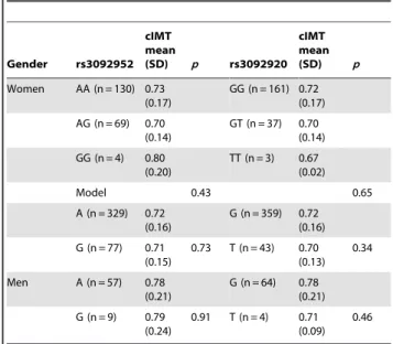
Since carotid intima-media thickness (IMT) has been found to predict the risk of CV events in the extended follow-up of patients with RA [42], in a further step we analyzed potential differences in the carotid IMT in 273 RA patients with no history of CV events stratified according to the genotype and allele distribution. In this regard, when we assessed the genotype distribution of theCD40 rs1535045 polymorphism, we observed that RA patients carrying the CC CD40rs1535045 genotype (n = 155) had higher carotid IMT (0.7560.19 mm) than those CTCD40rs1535045 heterozy-gous (n = 104; carotid IMT: 0.7260.16 mm) or the homozygous for the minor T allele CD40 rs1535045 (n = 14; carotid IMT: 0.7060.16 mm). However, these differences in the genotype distribution did not reach statistical significance. In keeping with this observation when we assessed allele distribution of theCD40 rs1535045 polymorphism, we observed that RA patients carrying the C allele of the CD40 rs1535045 gene variant (n = 414) had higher carotid IMT (0.7460.18 mm) than those carrying the less common T allele (n = 132; carotid IMT: 0.7160.16 mm). Nevertheless, differences were not statistically significant (p = 0.13) (Table 3). It was also the case for the remaining CD40 gene polymorphisms (Table 3), as well as CD154 gene variants assessed in the study (Table 4).
Interestingly, when the analysis of variance (ANOVA) model was adjusted for sex, age at the time of the ultrasonography assessment, follow-up time, and traditional CV risk factors (ANCOVA model), we did observe nominally significant evidence of association between carotid IMT and rs1535045 CD40 polymorphism (p= 0.023) (Table 5); therefore, we carried out a permutation test with 100000 replications in order to control type I error; in the permutation test, the relationship between carotid
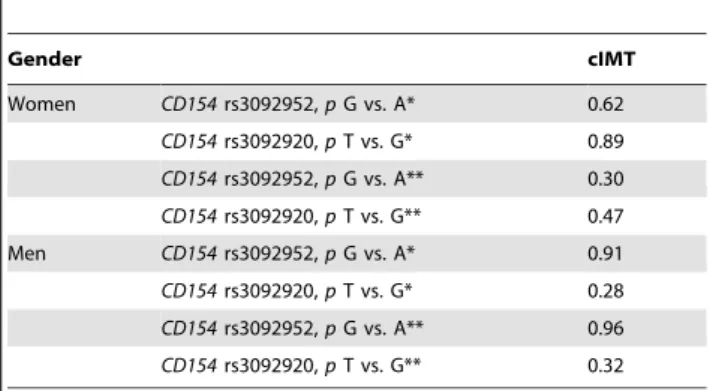
IMT and rs1535045 CD40 gene variant had p value = 0.048 (estimated with precision lower than 0.001). This result could be related to the fact that increased CD40 expression has been established in unstable atherosclerotic plaques [9], and carotid IMT is one of the parameters well known as a marker of early atherosclerosis. In addition,CD40rs1535045 SNP has previously been reported to be significantly associated with coronary artery calcification, which correlates with atherosclerosis and CV disease [39], although the SNP was not previously associated with carotid IMT. Since presence of anti-CCP antibodies had been previously associated with stronger evidence of CV disease in patients with RA [43], in a further step the ANCOVA model was also adjusted for anti-CCP status, that confirmed the statistically significant association between rs1535045CD40polymorphism and carotid IMT (p= 0.0047). Conditional analysis of the rs1883832 and rs1535045CD40gene variants in the adjusted ANCOVA model, rendered followingp-values: 0.82 and 0.0047 for rs1883832 and rs1535045, respectively. No association was found in case of CD154/CD40Lgene variants (Table 6). Our results indicate that a potential association between surrogate markers of atheroscle-rosis, in this case carotid IMT, andCD40 rs1535045 polymor-phism may exist. It may have clinical significance as carotid IMT has proved to be a good predictor of CV events in patients with
Table 3.Comparison of carotid artery intima-media thickness (cIMT) according toCD40rs1883832, rs4810485 and rs1535045 polymorphisms.
rs1883832
cIMT mm,
mean (SD) p rs4810485
cIMT mm,
mean (SD) p rs1535045
cIMT mm,
mean (SD) p
CC (n = 143) 0.73 (0.17) GG (n = 143) 0.73 (0.18) CC (n = 155) 0.75 (0.19)
CT (n = 107) 0.74 (0.18) GT (n = 106) 0.74 (0.18) CT (n = 104) 0.72 (0.16)
TT (n = 23) 0.75 (0.16) TT (n = 24) 0.75 (0.16) TT (n = 14) 0.70 (0.16)
Model 0.71 0.82 Model 0.29
C (n = 393) 0.73 (0.18) G (n = 392) 0.73 (0.18) C (n = 414) 0.74 (0.18)
T (n = 153) 0.74 (0.17) 0.41 T (n = 154) 0.74 (0.17) 0.52 T (n = 132) 0.71 (0.16) 0.13
doi:10.1371/journal.pone.0049214.t003
Table 4.Comparison of carotid artery intima-media thickness (cIMT) according toCD154rs3092920 and rs3092952 SNPs.
Gender rs3092952 cIMT mean
(SD) p rs3092920 cIMT mean (SD) p
Women AA (n = 130) 0.73 (0.17)
GG (n = 161) 0.72 (0.17) AG (n = 69) 0.70
(0.14)
GT (n = 37) 0.70 (0.14)
GG (n = 4) 0.80 (0.20)
TT (n = 3) 0.67 (0.02)
Model 0.43 0.65
A (n = 329) 0.72 (0.16)
G (n = 359) 0.72 (0.16) G (n = 77) 0.71
(0.15)
0.73 T (n = 43) 0.70 (0.13)
0.34
Men A (n = 57) 0.78 (0.21)
G (n = 64) 0.78 (0.21)
G (n = 9) 0.79 (0.24)
0.91 T (n = 4) 0.71 (0.09)
0.46
RA [42]. Association between endothelial dysfunction, another surrogate marker of atherosclerosis that is considered an early step in the atherogenesis process, and other genetic markers has been reported by other studies in RA patients [44,45,46]. These findings showing association between markers of subclinical atherosclerosis and genetic polymorphisms in RA have clinical relevance as they can help to identify patients at risk before CV events may occur.
Looking for a relation between rs1535045CD40gene variant and the presence of carotid plaques in patients with carotid IMT.1.0 mm, we did not find any significant association nor in the crude (allelicp= 0.71, 95% CI [0.23–1.81]) or adjusted by sex, age at RA diagnosis, follow-up time and traditional CV factors, model (allelicp= 0.65, 95% CI [0.23–1.83]), although number of patients involved was small (n = 19).
As in many other human genetic studies, potential limitations may exist in our study. In this regard, the phenotypes involved in the atherosclerosis disease, which some consider an inflammatory autoimmune disease [47], are complex and involve multiple small-to modest effects of multiple genes and environmental facsmall-tors. Since increased carotid IMT is a surrogate marker of atheroscle-rosis that may allow identifying high risk patients in a subclinical phase of the disease, longer duration of follow-up may be required to see the clinical relevance of the CD40 rs1535045 in the atherosclerosis disease of patients with RA. In keeping with our data showing association of subclinical atherosclerosis withCD40 rs1535045 gene variant, a recent study on 681 Swedish patients with RA disclosed an association between stroke/transient ischemic accident in men with RA that were homozygous for theCD40rs1535045 polymorphism major allele [48]. Regrettably, although our study encompassed a larger number of patients, we could not confirm association of any of the CD40 gene polymorphisms with clinically evident CV events in Spanish RA patients. There are a number of potential explanations that might enlighten these differences. First, RA is a complex disease and both the genetic backgrounds as well as environmental factors have clearly been shown to influence the phenotype expression of the disease and its potential complications. Second, the Swedish study included patients with longer follow-up (mean 15.5 years) from disease diagnosis than our cohort (median 10.8 years). Since the carotid IMT has been found to predict the development of CV events in the extended follow-up of patients with RA [42], a longer follow-up might be required to disclose a clinical association
between rs1535045 polymorphism inCD40and CV events in our population.
In conclusion, our data may indicate a potential association of the single nucleotide polymorphism located in the 59-UTR of the CD40gene (rs1883832,21C/T) with susceptibility to RA in the Spanish population. Also, based on our results, a potential influence of the CD40rs1535045 variant in the development of subclinical atherosclerosis in RA patients may exist. However, given that no other independent cohort was available for replication; our study should be regarded as a pilot one. Therefore, further studies in population with different genetic backgrounds are required to confirm the implication of CD40/CD154 system in the atherosclerosis process associated to RA.
Materials and Methods
Patients and Controls
Between March 1996 and September 2008,1575 consecutive patients that fulfilled the 1987 American College of Rheuma-tology classification criteria for RA [49] were recruited from the Rheumatology Outpatient Clinics of Hospital Xeral-Calde (Lugo), Hospital Clı´nico San Carlos (Madrid), Hospital Uni-versitario La Paz (Madrid), Hospital UniUni-versitario La Princesa (Madrid), Hospital Universitario Marque´s de Valdecilla (San-tander), and Hospital Universitario de Bellvitge (Barcelona), Spain. Patients and 1600 bone marrow and blood donors from National Repository DNA Bank (University of Salamanca, Spain), matched by age, sex and ethnicity, from the corre-sponding regions, were assessed for differences in the CD40 rs1883832, rs4810485 and rs1535045, and CD154 rs3092952 and rs3092920 gene variants.
Study Protocol
Ethics Statement. A subject’s written consent was obtained according to the declaration of Helsinki, and propose of the work was approved by the Ethics Committee of Galicia (Spain). The Ethics Committees of the Hospital Clı´nico San Carlos (Madrid), Hospital La Paz (Madrid), Hospital de La Princesa (Madrid), Hospital Universitario Bellvitge (Barcelona) and Hospital Universitario Marque´s de Valdecilla (Santander) also approved the study.
Between December 2009 and January 2010 patient’s clinical records were examined until patient’s death, loss of follow-up or
Table 5.Comparison of carotid intima-media thickness (cIMT) according toCD40rs1883832, rs4810485 and rs1535045 alleles in an adjusted ANCOVA model.
cIMT
CD40rs1883832,pT vs. C* 0.62
CD40rs4810485,pT vs. G* 0.63 CD40rs1535045,pT vs. C* 0.023
CD40rs1883832,pT vs. C**{ 0.59
CD40rs1535045,pT vs. C**{
0.023
*Analyses adjusted for sex, age at rheumatoid arthritis diagnosis, follow-up time, and traditional cardiovascular risk factors.
**Conditional analysis of the rs1883832 and rs1535045CD40gene variants.
{Analyses adjusted for sex, age at rheumatoid arthritis diagnosis, follow-up
time, traditional cardiovascular risk factors and anti-CCP status:p= 0.82 and 0.0047 for rs1883832 and rs1535045, respectively.
doi:10.1371/journal.pone.0049214.t005
Table 6.Comparison of cIMT according toCD154rs3092952 and rs3092920 alleles in an adjusted ANCOVA model, stratified by gender.
Gender cIMT
Women CD154rs3092952,pG vs. A* 0.62
CD154rs3092920,pT vs. G* 0.89 CD154rs3092952,pG vs. A** 0.30
CD154rs3092920,pT vs. G** 0.47 Men CD154rs3092952,pG vs. A* 0.91
CD154rs3092920,pT vs. G* 0.28
CD154rs3092952,pG vs. A** 0.96
CD154rs3092920,pT vs. G** 0.32
*Analyses adjusted for age at rheumatoid arthritis diagnosis, follow-up, and classic CV risk factors and anti-CCP status.
December 1st, 2009. Information on the main demographic characteristics, CV risk factors and CV events of RA patients is shown inTable 1. Clinical definitions for CV events and classic CV risk factors were described elsewhere [41,42,50]. A CV event was considered to be present if the patient had ischemic heart disease, heart failure, cerebrovascular accident or peripheral arteriopathy.
DNA was obtained from patients peripheral blood, using standard methods.
Genotyping
CD40andCD154genotyping. Subjects were genotyped to
determine:
*Three SNPs of CD40 associated with other autoimmune diseases: rs1883832 located in the Kozak consensus sequence of the 59-UTR, had been associated with GD [19]; while rs4810485, located in the first intron of the gene and in high linkage disequilibrium with rs1883832 (r2= 0.95), has been identified as a risk factor for RA [11]. Moreover, the major allele of rs1535045 SNP, located in the first intron of the gene, has been associated with subclinical atherosclerosis in diabetes families [39].
*Two genetic variants located in 59 UTR (rs3092952) and 39 UTR (rs3092920) ofCD40L(r2= 0.38) were selected. These SNPs are located in different haplotype blocks of CD40L [51]. The variant rs3092920 is located near to 39-UTR microsatellite, which was previously associated with RA and SLE [27,28]; while rs3092952 is a functional variant related with the levels of sCD40LG in plasma [26].
CD40 and CD154 variants genotyping were made using TaqMan Assays-on-Demand and TaqMan Genotyping Master Mix, and analyzed using the ABI 7900HT Fast Real-Time PCR System (Applied Biosystems, Foster City, CA, USA), following manufacturer instructions. Negative controls and duplicates were included to check the accuracy of the genotyping.
Shared Epitope determination. SeveralHLA-DRB1alleles (HLA-DRB1*0401, *0404, *0405, *0408, *0101, *0102, *1001, *1402) are associated with susceptibility to RA. These alleles encode a conserved amino acid sequence called the shared epitope, at position 70–74 in the third hypervariable region of the HLA-DRb1 molecule [52]. HLA-DRB1 typing was carried out using a reverse dot-blot kit with sequence-specific oligonucleotide (SSO) probes (Dynal RELITM SSO HLA-DRB1 typing kit; Dynal Biotech, Bromborough, UK).
Assessment of Carotid Plaque and Carotid Intima Media Thickness (IMT)
To determine the potential association between the CD40/ CD154 (CD40L) polymorphisms and the presence of subclinical atherosclerosis, between March 2007 and September 2010 a random subgroup of patients (n = 273) with no previous history of CV events was assessed by carotid ultrasonography to determine the carotid IMT and carotid plaques [53,54]. The common carotid arteries (CCAs) were evaluated with high resolution B-mode ultrasound (SONOS5500 and IE33, Philips Medical Systems, Andover, MA, USA) using a linear array 11-MHz probe with a standardized setup. Carotid IMT was measured at the posterior wall of the right and left CCAs, 10 mm from the carotid bifurcation, over the proximal 10-mm-long segment. The patients were placed in the supine position with their heads slightly bent in the opposite direction from the examination side. The right and left CCAs was first identified in B-mode, in a transverse view and followed from the proximal part to the bulb origin. Immediately afterwards, the CCAs and the most proximal part of the bulb were imaged in a
longitudinal view from a lateral approach. Both common carotid arteries were scanned longitudinally to visualize the intima-media complex of the far wall of the artery. The segments of the CCAs 10 mm proximal to the carotid bifurcation were scanned. The image was focused on the posterior carotid wall in longitudinal view. For measurement of the IMT, the distance between the leading edges of the lumen-intima interface and the media-adventitia interface of the B-mode frame was taken. The IMT was calculated for both the left and the right CCA, and the CCA-IMT was defined as the maximum of these. An ECG recording during the ultrasound examination was obtained for all patients. All measurements were performed in the end-diastole. Carotid plaque was considered to be present when there was a localized irregular thickening of at least 1.5 mm.
Statistical Analysis
All genotype data were checked for deviation from Hardy-Weinberg equilibrium (HWE) using http://ihg.gsf.de/cgi-bin/ hw/hwa1.pl.
Comparison of proportions was carried out using x2 test or Fisher test, when required. Strength of associations between CV events and genotypes or alleles ofCD40andCD154variants were estimated using odds ratios (OR) and 95% confidence intervals (CI), via multiple logistic regression; estimates were further adjusted by sex, age at RA diagnosis, time of follow-up, presence or absence of the rheumatoid shared epitope, and classic CV risk factors (hypertension, diabetes mellitus, dyslipidemia, obesity and smoking habit) as potential confounders. GivenCD40LGis located on the X-chromosome and the sex bias of RA disease, we performed the analysis separately to each gender for those gene variants.
The association between genotypes of the CD40 and CD154 SNPs and carotid IMT as surrogate marker of subclinical atherosclerosis were tested using unpaired t test to compare between 2 groups, and one-way analysis of variance (ANOVA) to compare among more than two groups. Moreover, we also tested the association between these parameters and alleles using analysis of covariance (ANCOVA) adjusting for gender, age and duration of the disease at the time of the ultrasonographic study, traditional CV risk factors, and anti-CCP status.
Nominal statistical significance was defined as p,0.05. Statis-tical significance, after applying a Bonferroni adjustment for 4 independent SNPs, was p,0.0125. We carried out a permutation test with 100,000 replications in order to control type I error, using the programme Stata 12/SE (StataCorp, College Station, TX, USA).
Statistical power for the study was calculated using ‘‘CaTS -Power Calculator for Two Stage Association Studies’’ (http:// www.sph.umich.edu/csg/abecasis/CaTS/) [55].
Supporting Information
Table S1 Logistic regression model to explain the presence of CV disease in patients with RA according to CD40rs1883832, rs4810485 and rs1535045 allele distribution.
(DOC)
Table S2 Logistic regression model to explain the presence of CV disease in RA patients stratified by gender according toCD154 rs3092952 and rs3092920 allele distribution.
Table S3 Conditional logistic regression analysis of CD40 rs1883832 and rs1535045 polymorphisms in the risk of cardio-vascular disease in RA patients.
(DOC)
Table S4 Conditional logistic regression analysis of CD154 rs3092952 and rs3092920 variants in CV disease risk stratified by gender (Xq26).
(DOC)
Table S5 Distribution of haplotypes ofCD40andCD154 gene variants in RA patients with and without CV disease.
(DOC)
Acknowledgments
We are indebted to Banco Nacional de ADN (University of Salamanca, Spain) which supplied part of the control DNA samples, and all patients and donors for their invaluable collaboration.
Author Contributions
Conceived and designed the experiments: MGB CGJ AC MT. Performed the experiments: MGB MT CGJ AC. Analyzed the data: MGB CGJ AC MT RLM SC JL. Contributed reagents/materials/analysis tools: MGB CGJ AC MT RLM SC JAMF AB BFG IGA CGV RB JL JM MAGG. Wrote the paper: MGB JM MAGG.
References
1. Bartoloni E, Shoenfeld Y, Gerli R (2011) Inflammatory and autoimmune mechanisms in the induction of atherosclerotic damage in systemic rheumatic diseases: two faces of the same coin. Arthritis Care Res (Hoboken) 63: 178–183. 2. Zinger H, Sherer Y, Shoenfeld Y (2009) Atherosclerosis in autoimmune rheumatic diseases-mechanisms and clinical findings. Clin Rev Allergy Immunol 37: 20–28.
3. Gonza´lez-Gay MA, Gonza´lez-Juanatey C, Martı´n J (2005) Rheumatoid arthritis: a disease associated with accelerated atherogenesis. Semin Arthritis Rheum 35: 8–17.
4. Montecucco F, Mach F (2009) Common inflammatory mediators orchestrate pathophysiological processes in rheumatoid arthritis and atherosclerosis. Rheumatology (Oxford) 48: 11–22.
5. Kawabe T, Matsushima M, Hashimoto N, Imaizumi K, Hasegawa Y (2011) CD40/CD40 ligand interactions in immune responses and pulmonary immunity. Nagoya J Med Sci 73: 69–78.
6. Peters AL, Stunz LL, Bishop GA (2009) CD40 and autoimmunity: the dark side of a great activator. Semin Immunol 21: 293–300.
7. Tousoulis D, Androulakis E, Papageorgiou N, Briasoulis A, Siasos G, et al. (2010) From atherosclerosis to acute coronary syndromes: the role of soluble CD40 ligand. Trends Cardiovasc Med 20: 153–164.
8. Kozera L, Andrews J, Morgan AW (2011) Cardiovascular risk and rheumatoid arthritis–the next step: differentiating true soluble biomarkers of cardiovascular risk from surrogate measures of inflammation. Rheumatology (Oxford) 50: 1944–1954.
9. Rizvi M, Pathak D, Freedman JE, Chakrabarti S (2008) CD40-CD40 ligand interactions in oxidative stress, inflammation and vascular disease. Trends Mol Med 14: 530–538.
10. Liu MF, Chao SC, Wang CR, Lei HY (2001) Expression of CD40 and CD40 ligand among cell populations within rheumatoid synovial compartment. Autoimmunity 34: 107–113.
11. Raychaudhuri S, Remmers EF, Lee AT, Hackett R, Guiducci C, et al. (2008) Common variants at CD40 and other loci confer risk of rheumatoid arthritis. Nat Genet 40: 1216–1223.
12. Stahl EA, Raychaudhuri S, Remmers EF, Xie G, Eyre S, et al. (2010) Genome-wide association study meta-analysis identifies seven new rheumatoid arthritis risk loci. Nat Genet 42: 508–514.
13. Orozco G, Eyre S, Hinks A, Ke X, Wilson AG, et al. (2010) Association of CD40 with rheumatoid arthritis confirmed in a large UK case-control study. Ann Rheum Dis 69: 813–816.
14. Plant D, Flynn E, Mbarek H, Dieude P, Cornelis F, et al. (2010) Investigation of potential non-HLA rheumatoid arthritis susceptibility loci in a European cohort increases the evidence for nine markers. Ann Rheum Dis 69: 1548–1553. 15. Hughes LB, Reynolds RJ, Brown EE, Kelley JM, Thomson B, et al. (2010) Most
common single-nucleotide polymorphisms associated with rheumatoid arthritis in persons of European ancestry confer risk of rheumatoid arthritis in African Americans. Arthritis Rheum 62: 3547–3553.
16. Vazgiourakis VM, Zervou MI, Choulaki C, Bertsias G, Melissourgaki M, et al. (2011) A common SNP in the CD40 region is associated with systemic lupus erythematosus and correlates with altered CD40 expression: implications for the pathogenesis. Ann Rheum Dis 70: 2184–2190.
17. Jacobson EM, Huber AK, Akeno N, Sivak M, Li CW, et al. (2007) A CD40 Kozak sequence polymorphism and susceptibility to antibody-mediated autoimmune conditions: the role of CD40 tissue-specific expression. Genes Immun 8: 205–214.
18. Kozak M (1987) At least six nucleotides preceding the AUG initiator codon enhance translation in mammalian cells. J Mol Biol 196: 947–950.
19. Jacobson EM, Concepcion E, Oashi T, Tomer Y (2005) A Graves’ disease-associated Kozak sequence single-nucleotide polymorphism enhances the efficiency of CD40 gene translation: a case for translational pathophysiology. Endocrinology 146: 2684–2691.
20. Blanco-Kelly F, Matesanz F, Alcina A, Teruel M, Diaz-Gallo LM, et al. (2010) CD40: novel association with Crohn’s disease and replication in multiple sclerosis susceptibility. PLoS One 5: e11520.
21. (2009) Genome-wide association study identifies new multiple sclerosis susceptibility loci on chromosomes 12 and 20. Nat Genet 41: 824–828.
22. Rodrı´guez-Rodrı´guez L, Castan˜eda S, Va´zquez-Rodrı´guez TR, Morado IC, Mari-Alfonso B, et al. (2010) Influence of CD40 rs1883832 polymorphism in susceptibility to and clinical manifestations of biopsy-proven giant cell arteritis. J Rheumatol 37: 2076–2080.
23. Gerdes N, Zirlik A (2011) Co-stimulatory molecules in and beyond co-stimulation - tipping the balance in atherosclerosis? Thromb Haemost 106: 804– 813.
24. Toubi E, Shoenfeld Y (2004) The role of CD40-CD154 interactions in autoimmunity and the benefit of disrupting this pathway. Autoimmunity 37: 457–464.
25. Tamura N, Kobayashi S, Kato K, Bando H, Haruta K, et al. (2001) Soluble CD154 in rheumatoid arthritis: elevated plasma levels in cases with vasculitis. J Rheumatol 28: 2583–2590.
26. Malarstig A, Lindahl B, Wallentin L, Siegbahn A (2006) Soluble CD40L levels are regulated by the -3459 A.G polymorphism and predict myocardial infarction and the efficacy of antithrombotic treatment in non-ST elevation acute coronary syndrome. Arterioscler Thromb Vasc Biol 26: 1667–1673. 27. Citores MJ, Rua-Figueroa I, Rodrı´guez-Gallego C, Durantez A,
Garcı´a-Laorden MI, et al. (2004) The dinucleotide repeat polymorphism in the 3’UTR of the CD154 gene has a functional role on protein expression and is associated with systemic lupus erythematosus. Ann Rheum Dis 63: 310–317.
28. Martı´n-Donaire T, Losada-Fernandez I, Perez-Chacon G, Rua-Figueroa I, Erausquin C, et al. (2007) Association of the microsatellite in the 3’ untranslated region of the CD154 gene with rheumatoid arthritis in females from a Spanish cohort: a case-control study. Arthritis Res Ther 9: R89.
29. Pollard KM (2012) Gender differences in autoimmunity associated with exposure to environmental factors. J Autoimmun 38: J177–186.
30. Lougaris V, Badolato R, Ferrari S, Plebani A (2005) Hyper immunoglobulin M syndrome due to CD40 deficiency: clinical, molecular, and immunological features. Immunol Rev 203: 48–66.
31. Silman AJ, Pearson JE (2002) Epidemiology and genetics of rheumatoid arthritis. Arthritis Res 4 Suppl 3: S265–272.
32. Stolt P, Bengtsson C, Nordmark B, Lindblad S, Lundberg I, et al. (2003) Quantification of the influence of cigarette smoking on rheumatoid arthritis: results from a population based case-control study, using incident cases. Ann Rheum Dis 62: 835–841.
33. Klareskog L, Stolt P, Lundberg K, Kallberg H, Bengtsson C, et al. (2006) A new model for an etiology of rheumatoid arthritis: smoking may trigger HLA-DR (shared epitope)-restricted immune reactions to autoantigens modified by citrullination. Arthritis Rheum 54: 38–46.
34. Bang SY, Lee KH, Cho SK, Lee HS, Lee KW, et al. (2010) Smoking increases rheumatoid arthritis susceptibility in individuals carrying the HLA-DRB1 shared epitope, regardless of rheumatoid factor or anti-cyclic citrullinated peptide antibody status. Arthritis Rheum 62: 369–377.
35. Innala L, Moller B, Ljung L, Magnusson S, Smedby T, et al. (2011) Cardiovascular events in early RA are a result of inflammatory burden and traditional risk factors: a five year prospective study. Arthritis Res Ther 13: R131.
36. Bartoloni E, Alunno A, Luccioli F, Moscatelli S, Biscontini D, et al. (2010) Atherosclerotic vascular damage and rheumatoid arthritis: a complex but intriguing link. Expert Rev Cardiovasc Ther 8: 1309–1316.
37. Kayrak M, Bacaksiz A, Ulgen MS, Vatankulu MA, Zengin K, et al. (2011) Plasma concentrations of soluble CD40 ligand in smokers with acute myocardial infarction: a pilot study. Heart Vessels 26: 131–137.
38. Carmona L, Villaverde V, Hernandez-Garcı´a C, Ballina J, Gabriel R, et al. (2002) The prevalence of rheumatoid arthritis in the general population of Spain. Rheumatology (Oxford) 41: 88–95.
39. Burdon KP, Langefeld CD, Beck SR, Wagenknecht LE, Carr JJ, et al. (2006) Variants of the CD40 gene but not of the CD40L gene are associated with coronary artery calcification in the Diabetes Heart Study (DHS). Am Heart J 151: 706–711.
40. Libby P (2008) Role of inflammation in atherosclerosis associated with rheumatoid arthritis. Am J Med 121: S21–31.
contribute to cardiovascular events and cardiovascular mortality in patients with rheumatoid arthritis. Arthritis Rheum 57: 125–132.
42. Gonza´lez-Juanatey C, Llorca J, Martı´n J, Gonza´lez-Gay MA (2009) Carotid intima-media thickness predicts the development of cardiovascular events in patients with rheumatoid arthritis. Semin Arthritis Rheum 38: 366–371. 43. Lo´pez-Longo FJ, Oliver-Minarro D, de la Torre I, Gonza´lez-Diaz de Rabago E,
Sa´nchez-Ramo´n S, et al. (2009) Association between anti-cyclic citrullinated peptide antibodies and ischemic heart disease in patients with rheumatoid arthritis. Arthritis Rheum 61: 419–424.
44. Rodrı´guez-Rodrı´guez L, Gonza´lez-Juanatey C, Garcı´a-Bermu´dez M, Va´zquez-Rodrı´guez TR, Miranda-Filloy JA, et al. (2011) CCR5Delta32 variant and cardiovascular disease in patients with rheumatoid arthritis: a cohort study. Arthritis Res Ther 13: R133.
45. Palomino-Morales R, Gonza´lez-Juanatey C, Va´zquez-Rodrı´guez TR, Rodrı´-guez L, Miranda-Filloy JA, et al. (2010) A1298C polymorphism in the MTHFR gene predisposes to cardiovascular risk in rheumatoid arthritis. Arthritis Res Ther 12: R71.
46. Lo´pez-Mejias R, Gonza´lez-Juanatey C, Garcı´a-Bermu´dez M, Castan˜eda S, Miranda-Filloy JA, et al. (2012) The lp13.3 genomic region -rs599839- is associated with endothelial dysfunction in patients with rheumatoid arthritis. Arthritis Res Ther 14: R42.
47. Sherer Y, Shoenfeld Y (2006) Mechanisms of disease: atherosclerosis in autoimmune diseases. Nat Clin Pract Rheumatol 2: 99–106.
48. Arlestig L, Rantapaa-Dahlqvist S (2012) Polymorphisms of the genes encoding CD40 and growth differentiation factor 15 and in the 9p21.3 region in patients with rheumatoid arthritis and cardiovascular disease. J Rheumatol 39: 939–945. 49. Arnett FC, Edworthy SM, Bloch DA, McShane DJ, Fries JF, et al. (1988) The American Rheumatism Association 1987 revised criteria for the classification of rheumatoid arthritis. Arthritis Rheum 31: 315–324.
50. Ho KK, Pinsky JL, Kannel WB, Levy D (1993) The epidemiology of heart failure: the Framingham Study. J Am Coll Cardiol 22: 6A–13A.
51. Chadha S, Miller K, Farwell L, Lightstone LB, Daly MJ, et al. (2005) Haplotype structure of TNFRSF5-TNFSF5 (CD40-CD40L) and association analysis in systemic lupus erythematosus. Eur J Hum Genet 13: 669–676.
52. Gregersen PK, Silver J, Winchester RJ (1987) The shared epitope hypothesis. An approach to understanding the molecular genetics of susceptibility to rheumatoid arthritis. Arthritis Rheum 30: 1205–1213.
53. Gonza´lez-Gay MA, Gonza´lez-Juanatey C, Va´zquez-Rodrı´guez TR, Martı´n J, Llorca J (2008) Endothelial dysfunction, carotid intima-media thickness, and accelerated atherosclerosis in rheumatoid arthritis. Semin Arthritis Rheum 38: 67–70.
54. Gonza´lez-Juanatey C, Llorca J, Garcı´a-Porrua C, Martı´n J, Gonza´lez-Gay MA (2006) Effect of anti-tumor necrosis factor alpha therapy on the progression of subclinical atherosclerosis in severe rheumatoid arthritis. Arthritis Rheum 55: 150–153.