BGD
6, 7083–7102, 2009Pysiology of
Ammonia tepida
L. J. de Nooijer et al.
Title Page Abstract Introduction Conclusions References Tables Figures
◭ ◮
◭ ◮
Back Close Full Screen / Esc
Printer-friendly Version Interactive Discussion Biogeosciences Discuss., 6, 7083–7102, 2009
www.biogeosciences-discuss.net/6/7083/2009/ © Author(s) 2009. This work is distributed under the Creative Commons Attribution 3.0 License.
Biogeosciences Discussions
Biogeosciences Discussionsis the access reviewed discussion forum ofBiogeosciences
Physiological controls on seawater
uptake and calcification in the benthic
foraminifer
Ammonia tepida
L. J. de Nooijer1, G. Langer1,2, G. Nehrke1, and J. Bijma1
1
Alfred Wegener Institute for Polar- and Marine Research (AWI), Am Handelshafen 12, 27570 Bremerhaven, Germany
2
Institute of Environmental Science and Technology, Universitat Aut `onoma de Barcelona (UAB), 08193 Bellaterra, Spain
Received: 7 July 2009 – Accepted: 11 July 2009 – Published: 16 July 2009
Correspondence to: L. J. de Nooijer ([email protected])
BGD
6, 7083–7102, 2009Pysiology of
Ammonia tepida
L. J. de Nooijer et al.
Title Page Abstract Introduction Conclusions References Tables Figures
◭ ◮
◭ ◮
Back Close Full Screen / Esc
Printer-friendly Version Interactive Discussion
Abstract
During the last decades conceptual models describing the calcification pathway of foraminifera and its physiological controls have been developed. These models are de-rived by combining data of tracer experiments and microscopic observations obtained from different species. Although vital for understanding their calcitic isotopic and trace 5
elemental composition, direct observational evidence on e.g. seawater vacuolization and intracellular Ca-cycling is lacking for most species. To analyse the relation be-tween seawater uptake and calcification, we incubated juveniles of the cosmopolitan benthic, intertidal foraminiferAmmonia tepida with various fluorescent probes. Visu-alizing the membranes of endocytosed vesicles was achieved by incubating speci-10
mens with the dye FM1-43, while Ca ions in the calcification vesicles were detected by the Ca2+-indicator Fluo3-AM. Uptake of fluorescent latex-beads (0.5 µm diameter) and subsequent transport to the site of chamber formation provided additional evi-dence that endocytosis is related to the calcification pathway and not merely involved in membrane cycling. Our results show for the first time that endocytosis of seawater 15
is part of the calcification process inAmmonia tepida. Data on the intracellular calcium ion-cycling allowed for calculating a preliminary cellular Ca-budget during foraminiferal calcification.
1 Introduction
Fossil foraminiferal calcite carries imprints from past oceanic conditions since incorpo-20
ration of many trace elements and stable isotopes is correlated to specific environmen-tal conditions. Among the more popular tools, the δ18O of foraminiferal calciumcar-bonate has been widely used to reconstruct seawater temperature (after correction for the global ice volume: Duplessy et al., 1970). More recently, foraminiferal Mg/Ca ra-tios have also been adopted to reconstruct past marine temperatures and are in good 25
BGD
6, 7083–7102, 2009Pysiology of
Ammonia tepida
L. J. de Nooijer et al.
Title Page Abstract Introduction Conclusions References Tables Figures
◭ ◮
◭ ◮
Back Close Full Screen / Esc
Printer-friendly Version Interactive Discussion for these relationships are based on culturing (e.g. N ¨urnberg et al., 1996) and field
stud-ies (e.g. Elderfield and Ganssen, 2000). Data presented in these studstud-ies have shown large differences to data from inorganic precipitation experiments (e.g. Mucci, 1996; Morse and Bender, 1990) and therefore imply that foraminiferal calcite is biologically controlled. This control is also reflected by differences in trace elemental and isotopic 5
composition within (Hathorne et al., 2003; Kunioka et al., 2006) and between species (Blackmon and Todd, 1959; Lear et al., 2000). In addition, for some benthic species it is shown that there is an ontogenetic offset in their calciticδ13C andδ18O (Schmiedl et al., 2004). The sources of these biological variabilities are commonly termed the “vital effect” and need to be accounted for to improve the accuracy of reconstructions based 10
on foraminiferal proxy-relationships (Erez, 2003).
Besides improving foraminiferal-based paleoreconstructions, another reason that un-derscores the necessity for analyzing their calcification pathway is the importance of the response of foraminifera to ocean acidification. Culture experiments have shown that shell weights of foraminifera decrease with decreasing carbonate ion concentra-15
tion of seawater (Bijma et al., 1999). Planktonic foraminifera from sediments deposited over glacial-interglacial cycles (Barker and Elderfield, 2002) and those deposited from pre-industrial to modern times (Moy et al., 2009) apparently confirm the trend found in cultured specimens. With a decreasing oceanic pH from a pre-industrial value of 8.2 to approximately 7.8 by the end of the century, changes in the amount of calcite 20
precipitated or in the export of CaCO3from the surface to the bottom of the ocean may
have consequences for the capacity of the ocean to take up atmospheric CO2(Heinze,
2004; Orr et al., 2005). To predict the response of foraminifera to ocean acidification, the sensitivity of the calcification pathway to changes in seawater alkalinity and pH need to be quantified.
25
BGD
6, 7083–7102, 2009Pysiology of
Ammonia tepida
L. J. de Nooijer et al.
Title Page Abstract Introduction Conclusions References Tables Figures
◭ ◮
◭ ◮
Back Close Full Screen / Esc
Printer-friendly Version Interactive Discussion a low Mg-content (2–20 mol % MgCO3) of most species’ calcite (Blackmon and Todd,
1959; Bentov and Erez, 2006). It is therefore assumed that foraminifera have Mg2+ -channels or pumps that remove magnesium from vacuolized seawater. An internal pool with a very low Mg/Ca ratio then remains and is used for calcite precipitation (Erez, 2003; Bentov and Erez, 2006). During calcification, the pH is increased to≥9.0. 5
This results in the conversion of bicarbonate into carbonate and thereby increases the calcite saturation state during chamber formation greatly (de Nooijer et al., 2009). The speed at which foraminifera can produce a new chamber suggests that all or most of the Ca2+ and inorganic carbon must be accumulated within the individual prior to chamber formation (Erez, 2003). This could be stored either as crystalline CaCO3 (as 10
in miliolids: Hemleben et al., 1986; Debenay et al., 1996), in separate pools (Anderson and Faber, 1984; Ter Kuile et al., 1989) or as amorphous CaCO3 (as suggested by
Erez, 2003; Bentov and Erez, 2006).
To understand trace elemental/isotopic fractionation in foraminiferal calcite, a number of conceptual models of the calcification pathways have been developed that are based 15
on experimental results and a suite of assumptions (Grossman, 1987; Ter Kuile et al., 1989; Ter Kuile and Erez, 1991; Elderfield et al., 1996; Erez, 2003). The ecological and physiological diversity among foraminiferal taxa spans symbiosis with and with-out photosynthetic organisms, benthic and planktic lifemodes, and precipitation of both calcitic and aragonitic test. This renders it very unlikely that the biomineralization pro-20
cess in foraminifera is similar among the taxa, which is well reflected in the substantial variety of Mg concentrations observed among foraminiferal CaCO3 (Bentov and Erez,
2006). Therefore, understanding biomineralization in foraminifera starts by obtaining all information on the calcification pathway from a single species, since it is not known to what extent observations from different species on e.g. seawater vacuolization can 25
be generalized.
BGD
6, 7083–7102, 2009Pysiology of
Ammonia tepida
L. J. de Nooijer et al.
Title Page Abstract Introduction Conclusions References Tables Figures
◭ ◮
◭ ◮
Back Close Full Screen / Esc
Printer-friendly Version Interactive Discussion various parts of their calcification pathway by using fluorescent probes and Confocal
Laser Scanning Microscopy (CLSM). The obtained results are used to construct a basic model and calculate Ca and CO3budgets for calcification inA. tepida.
2 Methods
2.1 Collection and maintainance
5
Sediment containing foraminifera was collected at the intertidal flats near Dorum, Northwestern Germany in fall 2008. Upon return in the laboratory, material was sieved over a 1 mm-screen to remove the largest macrofauna. The remaining sediment was stored at 10◦C and used to isolate living foraminifera. Before isolation, a small amount of sediment was sieved over a 250 µm-mesh and the remaining material was screened 10
for individuals of Ammonia tepida containing bright yellow protoplasm. Isolated, liv-ing adults ofA. tepida were incubated at 25◦C in 3 ml of filtered seawater (0.2 µm) in petridishes with a glass bottom and were fed livingDunaliella salina. Every two days the medium was replaced and once a week new food was provided. Regularly, adults underwent asexual reproduction, resulting in 50–200 juveniles consisting of a mega-15
lospheric proloculus (diameter≥40 µm). In the first week, these juveniles can grow a new chamber every day and were therefore selected to be incubated with several fluorescent probes and latex beads.
2.2 Fluorescent probes
Individuals with 3–5 chambers were incubated for 15 min with the fluorescent probe 20
Exicta-BGD
6, 7083–7102, 2009Pysiology of
Ammonia tepida
L. J. de Nooijer et al.
Title Page Abstract Introduction Conclusions References Tables Figures
◭ ◮
◭ ◮
Back Close Full Screen / Esc
Printer-friendly Version Interactive Discussion tion of the specimens was accomplished by a Kr/Ar laser, wavelength tuned to 488 nm.
Emission wavelengths between 600 and 650 nm were recorded.
Similar juveniles were placed in seawater with dissolved latex beads (Invitrogen, Molecular Probes). The beads had a diameter of 0.5 µm, an excitation optimum of 575 nm and an emission optimum of 610 nm. Individuals were allowed to take up the 5
beads for 24 h, washed with seawater and scanned under the CLSM after excitation at 568 nm.
Finally, juveniles were incubated with the fluorescent Ca2+-indicator Fluo3-AM (8 µM: Toyofuku et al., 2008) for 24 h. To track the Ca2+ utilization during chamber formation, individuals that were starting to make a new chamber were washed several times and 10
placed under the CLSM and excited by 488 nm every minute until chamber formation was finished.
3 Results
3.1 Cell membranes-vacuolization
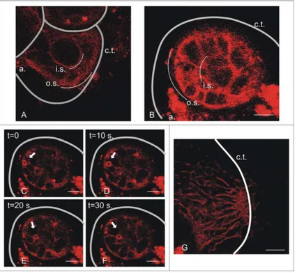
After 15 min of incubation with FM1-43, only cell membranes of the pseudopodia and 15
around the aperture are stained. In the hours following, fluorescence spreads through-out the individual, starting with membranes in the through-outer chambers. In a number of specimens, this reveals a quasi-circular organization of cell membranes in the final chamber (Fig. 1a,b). This structure consists of an outer and inner sphere, with a num-ber of strands of membranes connecting them. The shape of the outer sphere some-20
times resembles the form of the final chamber rather than a geometrical sphere. The spaces inbetween the membrane strands, as well as the space within the inner sphere appeared not to be filled with cytoplasm. Rather, the cytoplasm was confined to thin films bordered by labelled cell membranes. Occasionally, small vesicles (diameter app. 3 µm) are transported from the edge of the outer towards the inner sphere (Fig. 1c–f). 25
BGD
6, 7083–7102, 2009Pysiology of
Ammonia tepida
L. J. de Nooijer et al.
Title Page Abstract Introduction Conclusions References Tables Figures
◭ ◮
◭ ◮
Back Close Full Screen / Esc
Printer-friendly Version Interactive Discussion shows that pseudopodia extend radially from the former aperture towards the new
chamber wall. Around this new wall, these pseudopodia converge and form a zone of cytoplasm delining the site of calcification (Fig. 1g).
3.2 Fluorescent beads
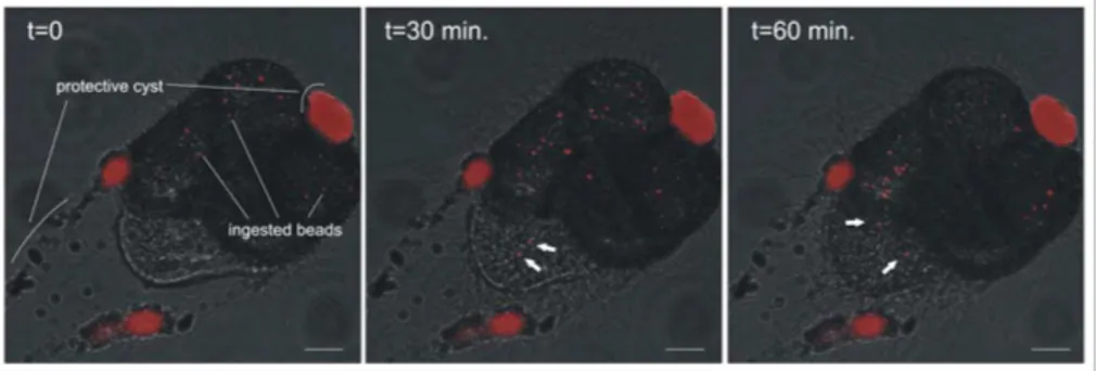
In the hours preceding chamber formation, foraminifera form a protective cyst around 5
themselves, in which many latex beads in the individual’s vicinity are incorporated (Fig. 2). A number of beads, however, are ingested prior to cyst formation and circulate through all chambers of the specimen. During calcification, some of these beads are transported from the cytoplasm into the final chamber towards the new chamber wall (Fig. 2).
10
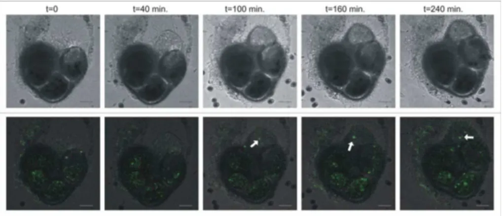
3.3 Ca2+ during chamber formation
Before and during chamber formation in juveniles of Ammonia tepida, two types of fluorescence by Fluo3-AM are present. As reported before (Toyofuku et al., 2008), a weak background fluorescence exists throughout the cytoplasm, and in a number of chambers brightly fluorescent vesicles circulate through the cytoplasm. From the start 15
until the end of chamber formation, a number of these bright vesicles are transported towards the site of calcification (Fig. 3). Over 90% of the Ca2+-containing vesicles do not participate in chamber formation and remain in the cytoplasm after the new chamber is completed. In several individuals that were observed, the amount of such vesicles that are transported to the site of calcification in a new chamber does not 20
BGD
6, 7083–7102, 2009Pysiology of
Ammonia tepida
L. J. de Nooijer et al.
Title Page Abstract Introduction Conclusions References Tables Figures
◭ ◮
◭ ◮
Back Close Full Screen / Esc
Printer-friendly Version Interactive Discussion
4 Discussion
4.1 Seawater uptake
Theoretically, the internal Ca- and carbonate-pools from which foraminifera produce new chambers can be formed by transporting Ca-ions and dissolved inorganic car-bon via trans-membrane transport from the medium into the cytoplasm (as in coc-5
colithophores; Brownlee and Taylor, 2004). It has been reported, however, that their internal pools are essentially modified seawater (e.g. Erez, 2003). Our results are the first direct evidence thatA. tepidavacuolizes seawater and that the internal pools containing Ca2+and CO2−3 are very likely modified seawater. The vacuolization of sea-water and its participation in the calcification pathway were inferred from the following 10
observations. The spherical organization of cell membranes in the final chamber and the transport of small vesicles (Fig. 1) strongly suggest that seawater vacuolization takes place. An alternative explanation for the occurrence of these vesicles could be membrane recycling unrelated to calcification. This, however, appears to be unlikely, because the fluorescent beads that cycle through the cytoplasm of incubated individu-15
als (Fig. 2) can only be taken up by vacuolization. This also indicates that the observed endocytosis serves the purpose of taking up ions from seawater and is supported by two observations. First, some of the vesicles containing beads were transported from the cytoplasm into the final chamber towards the new chamber wall (Fig. 2). This indicates that these vesicles transport material, i.e. Ca and/or carbonate ions for cal-20
cification. In addition, transport of high-Ca vesicles to the site of calcification (Fig. 3) shows that the Ca necessary for calcification comes from within the individual. This shows thatA. tepidauses endocytosis-vesicles to build up an internal Ca reservoir that is subsequently used for calcification: below we will use this mechanism to calculate a Ca-budget for this species.
BGD
6, 7083–7102, 2009Pysiology of
Ammonia tepida
L. J. de Nooijer et al.
Title Page Abstract Introduction Conclusions References Tables Figures
◭ ◮
◭ ◮
Back Close Full Screen / Esc
Printer-friendly Version Interactive Discussion
4.2 Ca-utilization during chamber formation
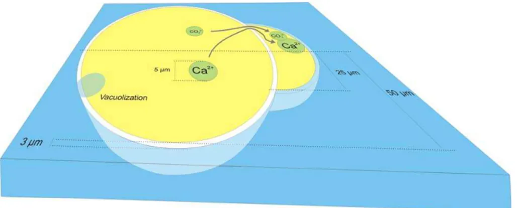
The observation of calcium-rich vesicles during chamber formation (Fig. 3) was also reported by Toyofuku et al. (2008), but the time-lapse recordings reported here allow estimating the fluxes of intracellular Ca2+ in relation to chamber formation. To do so, we propose a conceptual model for Rotallid foraminiferal chamber formation (Fig. 4). 5
In this simplification, a small individual consists of one spherical chamber and is build-ing a second, small chamber with the shape of half a sphere. Whether the first, inner chamber is really one chamber or consists of a number of chambers (as the individuals in Figs. 2 and 3), does not affect the volume ratios between the new and older cham-bers significantly. For simplicity, we assumed that pore volume (reducing the volume of 10
calcite in a new chamber) and the amount of calcite precipitated as a layer onto older chambers (increasing the volume of calcite precipitated; Erez, 2003) are in the same range and therefore do not need to be corrected for. We also assume that all Ca2+ for this new chamber is located in an intracellular Ca-pool formed as described above. In our simplified model, the new chamber has a wall thickness of 3 µm, as estimated from 15
embedded and cross-sectioned specimens (results not shown here).
The volume of the calcite precipitated to form this new chamber equals 1.33×10⁻⁸cm3CaCO3. With a density of 2.71 g/cm
3
, this new chamber con-tains 3.60×10⁻⁸g of CaCO3. With 40 wt% Ca, this means that 1.44×10⁻⁸g (or 3.6×10⁻¹⁰mol) of Ca2+ must be inside the foraminifer at the onset of chamber for-20
mation. Assuming that all calcium is ultimately derived from seawater and that 100% of the calcium taken up is utilized for calcification, a volume of 3.50×10⁻⁸l of seawater ([Ca2+]=10.3 mmol/l) is needed.
In our simplified model, the individual has a total volume of 5.24×10⁻¹⁰l (Fig. 4), excluding the volume of the new chamber. This means that this foraminifer has to pro-25
pre-BGD
6, 7083–7102, 2009Pysiology of
Ammonia tepida
L. J. de Nooijer et al.
Title Page Abstract Introduction Conclusions References Tables Figures
◭ ◮
◭ ◮
Back Close Full Screen / Esc
Printer-friendly Version Interactive Discussion vious study the intracellular Ca-pool ofA. lobiferawas estimated to contain Ca2+ with
a concentration of 2–20 M (Erez, 2003). Considering the uncertainties in the methods and assumptions used, our results can be regarded as complementing the previous estimations. The Ca-concentration of the putative pool cannot represent free Ca-ions, because Ca-ATPases are not able to build up the respective Ca-gradient between cy-5
tosol and vesicle-interior (for a more detailed discussion see: Langer et al., 2006). If we would assume that the Ca-pool is basically seawater, i.e. Ca in an aqueous solu-tion, the consequence is that the Ca-pool does not contain enough Ca-ions to produce a new chamber, implying that more Ca must be taken up during chamber formation. We have observed, however, that on average only 20 vesicles (with a diameter of 5 µm) 10
were consumed in the production of a new chamber. Supposing that all Ca2+ neces-sary for the new chamber is stored in these 20 vesicles, its concentration would equal approximately 50 000 M. This estimation clearly shows that Ca cannot be transported to the site of calcification in vesicles containing seawater (i.e.∼10 mM Ca).
This conclusion only holds if most of the Ca used for a new chamber can be de-15
tected by our method. For example, it could be that most of the Ca-transport is through very small vesicles that bud offfrom the larger ones, but are invisible even when us-ing confocal microscopy. Another possibility is that Ca is sequestered by specialized organic compounds in the Ca-pool that would lower the free calcium-concentration. Release, however, from these organics at the site of calcification should then be visible 20
as a fluorescent zone at the new chamber wall, which was not observed. It may also be that most calcium is stored in the form of amorphous calciumcarbonate (ACC) prior to chamber formation. The Ca in amorphous CaCO3would not bind to Fluo3 and thus not
cause fluorescence to be emitted from the Ca-containing vesicles. Furthermore, the ACC would not be detectable by examining these individuals with crosspolarized light. 25
BGD
6, 7083–7102, 2009Pysiology of
Ammonia tepida
L. J. de Nooijer et al.
Title Page Abstract Introduction Conclusions References Tables Figures
◭ ◮
◭ ◮
Back Close Full Screen / Esc
Printer-friendly Version Interactive Discussion aragonite grow by the addition of stable clusters of ACC. Foraminifera may be able to
control these conversions precisely so that very little free calcium is observed to par-ticipate in chamber formation. In addition, the size of these ACC clusters may explain the observation that most foraminifera’s tests consist of small crystallites (Debenay et al., 1996).
5
4.3 Implications for trace metal partitioning
Compared to most elements, magnesium is incorporated into the calcite of many foraminiferal species with a partitioning coefficient much lower than the one deter-mined for inorganic calcite formation (e.g. Blackmon and Todd, 1959; N ¨urnberg et al., 1996; Rosenthal et al., 1997). For Ammonia tepida/A. beccarii the Mg/Cacalcite is 1–
10
10 mmol/mol (Allison and Austin, 2003; de Nooijer et al., unpublished data), suggest-ing that this species precipitates its calcite from an internal Ca-reservoir with a much lower Mg/Ca than that of seawater. The mechanism that discriminates Mg2+from Ca2+ is not known, but our results imply that they are separatedafter mutual vacuolization. An obvious possibility is that Mg2+ is pumped out of the seawater vacuoles and trans-15
ported back into the surrounding medium. In that case the fluid of Ca-transporting vesicles would be essentially Mg-depleted seawater. Another possibility is that Ca2+ is pumped out of these vacuoles into an intracellular Ca-reservoir, i.e. another type of vesicle. In that case the fluid of Ca-transporting vesicles would have a composi-tion radically different from seawater. For many trace elements, such as Sr, Ba, and 20
Cd, the fractionation between seawater and foraminiferal calcite can be explained by Raleigh fractionation during CaCO3 precipitation from an internal reservoir (Elderfield
et al., 1996). This model implies that for these trace elements, the internal reservoirs are comparable to seawater and thus, the Mg-pumping mechanism, as opposed to the Ca-pumping mechanism, is more likely to be responsible for the low Mg/Ca ratio in 25
BGD
6, 7083–7102, 2009Pysiology of
Ammonia tepida
L. J. de Nooijer et al.
Title Page Abstract Introduction Conclusions References Tables Figures
◭ ◮
◭ ◮
Back Close Full Screen / Esc
Printer-friendly Version Interactive Discussion be transported back into the medium, most of the fluorescent beads would not remain
inside the cell.
Given that Sr partitioning can be explained by Raleigh fractionation (Elderfield et al., 1996), the question arises what that internal reservoir actually is and what amount of calcite is precipitated from it. Assuming that an entire chamber is precipitated from 5
this reservoir, one would expect an area of elevated Ca-concentration (in comparison to cytosolic background) in the vicinity of the primary organic sheet. Since we have not observed this during chamber formation (Fig. 3), we conclude that the reservoir is much smaller and that it is impossible to precipitate an entire chamber from a single reservoir. This conclusion fits the possibility of the involvement of ACC-cluster trans-10
formation in chamber formation (see above). Since the Sr partitioning coefficients for ACC precipitation are unknown, but may be higher than for calcite precipitation from an aqueous solution, a Raleigh fractionation does not necessarily be the mechanism of trace metal partitioning.
4.4 Inorganic carbon-utilization during chamber formation
15
To cover the amount of carbonate needed during chamber formation, our hypothetical individual (Fig. 4) would need an even greater volume of seawater. Assuming that all inorganic carbon in vacuolized seawater can be converted into carbonate by increasing the pH (de Nooijer et al., 2009), a juvenile would still need approximately 17.5×10⁻⁸l seawater (more than 300 times its own volume). This amount of seawater could be 20
reduced in case the intracellular carbonate-pool is enriched with metabolic CO2(upto 10%; Grossman, 1987; Ter Kuile, 1991). Tracer experiments with 14C have shown that the hyaline speciesAmphistegina lobiferahas an intracellular carbon pool with an estimated concentration of app. 2 M (Ter Kuile and Erez, 1988). This matches the estimated Ca-concentration of the intracellular Ca-pool (Erez, 2003). From inorganic 25
calcite precipitation experiments it is known that a 1:1 Ca:CO3ratio results in highest
BGD
6, 7083–7102, 2009Pysiology of
Ammonia tepida
L. J. de Nooijer et al.
Title Page Abstract Introduction Conclusions References Tables Figures
◭ ◮
◭ ◮
Back Close Full Screen / Esc
Printer-friendly Version Interactive Discussion Direct measurements on the concentrations of intracellular Ca and inorganic carbon
need to be made to improve the model for calcification as described above. In addition, the phase in which they are present in the internal pools (i.e. free, sequestered by organic compounds or as ACC) need to be complemented by our results to understand the effect that foraminiferal physiology has on trace element and isotope fractionation 5
during chamber formation inA. tepida.
References
Allison, N. and Austin, W. E. N.: The potential of ion microprobe analysis in detecting geo-chemical variations across individual foraminifera tests, Geochem. Geophy. Geosy., 4(2), 8403, doi:10.1029/2002GC000430, 2003.
10
Anderson, O. R. and Faber Jr., W. W.: An estimation of calcium carbonate deposition rate in a planktonic foraminiferGlobigerinoides sacculifer using45Ca as a tracer: A recommended procedure for improved accuracy, J. Foramin. Res., 14(4), 303–308, 1984.
Barker, S. and Elderfield, H.: Foraminiferal calcification response to glacial-interglacial changes in atmospheric CO2, Science, 297, 333–336, 2002.
15
Bentov, S. and Erez, J.: Impact of biomineralization processes on the Mg content of foraminiferal shells: a biological perspective, Geochem. Geophy. Geosy., 7(1), Q01P08, doi:10.1029/2005GC001015, 2006.
Bijma., J., Spero, H. J., and Lea, D. W.: Reassessing foraminiferal stable isotope geochemistry: Impact of the oceanic carbonate system (experimental results), in: Use of proxies in
pale-20
oceanography, edited by: Fischer, H. and Wefer, G., Springer, Berlin, Germany, 489–512, 1999.
Blackmon, P. D. and Todd, R.: Mineralogy of some foraminifera as related to their classification and ecology, J. Paleontol., 33(1), 1–15, 1959.
Brownlee, C. and Taylor, A. R.: Calcification in coccolithophores: A cellular perspective, in:
25
Coccolithophores-from molecular processes to global impact, edited by: Thierstein, H. R. and Young, J. R., Springer, Germany, 31–49, 2004.
BGD
6, 7083–7102, 2009Pysiology of
Ammonia tepida
L. J. de Nooijer et al.
Title Page Abstract Introduction Conclusions References Tables Figures
◭ ◮
◭ ◮
Back Close Full Screen / Esc
Printer-friendly Version Interactive Discussion
de Nooijer, L. J., Reichart, G. J., Due ˜nas-Boh ´orquez, A., Wolthers, M., Ernst, S. R., Mason, P. R. D., and van der Zwaan, G. J.: Copper incorporation in foraminiferal calcite: results from culturing experiments, Biogeosciences, 4, 493–504, 2007,
http://www.biogeosciences.net/4/493/2007/.
de Nooijer, L. J., Toyofuku, T., and Kitazato, H.: Foraminifera promote calcification by elevating
5
their intracellular pH, PNAS, submitted, 2009.
Dissard, D., Nehrke, G., Reichart, G. J., and Bijma, J.: Impact of seawater pCO2 changes on calcification and on Mg/Ca and Sr/Ca in benthic foraminifera calcite (Ammonia tepida): results from culturing experiments, Biogeosciences Discuss., 6, 3771–3802, 2009,
http://www.biogeosciences-discuss.net/6/3771/2009/.
10
Duplessy, J. C., Lalou, C., and Vinot, A. C.: Differential isotopic fractionation in benthic foraminifera and paleotemperatures reassessed, Science, 168(3928), 250–251, 1970. Elderfield, H., Bertram, C. J., and Erez, J.: A biomineralization model for the incorporation of
trace elements into foraminiferal calcium carbonate, Earth Planet. Sc. Lett., 142, 409–423, 1996.
15
Elderfield, H. and Ganssen, G.: Past temperature andδ18O of surface ocean waters inferred from foraminiferal Mg/Ca ratios, Nature, 405, 442–445, 2000.
Erez, J.: The source of ions for biomineralization in foraminifera and their implications for pa-leoceanographic proxies, in: Reviews in mineralogy and geochemistry, vol. 54, edited by: Dove, P. M., De Yoreo, J. J., and Weiner, S., Mineralogical Society of America, Geochemical
20
Society, Washington, USA, 115–149, 2003.
Gebauer, D., V ¨olkel, A., and C ¨olfen, H.: Stable prenucleation calcium carbonate clusters, Sci-ence, 322, 1819–1822, 2009.
Grossman, E. L.: Stable isotopes in modern benthic foraminifera: a study of vital effect, J. Foramin. Res., 17(1), 48–61, 1987.
25
Hathorne, E. C., Alard, O., James, R. H., and Rogers, N. W.: Determination of intratest variabil-ity of trace elements in foraminifera by laser ablation inductively coupled mass spectrometry, Geochem. Geophy. Geosy., 4(12), 8408, doi:10.1029/2003GC000539, 2003.
Heinze, C.: Simulating oceanic CaCO3 export production in the greenhouse, Geophys. Res. Lett., 31, L16308, doi:10.1029/2004GL020613, 2004.
30
BGD
6, 7083–7102, 2009Pysiology of
Ammonia tepida
L. J. de Nooijer et al.
Title Page Abstract Introduction Conclusions References Tables Figures
◭ ◮
◭ ◮
Back Close Full Screen / Esc
Printer-friendly Version Interactive Discussion
Clarendon, Oxford, UK, 237–249, 1986.
Kunioka, D., Shirai, K., Takahata, N., Sano, Y., Toyofuku, T., and Ujiie, Y.: Microdistribution of Mg/Ca, Sr/Ca, and Ba/Ca ratios inPulleniantina obliquilloculatatest by using a NanoSIMS: Implication for the vital effect mechanism, Geochem. Geophy. Geosy., 7(12), Q12P20, doi:10.1029/2006GC001280, 2006.
5
Langer, G., Gussone, N., Nehrke, G., Riebesell, U., Eisenhauer, A., Kuhnert, H., Rost, B., Trimborn, S., and Thoms, S.: Coccolith strontium to calcium ratios inEmiliania huxleyi: The dependence on seawater strontium and calcium concentrations, Limnol. Oceanogr., 51(1), 310–320, 2006.
Lear, C. H., Elderfield, H., and Wilson, P. A.: Cenozoic deep-sea temperatures and global ice
10
volumes from Mg/Ca ratios in benthic foraminiferal calcite, Science, 287(2), 269–272, 2000. Morse, J. W. and Bender, M. L.: Partition coefficients in calcite: Examination of factors
influenc-ing the validity of experimental results and their application to natural systems, Chem. Geol., 82, 265–277, 1990.
Moy, A. D., Howard, W. R., Bray, S. G., and Trull, T. W.: Reduced calcification in modern
15
Southern ocean plank tonic foraminifera, Nat. Geosci., 2, 276–280, 2009.
Mucci, A.: Growth kinetics and composition of magnesian calcite overgrowths precipitated from seawater: Quantitative influence of orthophosphate ions, Geochim. Cosmochim. Ac., 50(10), 2255–2265, 1986.
Nehrke, G., Reichart, G.-J., Van Capellen, P., Meile, C., and Bijma, J.: Dependence of calcite
20
on growth rate and Sr partitioning on solution stoichiometry: Non-Kossel crystal growth, Geochim. Cosmochim. Ac., 71, 2240–2249, 2007.
N ¨urnberg, D., Bijma, J., and Hemleben, C.: Assessing the reliability of magnesium in foraminiferal calcite as a proxy for water mass temperatures, Geochim. Cosmochim. Ac., 60(5), 803–814, 1996.
25
Orr, J. C., Fabry, V. J., Aumont, O., Bopp, L., Doney, S. C., Feely, R. A., Gnanadesikan, A., Gruber, N., Ishida, A., Joos, F., Key, R. M., Lindsay, K., Maier-Reimer, E., Matear, R., Mon-fray, P., Mouchet, A., Najjar, R. G., Plattner, G.-K., Rodgers, K. B., Sabine, C. L., Sarmiento, J. L., Schlitzer, R., Slater, R. D., Totterdell, I. J., Weirig, M.-F., Yamanaka, Y., and Yool, A.: Anthropogenic ocean acidification over the twenty-first century and its impact on calcifying
30
organisms, Nature, 437, 681–686, 2005.
BGD
6, 7083–7102, 2009Pysiology of
Ammonia tepida
L. J. de Nooijer et al.
Title Page Abstract Introduction Conclusions References Tables Figures
◭ ◮
◭ ◮
Back Close Full Screen / Esc
Printer-friendly Version Interactive Discussion
Cryo-TEM, Science, 323, 1455–1458, 2009.
Rosenthal, Y., Boyle, E. A., and Slowley, N.: Temperature control on the incorporation of magnesium, strontium, fluorine, and cadmium into benthic foraminiferal shells from Little Bahama Bank: prospects for thermocline paleoceanography, Geochim. Cosmochim. Ac., 61(17), 3633–3643, 1997.
5
Schmiedl, G., Pfeilsticker, M., Hemleben, C., and Mackensen, A.: Environmental and biological effects on the stable isotope composition of recent deep-sea benthic foraminifera from the western Mediterranean Sea, Mar. Micropaleontol., 51, 129–152, 2004.
Ter Kuile, B. and Erez, J.: The size and function of the internal inorganic carbon pool of the foraminiferAmphistegina Iobifera, Mar. Biol., 99(4), 481–487, 1998.
10
Ter Kuile, B., Erez, J., and Padan, E.: Mechanisms for the uptake of inorganic carbon by two species of symbiont-bearing foraminifera, Mar. Biol., 103, 241–251, 1989.
Ter Kuile, B. H. and Erez, J.: Carbon budgets for two species of benthonic symbiont-bearing foraminifera, Biol. Bull., 180, 489–495, 1991.
Ter Kuile, B.: Mechanisms for calcification and carbon cycling in algal symbiont-bearing
15
foraminifera, in: Biology of Foraminifera, edited by: Lee, J. J. and Anderson, O. R., Aca-demic Press, London, 73–89, 1991.
Toyofuku, T., de Nooijer, L. J., Yamamoto, H., and Kitazato, H.: Real-time visualization of cal-cium ion activity in shallow benthic foraminiferal cells using the fluorescent indicator Fluo-3 AM, Geochem. Geophy. Geosy., 9(5), Q05005, doi:10.1029/2007GC001772, 2008.
BGD
6, 7083–7102, 2009Pysiology of
Ammonia tepida
L. J. de Nooijer et al.
Title Page Abstract Introduction Conclusions References Tables Figures
◭ ◮
◭ ◮
Back Close Full Screen / Esc
Printer-friendly Version Interactive Discussion Fig. 1.Distribution of cell membranes in the final chamber of two juveniles ofAmmonia tepida.
(A)Location of the two spheres with a number of cytoplasmic strands inbetween them. (B)
BGD
6, 7083–7102, 2009Pysiology of
Ammonia tepida
L. J. de Nooijer et al.
Title Page Abstract Introduction Conclusions References Tables Figures
◭ ◮
◭ ◮
Back Close Full Screen / Esc
Printer-friendly Version Interactive Discussion Fig. 2.Distribution of fluorescent beads in a calcifying juvenile ofAmmonia tepida. Many beads
BGD
6, 7083–7102, 2009Pysiology of
Ammonia tepida
L. J. de Nooijer et al.
Title Page Abstract Introduction Conclusions References Tables Figures
◭ ◮
◭ ◮
Back Close Full Screen / Esc
Printer-friendly Version Interactive Discussion Fig. 3.Distribution of Ca2+in a calcifying juvenile ofAmmonia tepida. The upper row shows the
BGD
6, 7083–7102, 2009Pysiology of
Ammonia tepida
L. J. de Nooijer et al.
Title Page Abstract Introduction Conclusions References Tables Figures
◭ ◮
◭ ◮
Back Close Full Screen / Esc
Printer-friendly Version Interactive Discussion
Fig 4: Simplified representation of calcification in a Rotallid juvenile, based on our