O
h
r
c
i
r
g
a
in
e
a
s
l
R
e
Emrah Kovalak, Tolga Atay, Y. Barbaros Baykal, Özgür Başal Department of Orthopaedics and Traumatology, Süleyman Demirel University Medical Faculty, Isparta, Turkey Proximal Humerus Fractures
Surgical Management of 3 and 4-Part Proximal
Humerus Fractures with Locking Plates in Elderly
Yaşlı Hastalarda 3 ve 4 Parçalı Proksimal
Humerus Kırıklarının Kilitli Plak ile Tedavisi
DOI: 10.4328/JCAM.4834 Received: 12.10.2016 Accepted: 13.11.2016 Printed: 01.05.2017 J Clin Anal Med 2017;8(3): 243-7 Corresponding Author: Emrah Kovalak, Ortopedi ve Travmatoloji Anabilim Dalı, Süleyman Demirel Üniversitesi Tıp Fakültesi, 32260 Çünür, Isparta, Türkiye. GSM: +905332346280 E-Mail: [email protected].
Özet
Amaç: Proksimal humerus kırıkları tüm kırıkların yaklaşık %5’ini oluşturur ve bunların da %15-20’si deplase ve dengesizdir. Kilitli plakların ortaya çıkma-sından sonra bu kırıkların osteosentez ile tedavisinde artış olmuştur. Ancak, hala bu kompleks kırıkların tedavisinde bir fikir birliği oluşmamıştır. Bu ret-rospektif çalışma ile yaşlılarda 3 ve 4- parçalı proksimal humerus kırıklarının kilitli plakla osteosentezinin fonksiyonel sonuçları ve prognozu etkileyen fak-törleri değerlendirmeyi amaçladık. Gereç ve Yöntem: Çalışmaya 2010- 2015 yılları arasında deplase, 3 ve 4 parçalı proksimal humerus kırığı tanısıyla kilit-li plak ile osteosentez yapılan 53 hasta dahil edildi. Kırıkların sınılaması Neer Sınılama sistemine göre yapıldı. Sonuçlar, Constant-Murley Skorlama siste-mi (CMS), görsel analog ağrı ölçeği, ve düz radyografi ile değerlendirildi. Ha-reket açıklığının değerlendirilmesi amacı ile kolun öne elevasyonu ve abduk-siyonu ölçüldü. Bulgular: 3 ve 4 parçalı kırıklar arasında, CMS, öne elevasyon ve abdüksiyonda istatiksel olarak bir fark saptanmadı (p>0.05). Ağrı 4 parçalı kırıklarda belirgin olarak daha fazla idi (p=0.035). CMS, öne elevasyon ve ab-duksiyon, yaş ve cerrahi süresinde gecikme ile ters yönde korale idi. Kompli-kasyon gelişen hastalar ile gelişmeyenler arasında CMS, öne elevasyon ve ab-duksiyonda istatiksel olarak anlamlı farklılık saptandı (p=0.029, p=0.017 and p=0.024). Tartışma: Proksimal humerus kırıklarının kilitli plak ile tespiti son-rasında fonksiyonel sonuçlar; hastaya ait faktörler, kırık yapısı, cerrah ve imp-lanta bağlı olmak üzere birçok faktörle ilişkilidir. Endikasyonlar dikkatli seçil-diği takdirde 3 ve 4 parçalı proksimal humerus kırıklarının kilitli plaklarla os-teosentezi iyi sonuçlar vermektedir.
Anahtar Kelimeler
Humerus Kırıkları; Proksimal; Omuz Kırıkları; Kırık Sabitlenmesi; İnternal
Abstract
Aim: Proximal humeral fractures are approximately 5% of all fractures and, %15-20 is displaced and unstable. By the introduction of locking plates there used to be a substantial rise in the osteosynthesis of the 3 and 4-part proxi-mal humeral fractures. But there is still a lack of consensus for the optiproxi-mal treatment of these complex fractures. In this retrospective study, we aimed to evaluate the functional outcomes and prognostic factors of 3 and 4-part proximal humerus fractures treated with locking plate osteosynthesis in el-derly. Material and Method: 53 patients with displaced 3 and 4-part proximal humeral fractures treated with locking plate osteosynthesis between 2010 and 2015 were included. The fractures were classiied according to Neer classiication system. Outcomes were assessed by Constant-Murley scoring system (CMS), visual analog pain scale and plain radiographs. In reference to range of motion, forward elevation and abduction of the arm were measured. Results: No statistically signiicant diferences found between the 3- part and 4- part fractures in CMS, forward elevation and, abduction (p>0.05). Pain was signiicantly higher in 4-part fractures (p=0.035). CMS, forward eleva-tion, and abduction were inversely correlated with age and delay in surgery. There was statistical signiicance between the patients had complications and those not in terms of CMS, forward elevation and, abduction (p=0.029, p=0.017 and p=0.024). Discussion: Functional outcomes of locking plate ixa-tion of proximal humerus fractures are associated with patient related fac-tors, fracture pattern, surgeon and, the implant. When indications are care-fully selected, locking plate osteosynthesis yield good outcomes in surgical treatment of 3 or 4-part proximal humerus fractures.
Keywords
Introduction
Proximal humeral fractures account for approximately 5% of all fractures and usually afect women over 50 years old with osteoporosis [1-4]. The 15% to 20% of these fractures are displaced, unstable and may negatively afect vascular supply of humeral head [1,2]. In these cases, operative ixation is in-dicated and the surgical management is usually based on the personal experience and preference of the surgeon [1,5,6]. Various ixation options such as tension bending, intramedullary nailing and plate ixation or hemiarthroplasty had been recom-mended for the treatment of three, and 4-part fractures of the proximal humerus [4,7-10].
There is a certain consensus on prosthetic replacement of head - split fractures, but out of these, in 3 and 4-part fractures the surgical management based on personal experience [5]. By the introduction of locking plates there used to be a sub-stantial rise in the osteosynthesis of the 3 and 4-part proximal humeral fractures [2,7,10-14]. These plates have some advan-tages over conventional plates such as, providing high stability allowing early rehabilitation because of angular stable construc-tion and multidirecconstruc-tional locking screws anchored in humeral head, with less dissection of sot tissue and less compromising of periosteal vascularization [2,15-17]. Also, locking plates have superior biomechanical properties under rotational loads than locking intramedullary nails [12,14,18,19]. These speciications made them the preferred choice for the treatment of proximal humeral fractures in elderly, particularly those with osteoporo-sis [3,4,15,16]. Clinical series have demonstrated some success with the use of locking plates for two part fractures but their clinical utility for 3 and 4-part fractures remain unclear [10]. Still there is a lack of consensus for the optimal treatment of these complex fractures in the written literature [1,5,10,13]. Also, debate goes on patient’s age or timing of the surgery on functional results that are managed with osteosynthesis [6,12,20-22].
With this retrospective study, we aimed to evaluate the func-tional outcomes and prognostic factors of 3 or 4-part proximal humerus fractures treated with locking plate osteosynthesis.
Material and Method
The retrospective analysis was undertaken on the patients who presented to our hospital between January 2010 and January 2015 with displaced, unstable 3-part and, 4-part proximal hu-meral fractures treated surgically with locking plate osteosyn-thesis. All fractures were classiied according to Neer [9] clas-siication system. Patient demographics such as age, gender, pre-operative hospitalization time, type of fracture and, union time were gathered from the patient records. Informed consent was obtained from all individual participants included in the study.
The method of surgical treatment was chosen according to the preoperative radiographs and CT images. Osteosytnhesis was preferred for the patients not including the following param-eters; articular surface fracture, head-split fracture, anatomic neck displacement > 2 cm, impaction of the head.
Patients were excluded if they had the following: multiple inju-ries to the same upper extremity or pre-existing upper extrem-ity disabilextrem-ity, pathologic fractures, American Society of
Anes-thesiologists (ASA) grades IV-V and age <50 years old.
All procedures were performed via the standard deltopectoral approach in the beach chair position by two trauma surgeons experienced on shoulder surgery.
Ater surgery, all patients were treated with same postoperative protocol. Patients were placed in a sling and were encouraged to start early passive range of motion (ROM) exercises and isometric deltoid, biceps and triceps strengthening on postop-erative day 1 for 6 weeks. Ater 6 weeks patients began active ROM exercises in a formal physiotherapy program. Strengthen-ing exercises began 3 months ater the operation.
Patients were seen in follow-up at 3, and 6 weeks, 3, 6, and 12 months and assessed on their postoperative outcome by physical and radiological examination. Physical examination was used to determine ROM, pain and discomfort. AP shoulder and axillary views were obtained at each follow-up visit and evaluated for fracture healing, hardware positioning, and os-teonecrosis.
Clinical outcomes were assessed at last follow-up visit using Constant-Murley scoring system (CMS; 0-100) [23] without cor-rection for sex and age, and pain via visual analog scale (VAS). In reference to ROM, forward elevation and abduction were measured with long-arm goniometer.
Data were statistically analyzed using SPSS sotware (v15.0; SPSS Inc. Chicago, IL, USA). Categorical variables were re-ported as frequencies (percent), and continuous variables were reported as means ± standard deviations (SD). The groups com-pared for equality by means of an independent samples T-test for continuous variables. Mann-Whitney U test for two unpaired groups were used. Fisher’s exact probability test was used for comparing categorical variables. Spearman’s rank correlation was used when looking for statistical dependence between two variables. A p value <0.05 was considered to be statistically signiicant.
Results
Fity- three patients were included in the study with an aver-age follow –up time of 23 (15-60) months. The 38 (71.69%) of the patients were female with a mean age of 68.3±10.3, and, 15 (28.31%) were male with a mean age of 62.0±8.2. Average union time was 12 (10-16) weeks. Patients’ demographics, pre-operative hospitalization and, union time are given in table 1. There were no statistically signiicant diferences between the 3- part and 4-part fractures in terms of CMS, forward elevation
Table 1. Demographics of the patients
Patients’ Osteosynthesis (n=53)
Male 62.0±8.2
Age† Female 68.3±10.3
Male 15 (28.31)
Sex n (%) Female 38 (71.69)
Right 29 (54.71)
Side n (%) Let 24 (45.29)
Neer classiication n (%) 3 37 (69.82)
and, abduction (Table 2). Pain was signiicantly higher, in 4-part fractures (p=0.035) (Table 2). CMS, forward elevation, and ab-duction were inversely correlated with age and pre-operative hospitalization time (Table 3).
Thirteen (24.5%) patients were sustained various complica-tions; osteonecrosis of the humeral head in 3, screw perfora-tion of the humeral head in 3, nonunion in 2, malunion in 3, subacromial impingement in 2 (Table 4).
Mean CMS of the patients who had complications was 58.72±5.60. When overall complications were enrolled there was statistical signiicance between the patients had complica-tions and those not in terms of CMS (p=0.029).
The mean forward elevation of the patients who had compli-cation was 128±23.4 and abduction was 87±21.6. There were statistical signiicance between the patients had complications and those not in terms of forward elevation and abduction (p=0.017 and p=0.024).
There was no dominance of any complication in regards to the fracture type.
A 65 years male old patient with a 3- part fracture had non-union that required conversion to hemiarthroplasty 7 months ater the operation, and the other 72 years old female patient with a 4-part fracture did not accept the revision surgery. These 2 patients were considered to be the part of the osteosynthe-sis group. Of the 3 patients (one 3-part, two 4- part) who had screw perforation, were underwent a second operation to repo-sition or remove the screw ater the initial surgery. The patients
who had osteonecrosis had no secondary operation.
The implant failure, screw breakage, infection, or nerve injury was not seen in the study.
Discussion
Surgical treatment of proximal humeral fractures are quite fre-quently performed procedure in clinical procedure [16]. These fractures usually occur by low-energy trauma in elderly and, manage surgically but generally considered as ‘’surgery of fail-ure’’ due to poor bone quality [4,16]. Additionally, poor bone quality arises arguments over the optimal treatment of these fractures, where as the functional outcome ater treatment de-termines patient’s level of independence [10,21].
In the present study it was found that CMS and ROM were in-versely afected by age and longer pre-operative hospitalization time in both 3 and 4- part fractures and, complications were re-lated to the worse functional outcomes. However, there were no signiicant diferences in functional results regard to fracture type, but pain was higher in 4- part fractures.
The afects of the fracture type on functional outcomes are var-ious and, the complications are the major cause of decreased functional status in treatment of proximal humeral fractures [4,16,17,22]. Even though the fracture type not afecting the functional status in non-complicated patients, complication rate seems to be increased by fracture type (more complica-tions in Neer type 4) [22]. Fracture types did not signiicantly inluence the incidence of implant-related complications [24]. The 40% of the complications are seems to be related to the incorrect surgical technique that is mostly related to the experi-ence [4]. Because of high complication rates in 4-part fractures, some authors recommend hemiarthroplasty to avoid second-er surgsecond-ery despite to lowsecond-er functional outcomes than locking plates [16,25].
Patient’s age negatively afects the functional results that are managed with osteosynthesis [6,20-22]. Anatomic reduc-tion and restorareduc-tion of the medial cortical support is harder and found related to the failure in elderly [26]. In the present study, older age and co- morbidities were related to the delay of the surgery and delayed surgery was found positively corre-lated with poor functional outcomes. Indirect efects of age on fracture such as lower bone mineral density, multifragmentary fracture pattern and age related patient compliance was also stated by Krappinger et al. [26].
Locking plate ixation is associated with some considerable complications [5]. Where as the complications such as avascu-lar necrosis, primary screw perforation, secondary impaction, and secondary dislocation of greater tuberosity are not related to the plate, the complications such as secondary loss of re-duction, secondary screw perforation, loosening, screw backing out, and breakage are stated as related to the plate and inci-dence of implant related complications increases in patients older than 70 years [24].
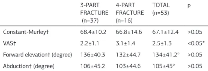
Avascular necrosis was reported as major and much feared complication in plate ixation, which was related to the worse outcomes and, leading major reason for further revision with secondary arthroplasty [2,5,10,15,17,27]. Locking plate conig-uration, the surgical technique and, sot tissue preservation al-lowed by the ixed angled construct lowers the AVN rates when Table 2. Functional results of the fractures according to the fracture types.
3-PART FRACTURE (n=37)
4-PART FRACTURE (n=16)
TOTAL (n=53)
p
Constant-Murley† 68.4±10.2 66.8±14.6 67.1±12.4 >0.05 VAS† 2.2±1.1 3.1±1.4 2.5±1.3 <0.05* Forward elevation† (degree) 136±40.3 132±44.7 134±41.2° >0.05 Abduction† (degree) 106±45.2 103±44.6 105±45° >0.05 † Mean ±SD, *p<0.05 is statistically signiicant.
Table 3. Correlation of preoperative hospitalization time and age with func-tional outcomes and pain.
Age Preoperative hospitalization time
rho p rho p
Constant- Murley -0.507 0.003* -0.410 0.005* VAS -0.170 0.253 -0.094 0.456 Forward elevation -0.402 0.005* -0.378 0.014* Abduction -0.390 0.007* -0.410 0.004* * p<0.05 is statistically signiicant
Table 4. Complications according to types of the fractures.
COMPLICATIONS 3-PART
FRACTURE (n=5)
4- PART FRACTURE (n=8)
TOTAL (n:13)
Osteonecrosis 1 2 3 (5.66%) Screw perforation 1 2 3 (5.66%)
Nonunion 1 1 2 (3.77%)
Malunion 1 2 3 (5.66%)
compared with the patients managed with conventional plates [1,24,27]. In some series, the patients in whom osteonecrosis developed had reasonable clinical outcomes and suggest that AVN was well tolerated in elderly population [5,10]. AVN is also well tolerated than malunion or nonunion [28].
Perforation of head screws primarily was one of the most fre-quent complications in this study with a rate of 5.66%. All of them were related to the initial surgery even with meticulous placement by intraoperative luoroscopy. In the written litera-ture, perforation of the head screws reported as the most com-mon complication with a range of 2 to 40% with high revi-sion rates [1,13,15,27]. Primarily perforation of head screws is probably related to purchase as much bone as possible coupled with spherical shape of the humeral head [27]. Egol et al. re-ported that, patients who had screw perforation were on av-erage 6 years older than who had not, without any statistical diference [1]. In our serie, we did not have a correlation like this. Solberg et al. reported that the all screw perforations oc-curred in the superoposterior quadrant and resulted screw con-tact with the glenoid but, did not afected the functional results worse than the patients had no screw perforation in contrast with other series [10]. In the present study, we performed screw repositioning in 3 patients immediately in 48 hours ater initial surgery, and according to us, they did not afect the functional outcomes.
Secondary screw perforation due to loss of reduction is another complication related to angular stable locking proximal humeral fractures and highly related to reoperations even though slight varus is accepted [5,24,27]. It is reported that missing medial support led to 30% screw perforations compared with 6% in-tact medial support [24]. It is stated that, the angular stable implant was responsible for screws cutting through osteopo-rotic humeral heads in elder patients and, was stated as 46% over 65 years old patients [5,21,24]. Anatomic reduction and restoration of the medial cortical support are crucial in order to prevent secondary varus angulation [14,26,27]. In the present study secondary varus angulation occurred in 2 without screw cut-out (Figure 1), where anatomic reduction was achieved and medial support screws were placed but tension band wiring was not performed. In fact tension band wiring was not used in any of the cases. Medial support screws have important contribu-tions to the strength of the medial comminution and, also using of tension band wiring is recommended to neutralize the trac-tion forces of rotator cuf when medial support is insuicient [24,29].
The non-union is another major complication in 3 or 4 -part
humeral head fractures [17]. In our series, non-union occurred in 2 (3.77%) patients and required to conversion to hemiarthro-plasty and performed in one. In the written literature the rate of non-union is 2,7%- 8% and, related to sot tissue preservation, surgical technique [1,17]. And also complex structure of the fracture is another reason of non-union [21].
Subacromial impingement occurred with a rate of 3.77% in the present study due to high positioning of the plate. Patients did not accept revision surgery. In order to avoid this complication meticulous attention must be paid to correct placement, and use of positioning K-wires is recommended [24].
Retrospective design and, some lack of knowledge such as, the rotator cuf pathologies and functional status of the patients prior to surgery and the physiotherapy performed by the pa-tients by themselves at home are the weak points of the pres-ent study.
In conclusion, functional outcomes of locking plate ixation of proximal humerus fractures are associated with many factors, which are related to the patient, fracture pattern, surgeon and the implant. According to our study and in the light of the lit-erature when indications are carefully selected, locking plate osteosynthesis yield good outcomes in surgical treatment of 3 or 4-part proximal humerus fractures.
Competing interests
The authors declare that they have no competing interests.
References
1. Egol KA, Ong CC, Walsh M, Jazrawi LM, Tejwani NC, Zuckerman JD. Early compli-cations in proximal humerus fractures (OTA Types 11) treated with locked plates. J Orthop Trauma 2008;22:159-64.
2. Ong CC, Kwon YW, Walsh M, Davidovitch R, Zuckerman JD, Egol KA. Outcomes of open reduction and internal ixation of proximal humerus fractures managed with locking plates. Am J Orthop 2012;41(9):407-12.
3. Strohm PC, Helwig P, Konrad G, Südkamp NP. Locking plates in proximal hu-merus fractures. Acta Chir Orthop Traumatol Cech 2007;74:410-5.
4. Ye T, Wang L, Zhuang C, Wang Y, Zhang W, Qiu S. Functional outcomes fol-lowing locking plate ixation of complex proximal humeral fractures. Orthopedics 2013;36:715-22.
5. Dai J, Chai Y, Wang C, Wen G. Meta-analysis comparing locking plate ixation with hemiarthroplasty for complex proximal humeral fractures. Eur J Orthop Surg Traumatol 2014;24(3):305-13.
6. Giovale M, Mangano T, Rodà E, Repetto I, Cerruti P, Kuqi E et al. Shoulder hemi-arthroplasty for complex humeral fractures: a 5 to 10-year follow-up retrospective study. Musculoskelet Surg 2014;98(suppl.1):S27-33.
7. Demirhan M, Kilicoglu O, Altinel L, Eralp L, Akalın Y. Prognostic factors in prosthetic replacement for acute proximal humerus fractures. J Orthop Trauma 2003;17:181-8.
8. Mighell MA, Kolm GP, Collinge CA, Frankle MA. Outcomes of hemiarthroplasty for fractures of the proximal humerus. J Shoulder Elbow Surg 2003;12: 569-77. 9. Neer CS 2nd. Displaced proximal humeral fractures I. Classiication and evalua-tion. J Bone Joint Surg Am 1970;52:1077-89.
10. Solberg BD, Moon CN, Franco DP, Paiement GD. Surgical treatment of three and four-part proximal humeral fractures. J Bone Joint Surg Am 2009;91:1689-97. 11. Fialka C, Stampl P, Arbes S, Reuter P, Oberleitner G, Vecsei V. Primary hemiar-throplasty in four-part fractures of the proximal humerus: randomized trial of two diferent implant systems. J Shoulder Elbow Surg 2008;17:210-5.
12. Min W, Davidovitch RI, Tejwani NC. Three-and four-part proximal humerus fractures: evolution to operative care. Bull NYU Hosp Jt Dis 2012;70:25-34. 13. Maier D, Jaeger M, Izadpanah K, Strohm PC, Suedkamp NP. Proximal humeral fracture treatment in adults. J Bone Joint Surg Am 2014;96:251-61.
14. Ponce BA, Thompson KJ, Raghava P, Eberhardt AW, Tate JP, Volgas DA et al. The role of medial comminution and calcar restoration in varus collapse of proximal humeral fractures treated with locking plates. Bone Joint Surg Am DOI:10.2106/ JBJS.K.00202.
15. Jost B, Spross C, Grehn H, Gerber C. Locking plate ixation of fractures of the proximal humerus: analysis of complications, revision strategies and outcome. J Shoulder Elbow Surg 2013;22: 542-9.
16. Matejcić A, Vidović D, Ivica M, Durdević D, Tomljenović M, Bekavac-Beslin M et al. Internal ixation with locking plate of 3- and 4-part proximal humeral frac-tures in elderly patients: complications and functional outcome. ActaClin Croat Figure 1. Plain radiograph of a 72 years old female patient with Neer Type 3
2013;52:17-22.
17. Parmaksizoglu AS, Sökücü S, Ozkaya U, Kabukçuoğlu Y, Gül M. Locking plate ixation of three- and four-part proximal humeral fractures. Acta Orthop Trauma-tol Turc 2010;44:97-104.
18. Foruria AM, Carrascal MT, Revilla C, Munuera L, Sanchez- Sotelo J. Proximal humerus fracture rotational stability ater ixation using a locking plate or a ixed-angle locked nail: the role of implant stifness. Clin Biomech 2010;25:307-11. 19. Micic ID, Kim KC, Shin DJ, Shin SJ, Kim PT, Park IH et al. Analysis of early failure of the locking compression plate in osteoporotic proximal humerus fractures. J Orthop Sci 2009;14:596-601.
20. Bastian JD, Hertel R. Osteosynthesis and hemiarthroplasty of fractures of the proximal humerus: outcomes in a consecutive case series. J Shoulder Elbow Surg 2009;18:216-9.
21. Leonard M, Mokotedi L, Alao U, Glynn A, Dolan M, Feminge P. The use of lock-ing plates in proximal humeral fractures: Comparison of outcome by patient age and fracture pattern. Int J Shoulder Surg 2009;3:85-9.
22. Shahid R, Mushtaq A, Northover J, Maqsood M. Outcome of proximal humerus fractures treated by PHILOS plate internal ixation. Experience of a district gen-eral hospital. Acta Orthop Belg 2008;74:602-8.
23. Constant CR, Murley AH. A clinical method of functional assessment of the shoulder. Clin Orthop Relat Res 1987;214:160-4.
24. Brunner F, Sommer C, Bahrs C, Heuwinkel R, Hafner C, Rillmann P et al. Open reduction and internal ixation of proximal humerus fractures using a proxi-mal humeral locked plate: a prospective multicenter analysis. J Orthop Trauma 2009;23:163-72.
25. Zhang AL, Schairer WW, Feeley BT. Hospital readmissions ater surgical treat-ment of proximal humerus fractures: Is arthroplasty safer than open reduction internal ixation? Clin Orthop Relat Res DOI: 10.1007/s11999-014-3613-y. 26. Krappinger D, Bizzotto N, Riedmann S, Kammerlander C, Hengg C, Kralinger FS. Predicting failure ater surgical ixation of proximal humerus fractures. Injury 2011;42:1283-8.
27. Thanasas C, Kontakis G, Angoules A, Limb D, Giannoudis P. Treatment of proxi-mal humerus fractures with locking plates: a systematic review. J Shoulder Elbow Surg 2009;18:837-44.
28. Panagopoulos A, Tsoumpos P, Evangelou K, Georgiou C, Triantaillopoulos I. Late prosthetic shoulder hemiarthroplasty ater failed management of complex proximal humeral fractures. Adv Orthop DOI: 10.1155/2013/403580.
29. Maddah M, Prall WC, Geyer L, Wirth S, Mutschler W, Ockert B. Is loss of ixa-tion following locked plating of proximal humeral fractures related to the number of screws and their positions in the humeral head? Orthop Rev DOI: 10.4081/ or.2014.5336.
How to cite this article: