Nanomed. J., 2(3):195-202, Summer 2015
AB STRACT:
Objective(s):Copper oxide nanoparticles have different industrial applications so it is inevitable that nanoparticulate products finally find their way into aquatic ecosystems. Nevertheless there is little information available about their effects on some of edible fish. The present study aims to determine the acute toxicity and evaluate the effect of two sub-acute concentrations (50 and 70% 96 h LC50) of CuO-NPs on some hematological and biochemical parameters ofR. rutilus.
Materials and Methods: 225 healthy specimen ofR. rutilus (mean weight 5.52±1.2 g; mean length 6.20±0.2 cm) were transported to the laboratory. In order to prepare the stock solution, CuO-NPs was dispersed in pure water with ultrasonication (50-60 kHz) for 15 min every day before dosing. At first,R. rutilus was exposed to CuO-NPs to determine the lethal concentration (LC50) value. Following acute test, fish were treated with sub-acute concentrations of CuO-NPs (50 and 70% 96 h-LC50 at) with one control group (no CuO-NPs) for a week to determine the changes in the level of some plasma hematological and biochemical parameters.
Results:The 96 h-LC50 values of CuO-NPs was 2.19±0.003 mg/l.R. rutilus exhibited significantly lower RBC count, Hb and Hct values and a significant increase in the WBC numbers, MCH, MCHC and MCV indices (p<0.05). Low glucose and higher cortisol content in plasma were observed in the fish exposed to CuO nanoparticles than those in control group (p<0.05).
Conclusion:These alterations indicateR. rutilus sensitivity to CuO-NPs and changes in blood parameters would be a useful tool for measurement early exposure to CuO nanoparticles.
Keywords:Acute toxicity, CuO-NPs, Fish, Glucose, Hematological, R. Rutilus, Sub-acute toxicity
Determination of acute toxicity and the effects of sub-acute
concentrations of CuO nanoparticles on blood parameters in Rutilus
rutilus
*A. Jahanbakhshi; A. Hedayati; A. Pirbeigi
Department of Fishery, Faculty of Fisheries and Environment, Gorgan University of Agricultural Science and Natural Resources, Gorgan, Iran
Nanomed. J., 2(3):195-202, Summer 2015 DOI: 10.7508/nmj. 2015.03.004
*Corresponding Author Email:[email protected] Tel: (+98)3223257
Note. This manuscript was submitted on January 15, 2015; approved on June 5, 2015
Received; 15 January 2015 Accepted; 5 June 2015
INTRODUCTION
As nanotechnology develops, its impacts on the environment and living organisms are becoming an important issue [1]. Nano-particles have many valuable properties and gained increasing attention because of their extensive surface area and tiny size, differed from those of the same materials in large scales [2]. Copper is
an essential trace element vital to the health of all living organisms as it is involved in several fundamental biological processes [3]. CuO-NPs have been applied in different industrial applications such as ceramic, glass [4]. In addition, they commonly used as bactericides because they are promising against microorganism
Escherichia coli[5]. In recent years, it has been cleared
their life cycles, causing problems for non-target organisms [7]. Recently, CuO-NPs have been shown to have adverse effects on the survival and growth of living organisms [6]. So many authors have studied the effects of copper oxide nanoparticles on various organisms especially those in aqueous environments [8]. However there is little information available about their effects on fish as a model organism [9].Therefore, there is an urgent need for studies on metal oxides like CuO-NPs impacts on different fish species because fish is considered as one of the main non-target aquatic organism affected by pollution [10]. Blood parameters could be measured easily and they are noteworthy indicators determining the condition of aquatic organisms [11]. For this, blood is a useful tool for prediction and diagnosis of chemicals toxicity [12]. Moreover, changes in stress indicators such as glucose and cortisol are widely used to detect physiological or environmental changes and can be considered as integrated measure of the physiological responses in organisms [13]. Glucose is a parameter having an important function in bioenergetics of animals [14]. Some authors have reported the levels of glucose changed under stress condition [15, 16,17]. Cortisol level of plasma is typically used as a general indicator of stressful conditions in fish [18]. In this regard, Köprücü et al. [19], Ribeiro et al. [20], Hedayati and Jahanbakhshi [21], Hedayati et al. [22] and Shaluei et al. [23] studied the effects of some pollutants on hematological indices of various fish species. Whereas literature is full with several studies on the impact of pollutants on blood parameters of fish, there is no data published on the sub-acute toxicity of the CuO-NPs on hematological and bioche-mical parameters ofR. rutilusas a suitable organism to evaluate
the impact of pollution in fresh-water ecosystems (24).In the light of foregoing ,the present study aims to determine the median lethal concentration (LC50) values (24, 48, 72 and 96 h) of CuO-NPs and the effects of the 50 and 70% of 96 h-LC50 of the nanoparticle on some hematological and biochemical parameters of the fish.
MATERIALS AND METHODS
For this experiment 225 healthy specimen of Roach (Rutilus rutilus) (mean weight 5.52±1.2 g; mean length
6.20±0.2 cm) were obtained from a fish farm. The animals were transported to aquaculture research center of Gorgan University of agricultural sciences and natural resources in a many containers equipped with an oxygen capsule and were acclimatized for a period of seven days under laboratory conditions prior to the exposures commenced
in several 200-L glass aquaria supplied with dechlorinated aerated tap water. Some of water quality characteristics of aquaria during the test were recorded as follows; temperature (25.4±1°C), dissolved oxygen (6.8 mg/L±0.9), pH (7.6±0.13) (mean+SD) and photoperiod was a 12:12 light-dark cycle. During the acclimation period fish were fed twice a day.
CuO-NPs suspension preparation
CuO nanoparticles (particle size in 20-30 nm with a purity > 98.0%) were purchased from NanoNasb Company, Tehran, Iran as uncoated nanoparticles. In order to prepare the stock solution, according to Zhao et al. (25), CuO-NPs were dispersed in pure water with ultrasonication (50-60 kHz) for 15 min every day before dosing.
Acute toxicity test
Groups of 21 (each concentration was composed of three replicates aquaria and each aquarium contained 7 fish) fish were exposed to 1, 10,100, 1000, 2000 and 4000 mg/l CuO NPs for 96h. Mortalities rate was measured at 24, 48, 72 and 96 h, and dead fish were immediately removed by dip net to avoid possible deterioration of the water quality. Test water was not changed during the 96-h time period and exposed fish were not fed. The LC50 values for 24, 48, 72 and 96h were calculated by Probit analysis and spss18 software (26).
Sub-acute toxicity test
Nanomed. J., 2(3):195-202, Summer 2015
Blood collection
At the end of the experiment (7 days),21.fish per treatment were removed for hematological and biochemical studies. Fish were immediately anesthetized into a 200 ppm solution of clove powder [21]. Blood collected from each fish by cutting the caudal peduncle was decanted into heparinized tubes and placed immediately on ice for the estimation of red blood cells count, white blood cell count, hemoglobin percentage, hematocrit, MCV, MCH and MCHC [20, 13]. The rest of the samples were kept for plasma measurements.
Hematological analyses
Examination of red blood cell (RBC) and total white blood cell (WBC) were carried out according to the hemocytometer method under the light microscope [29]. The micro-hematocrit method of Hesser [30] was used to estimate the hematocrit (Hct). Hemoglobin values (Hb, milligrams per liter) were immediately assessed calorimetrically according to Lee et al. [31] by determining the formation of cyanomethemoglobin. Red cell indices include mean corpuscular hemoglobin (MCH: pg/cell), mean corpuscular volume (MCV: ì3/cell) and
meancorpuscular hemoglobin concentration (MCHC in g/dl) were calculated from red blood cell count, hemoglobin and hematocrit according to formula were suggested by Lee et al. [31].
Glucose and cortisol measurement
In order to glucose assessment after assessing hematological parameters, the rest of the blood samples were allowed to clot at approximately 22°C (room temperature) for half an hour before centrifugation. The blood was centrifuged at 3500 rpm over 5 minutes for the collection of plasma. The supernatants were stored at -80°C until analysis. The plasma obtained was assayed for determine glucose and cortisol values. The plasma glucose was quantified using spectrophotometry method as described by Shaluei et al. [26]. Cortisol level of plasma was determined using ELISAkit (DRG Diagnostics, Mountainside, NJ, USA) as described by Shaluei et al. [26].
Statistical analysis
LC50 values and confidence intervals were calculated using EPA Probit Analysis Program V. 1.5 for each group, separately. All data were accepted if calculated chi-square for heterogeneity was lower than the tabular value at the
0.05 level. After determining LC50 values, statistical analysis was used to compare the significant difference between treatments. In order to estimate the mean values of hematological indices and biochemical parameters was used of all replicates. Each parameter was statistically analyzed for normality and homogeneity. Analysis of variance (One-way ANOVA) with Duncan post hoc test was applied to detect the significance of CuO-NPs levels on hematological and biochemical para-meters. Values were expressed as means ± standard deviation (X _ SD). Differences were considered to be significant at p<0.05
RESULTS Acute test
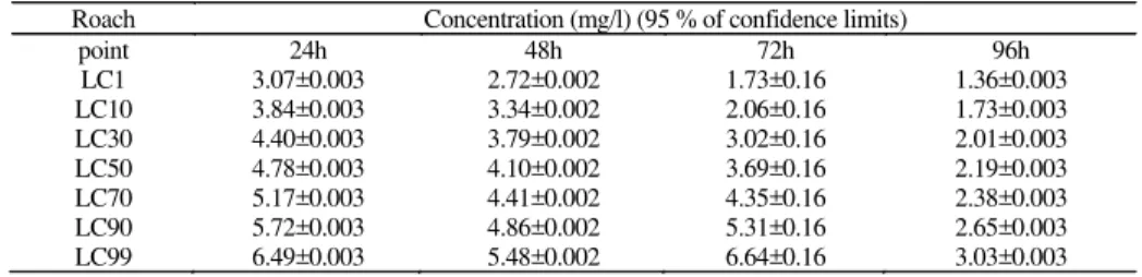
The number of dead fish was increased significantly with increasing concentration and time during the acute toxicity test (24-96 h). Therefore the highest mortality was recorded in the fish exposed to highest amount of CuO nanoparticles for 96h. The 96 h-LC50 of the material were found to be 2.19±0.003 mg/l. Obtained results for LC1-99 values tests are shown in Table 1.
Hematological parameters
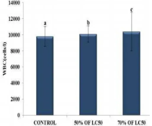
Results of hematological parameters (RBC, Hb, Hct, WBC, MCH, MCHC and MCV) of the test and controlR. rutilusexposed to 50 and 70% of LC50 are shown in Figures
1-7. At seventh day, R. rutilus specimens exhibited
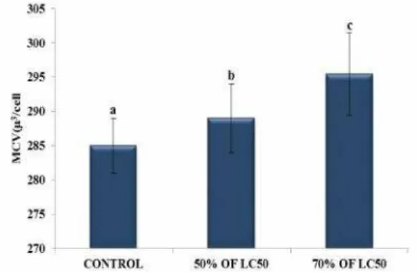
significantly lower RBC count, Hb and Hct values (p< 0.05). While, a significant increase in the WBC numbers, MCH, MCHC and MCV indices was found after exposure to 50 and 70% LC50 of CuO-NPs for seven days (p< 0.05).
Biochemical parameters
DISCUSSION
Release of metal oxide nanoparticles into the environment as a consequence of increasing production and exploitation, make it necessary to assess the environmental and health hazards that these compounds could exert [32, 33]. In toxicity studies, the LC50 values of new materials should be determined in the first stage [34]. LC50 is the most widely accepted basis for acute toxicity test kills 50% of the test organisms after a particular period of exposure, usually 96 h [23]. The individual variability in acute toxicity even within a species and with the same toxicant depends on the size, age, and condition of the tested organism as well as on experimental factors [35]. To the best of our knowledge, this study is one of the first reports detailing the effects of CuO-NPs onR. rutilus.
The results obtained from acute toxicity test showed that the 96 h-LC50 value was 2.19±0.003 mg/l. Griffitt et al [36].
Table 1. Lethal Concentrations (LC1-99) of CuO-NPs (mean ± Standard Error) depending on time (24-96h) inR. rutilus
Roach Concentration (mg/l) (95 % of confidence limits)
point 24h 48h 72h 96h
LC1 3.07±0.003 2.72±0.002 1.73±0.16 1.36±0.003
LC10 3.84±0.003 3.34±0.002 2.06±0.16 1.73±0.003
LC30 4.40±0.003 3.79±0.002 3.02±0.16 2.01±0.003
LC50 4.78±0.003 4.10±0.002 3.69±0.16 2.19±0.003
LC70 5.17±0.003 4.41±0.002 4.35±0.16 2.38±0.003
LC90 5.72±0.003 4.86±0.002 5.31±0.16 2.65±0.003
LC99 6.49±0.003 5.48±0.002 6.64±0.16 3.03±0.003
Studied the acute toxicity Cu-NPs to adult zebrafish (Danio rerio) and reported 48 h-LC50 value as 1.56 mg/l, and 7.20
mg/l in larvae. Das and Das [37] recorded 1.40 mg/l Cu as 96 h-LC50 to fry of common carp (Cyprinus carpio),
explantation, the LC50 values indicated that Cu was highly toxic to the organisms. Moreover, the acute toxicity of copper to some types of fish was reported by Richey and Roseboom [38]. They illustrated that the 48 h-LC50 of Cu to rainbow trout (Oncorhynchus mykiss) in size of a
yearling and alkalinity of 250, was 0.75 mg/l. In addition, the 96 h-LC50 value of Ag-NPs (Nanocid) in Caspian roach (Rutilus rutilus caspicus) was found to be 0.028 mg/l
[39]. As described by Kalbassi et al. [40], CuO NPs in the present survey can be classified as moderately toxic for
R. rutiluz (96 h-LC 50 in the of range 1 to 10 ppm).
Fig. 1. Changes in number of red blood cells (106 cell /L) inR. rutilus after exposure to 50-70% 96 h-LC50 of CuO-NPs after 7 days. Statistical significance was determined using aone-way analysis of variance (ANOVA) and a Duncan post hoc test (α= 0.05). Values are means ± SD (n = 21). Different letters show
significant differences among exposure concentrations
Fig. 2. Changes in values of hemoglobin (mg /L) inR. rutilus after exposure to 50-70% 96 h-LC50 of CuO-NPs after 7 days.
Statistical significance was determined using a one-way analysis of variance (ANOVA) and a Duncan post hoc test (α= 0.05).
Nanomed. J., 2(3):195-202, Summer 2015
Fig. 3. Changes in values of hematocrit (%) inR. rutilusafter exposure to 50-70% 96 h-LC50 of CuO-NPs after 7 days. Statistical significance was determined using a one-way analysis
of variance (ANOVA) anda Duncan post hoc test (α= 0.05). Values are means ± SD (n = 21). Different letters show significant differences among exposure concentrations
Fig. 4. Changes in number of white blood cells (cell /L) inR. rutilus after exposure to 50-70% 96 h-LC50 of CuO-NPs after
7 days.Statistical significance was determined using a one-way analysis of variance (ANOVA) and a Duncan post hoc test (α = 0.05). Values are means ± SD (n = 21). Different letters show
significant differences among exposure concentrations
Sub-acute toxicity test
The effects of CuO-NPs on hematological and biochemical parameters
Blood is a very good indicator of toxic stress and analysis of hematological parameters in fish is greatly used to estimate toxic stress and practical status of the
animals’ health [41]. Hematological data showed that
CuO-NPs exerted a certain influence on afore-mentioned blood indices in this study. Condition of the specimens during a long period could be reflected by erythrocytes [42]. Decreased RBC, Hb and Hct content inR. rutiluswere
observed in our study. Similar results were also reported in fish exposed to metals [43], pesticides [44] and other toxicant [45].A reduction in the number of RBC of trout (Salmo gairdneri) was observed after exposure to 0.301
mg/l of copper for a day [46]. Moreover, the analysis of hematological indices of rainbow trout (Oncorhynchus mykiss) treated with CuO-NPs for a period of 96 h showed
that CuO-NPs stimulated red and white blood cells, hematocrit, MCH,MCHC and MCV, and did not have any effects on hemoglobin content [47].The decrease in RBC count, Hb and Hct levels observed in this study may be due to anemia or erythropoiesis disorder [23]. As mentioned by Thomas and Egee [48], the transport of carbon dioxide and oxygen within the blood is related to the electrolytes and the acid-base status of the red blood cells (RBCs). Therefore respiratory disorders may occur following exposure to CuO-NPs which in turn led to changes in the number of RBCs [47]. Hemolysis also can be expressed as a reason for reduction observed in RBC count in the affected fish [49]. Significant reduction in
hematocrit and hemoglobin has been recorded in various toxicant-treated fish by some authors. Decreased Hb and hematocrit content may be attributed to the stress induced by innutrition during the test, collapse of erythrocytes because of toxicant stress [23, 50] and or lysing of RBC owing to stressor [51]. Therefore, decreased RBC count and Hb content seen in the current survey may be as a result of disarranging action of the used material on the erythro-poietic tissue [19].
Average volume of red blood cells by dividing the hematocrit is expressed as MCV. Since The MCV, MCH and MCHC values are exactly calculated based on hemoglobin and hematocrit content and RBC number, changes in these parameters will lead to changes in MCV, MCH and MCHC values [52]. In this regard, a marked increase in MCV value might be resulted from decrease in RBC number induced by hypoxic condition [53]. According to Barton et al. [54], alternations in WBC numbers can be used as a susceptible indicator of stress in fish. The increase in white blood cells ofR. rutilusis in
accordance with those of Oliveira Riberio et al. [20] who noted disorders of hematological tissues (spleen and kidney) and Remyla et al. [13] who stated increase in WBC numbers may be occur in order to overcome stressful condition. Major reduction in plasma glucose levels after exposure to stressors has reported by few authors. noticeable reduction in glucose content of pesticide treatedChanna punctatus[55],Catla catla affected by
acute of arsenate [56] and rainbow trout (Oncorhynchus mykiss) subjected to waterborne copper nanoparticles
decline in glucose content of plasma during the study may be associated to excessive consumption of stored carbohydrates in the body followed by hypoxic condition caused by nanomaterials [56], which in turn may be a reason for hypoglycemic condition [55].
Cortisol is the major corticosteroid produced by teleostean
fish. Different stressors activate the hypothalamus– pituitary–inter-renal (HPI) axis, resulting in a cortisol release,
which causes secondary stress responses [23]. Cortisol maintenance the homeostasis through mobilizing some factors such as fatty acids and glucose and so cortisol has an important role in exerts direct and indirect effects on intermediary metabolism, particularly in response to stress [58]. Increased cortisol of plasma can be considered as the reaction of the species to recognize the presence of a lethal or potential harmful substance in the environment [59].
Fig.5. Changes in values of MCH (pg/cell) inR. rutilusafter exposure to 50-70% 96h-LC50 of CuO-NPs after 7 days. Statistical significance was determined using a one-way analysis
of variance (ANOVA) and a Duncan post hoc test (α= 0.05). Valuesare means ± SD (n = 21). Different letters show significant differences among exposure concentrations
Fig. 7. Changes in values of MCHC (g/dl) inR. rutilusafter exposure to 50-70% 96 h-LC50 of CuO-NPs after 7 days. Statistical significance was determined using a one-way analysis
of variance (ANOVA) and a Duncan post hoc test (α= 0.05). Values are means ± SD (n = 21). Different letters show significant differences among exposure concentrations
Fig. 6. Changes in values of MCV (μ 3/cell) inR. rutilusafter exposure to 50-70% 96h-LC50 of CuO-NPs after 7 days.
Statistical significance wasdetermined using a one-way analysis of variance (ANOVA) and a Duncan post hoc test (α= 0.05). Values are means ± SD (n = 21). Different letters show significant differences among exposure concentrations
Fig. 8. Changes in glucose levels (mg/dl) inR. rutilusafter exposure to 50 and 70% of LC50 after 7 days. Statistical significance was determinedusing a one-way analysis of variance (ANOVA) and a Duncan post hoc test (α = 0.05).
Values are means ± SD (n = 21). Different letters show significant differences among exposure concentrations
Fig. 9. Changes in cortisol levels (mg/ml) inR. rutilusafter exposure to 50 and 70% of LC50 after 7 days. Statistical
significance was determined using a one-way analysis of variance (ANOVA) and a Duncan post hoc test (á = 0.05).
Nanomed. J., 2(3):195-202, Summer 2015
CONCLUSION
Though little surveys have focused on the effects of copper oxide nanoparticles on blood parameters of various fish species, the current investigation proves that exposure to lower concentrations than LC50 of copper oxide nanoparticles leads to change in hematological and biochemical parameters ofR. rutilus. Since the species is
among edible species, infection in turn affects human health. Furthermore, the results show that alternation in blood parameters would be a useful tool for measurement early exposure to CuO nanoparticles.
ACKNOWLEDGMENTS
The authors thankful to Gorgan University of Agricultural Science and Natural Resources, Gorgan, Iran, for the continuous support in providing the research facilities.
REFERENCES
1.Adhikari T, Kundu S, Biswas A.K, Tarafdar J.C, Rao A.S. Effect of Copper Oxide Nano Particle on Seed Germination of Selected Crops. J Agri Sci Tech. 2012; 2: 815-823.
2.Biswas P, Wu C.Y. Nanoparticles and the environment. J Air Waste Manag Assoc. 2005; 55(6): 708-746.
3.Isani G, Falcioni M.L, Barucca G, Sekar D, Andreani G, Carpenè E, Falcioni G. Comparative toxicity of CuO nanoparticles and CuSO4 in Rainbow trout. Ecotoxicol Environ Saf. 2013; 97: 40-46. DOI:10.1016/j.ecoenv.2013.07.001.
4.Jammi S, Sakthivel S, Rout L, Mukherjee T, Mandal S, Mitra R, Saha P, Punniyamurthy T.. CuO nanoparticles catalyzed C - N, C - O, and C - S cross-coupling reactions: Scope and mechanism. J organ chem. 2009; 74(5): 1971-1976. DOI: 10.1021/ jo8024253.
5.El-Nahhal M, Zourab S. M, Kodeh F.S, Selmane M, Genois I, Babonneau F. Nanostructured copper oxide-cotton fibers: synthesis, characterization, and applications. Internat Nano Let. 2012; 2(1): 1-5.
6.Nations, S, Wages M, Cañas J.E, Maul J, Theodorakis C, Cobb G. Acute effects of Fe2O3, TiO2, ZnO and CuO nanomaterials on Xenopus laevis. Chemosphere. 2011; 83(8): 1053-1061. 7.Ates M, Dugo M.A, Demir V, Arslan Z, Tchounwou P.B.. effect
of copper oxide nanoparticles to sheepshead minnow (Cyprinodon variegatus) at different salinities. Digest J Nanomat Biostruct. 2014; 9: 369-377.
8.Chang Y.N, Zhang M, Xia L, Zhang J, Xing G. The toxic effects and mechanisms of CuO and ZnO nanoparticles. Materials. 2012; 5(12): 2850-2871.
9.Karthikeyeni S, Siva Vijayakumar T, Vasanth S, Arul Ganesh M.M, Subramanian P. Biosynthesis of Iron oxide nanoparticles and its haematological effects on fresh water fishOreochromis mossambicus. J Acad Indus Res. 2013; 10: 645- 649. 10.Ogundele O, Ihuahi J.A, Omojowo F.S, Bitrus P. Toxicity of
linear alkylbenene sulphonate (LAS) detergent, to Clarias gariepinus fingerlings. Pan-American J Aquat Sci. 2005; 273-276.
11.Bahmani M, Kazemi R, Donskaya, P. A comparative study of some hematological features in young reared sturgeons (Acipenser persicus andHuso huso). Fish Physiol Biochem. 2012; 4(2): 135-140.
12.Banaee M, Mirvagefei A, Rafei G, Amiri, B.M. Effect of sub-lethal diazinon concentrations on blood plasma biochemistry. Internation J Environ Res. 2008; 2(2): 189-198.
13.Remyla S.R, Ramesh M, Sajwan K.S, Kumar K.S. Influence of zinc on cadmium induced haematological and biochemical responses in a freshwater teleost fishCatla catla. Fish Physiol Biochem. 2008; 34(2): 169-174.
14.Lucas A. Physical concepts of bioenergetics. Bioenergetics of aquatic animals, Englishth edn. Taylor & Francis, France, 1996. 15.Camargo M.M.P, Fernandes M.N, Martinez C.B.R. How aluminium exposure promotes osmoregulatory disturbances in the neotropical freshwater fishProchilus lineatus. Aquat toxicol. 2009; 94(1): 40-46. DOI:10.1016/j.aquatox.2009.05.017. 16.David M, Kumar R.S, Mushigeri S.B, Kuri R.C. Blood glucose
and glycogen levels as indicators of stress in the freshwater fish, Labeo rohita under fenvalerate intoxication. J Ecotoxicol Environ Monitor. 2005; 15(1): 1-5.
17.Jahanbakhshi A, Baghfalaki M, Imanpour M.R, Nodeh A.J, Shaluei F. Effects of different concentrations of 2-phenoxyethanol on primary and secondary stress responses in persian sturgeon,Acipenser persicus. J Appl Ichthyol. 2012; 29(3): 499-502. DOI: 10.1111/jai.12112.
18.Pickering A.D, Pottinger T.G. Stress responses and diseases response in salmonid fish: effects of chronic elevation of plasma cortisol. Fish Physiol Biochem. 1989; 7: 253-258. DOI: 10.1007/BF00004714.
19.Köprücü S.Þ, Köprücü K, Ural M.Þ, Ýspir Ü, Pala M.. Acute toxicity of organophosphorous pesticide diazinon and its effects on behavior and some hematological parameters of fingerling European catfish (Silurus glanisL.). Pest biochem physiol. 2006; 86(2): 99-105.
20.Oliveira Ribeiro C.A, Filipak Neto F, Mela M, Silva P.H, Randi M.A.F, Rabitto I.S, Alves Costa J.R.M, Pelletier E. Hematological findings in neotropical fishHoplias malabaricus exposed to subchronic and dietary doses of methylmercury, inorganic lead, and tributyltin chloride. Environ res. 2006; 101(1): 74-80. 21.Hedayati A, Jahanbakhshi A. The effect of water-soluble
fraction of diesel oil on some hematological indices in the great sturgeonHuso huso. Fish Physiol Biochem. 2012; 38(6): 1753-1758.
22.Hedayati A, Kolangi H, Jahanbakhshi A, Shaluei F. Evaluation of silver nanoparticles ecotoxicity in silver carp (Hypophthalmicthys molitrix) and goldfish (Carassius auratus). l J Vet Med. 2012; 15(3): 172-177.
23.Shaluei F, Hedayati A, Jahanbakhshi A, Kolangi H, Fotovat M.. Effect of subacute exposure to silver nanoparticle on some hematological and plasma biochemical indices in silver carp (Hypophthalmichthys molitrix). Hum Experim Toxicol. 2013; 32(12): 1270-1277.
24.Tyler C.R, Lange A, Paull G.C, Katsu Y, Iguchi T. The roach (Rutilus rutilus) as a sentinel for assessing endocrine disruption. Environmental sciences: internat j environ physiol toxicol. 2006;14(5): 235-253.
25.Zhao J, Wang Z, Liu X, Xie X, Zhang K, Xing B.. Distribution of CuO nanoparticles in juvenile carp (Cyprinus carpio) and their potential toxicity. J hazard mater. 2011; 197: 304-310. DOI:10.1016/j.jhazmat.2011.09.094.
26.Shaluei F, Hedayati A, Jahanbakhshi A, Baghfalaki M. Effects of nanometer-sized silver materials on survival response of Caspian roach (Rutilus rutilus caspicus). Toxicol Ind Health. 2012; 3: 207-211.
How to cite this article:
Jahanbakhshi A, Hedayati A, Pirbeigi A. Determination of acute toxicity and the effects of sub-acute concentrations of CuO nanoparticles on blood parameters in Rutilus rutilus. Nanomed. J., 2015; 2(3): 195-202.
28.Shalaby A.M. Sublethal effects of Heavy metal copper, Cadmium and zinc alone in combinations on enzymes activities of common carp Cyprinus carpio L. Egypt. J. Aquat. Biol., 2000; 4(2): 229-246.
29.Stevens M.L. 1997. Fundamentals of clinical hematology. Saunders Philadelphia, PA.
30.Hesser E.F. Methods for routine fish hematology. The Progressive Fish-Culturist. 1960; 22: 164-171.
31.Lee G.R, Foerster J, Lukens J, Paraskevas F, Greer J.P, Rodgers G.M. Wintrobe’s clinical hematology 10th. Bethseda, Maryland: Lippincort Williams and Wilkins, 1999.
32.Lee J, Mahendra S, Alvarez PJJ. Nanomaterials in the construction industry: a review of their applications and environmental health and safety considerations. ACS Nano. 2010; 4: 3580–3590.
33.Wang J, Gerlach J.D, Savage N, Cobb G.P. Necessity and approach to integrated nanomaterial legislation and governance. Sci Tot Environ. 2013; 442: 56–62.
34.Di Giulio R.T, Hinton D.E. The toxicology of fishes. 2008; CRC Press.
35.Hedayati A, Kolangi H, Jahanbakhshi A, Shaluei F. Evaluation of silver nanoparticles ecotoxicity in silver carp (hypophthalmicthys molitrix) and goldfish (carassius auratus). Bulg. J. Vet. Med., 2012; 15(3): 172"177.
36.Griffitt R.J, Weil R, Hyndman K.A, Denslow N.D, Powers K, Taylor D, Barber D.S.Exposure to copper nanoparticles causes gill injury and acute lethality inzebrafish (Danio rerio). Environ Sci Technol. 2007; 41: 8178–8186.
37.Das B.K, Das N. Impacts of quicklime (CaO) on the toxicity of copper (CuSO4, 5H(2)O) to fish and fish food organisms. Chemosphere. 2005; 61: 186-191. DOI:10.1016/ j.chemosphere.2005.02.064.
38.Richey D, Roseboom D. Acute toxicity of copper to some fishes in high alkalinity water. Illinois state water survey, Urbana,Circular. 1978; 131: 24.
39.Shaluei F, Hedayati A, Jahanbakhshi A, Baghfalaki M. Effects of nanometer-sized silver materials on survival response of Caspian roach (Rutilus rutilus caspicus). Toxicol Ind Health. 2012. DOI: 10.1177/0748233712457445.
40.Kalbassi M.R, Salari-joo H, Johari A. Toxicity of Silver Nanoparticles in Aquatic Ecosystems Salinity the Main Cause in Reducing Toxicity. Iran. J. Toxicol., 2011; 5: 436-443. 41.Hedayati A, Ghaffari Z. Evaluation of the Effects of Exposure
to Copper Sulfate on some Eco-physiological Parameters in Silver Carp (Hypophthalmichthys molitrix). Iran. J. Toxicol., 2013; 7(22): 887-893.
42.Haley P.J, Weiser M.G. Erythrocyte volume distribution in rainbow trout. American j vet res. 1985; 46(10): 2210-2212. 43.Laban G, Nies L.F, Turco R.F, Bickham J.W, Sepu´lveda M.S. The effects of silvernanoparticles on fatheadminnow (Pimephales promelas) embryos. Ecotoxicology. 2010; 19: 185–195.
44.Webb N.A,Wood C.M. Physiological analysis of the stress response associated with acute silver nitrate exposure in freshwater rainbow trout (Oncorhynchus mykiss). Environ Toxicol Chem. 1998; 17: 579–588.
45.Benna S,Viswaranjan S. Effect of cadmium andmercury on the hematological parameter of the fishCyprinus carpio. Environ Ecol.1987; 5: 726–732.
46.Dick P.T, Dixon D.G. Changes in circulating blood cell levels of rainbow trout,Salmo gairdneri, Richardson, following acute and chronic exposure to copper. J fish biol. 1985; 26(4): 475-481.
47.Khabbazi M, Harsij M, Hedayati A, Gholipoor H, Gerami M,Ghafari H.Effect of CuO nanoparticles on some hematological indices of rainbow trout,Oncorhynchus mykiss and their potential toxicity. Nanomed J. 2015;2(1): 67-73. 48.Thomas S, Egee S. Fish Red Blood Cells: Characteristics and
Physiological Role of the Membrane Ion Transporters. Comp Biochem Physiol A Mol Integr Physiol. 1998; 119(1): 79-86. 49.Johansson-Sjöbeck M.L, Larsson A. The effect of cadmium on the hematology and on the activity of ä-aminolevulinic acid dehydratase (ALA-D) in blood and hematopoietic tissues of the flounder,Pleuronectes flesus L. Environ Res. 1978; 17: 191-204.
50.Lavanya S, Ramesh M, Kavitha C, Malarvizhi A. Hematological, biochemical and ionoregulatory responses of Indian major carp Catla catla during chronic sublethal exposure to inorganic arsenic. Chemosphere. 2011; 82: 977-985.
51.Ololade I.A, Oginni O. Toxic stress and hematological effects of nickel on African catfish,Clarias gariepinus, fingerlings. J Environ Chem Ecotoxicol. 2010; 2: 014-019.
52.Desai B, Parikh P. Impact of Curzate (fungicide) on Hematological Parameters of Oreochromis mossambicus. Internat J Sci Engineer Res. 2012; 3(7): 1-6.
53.Wepener V, Van Vuren J.H.J, Du Preez H.H. The effect of hexavalent chromium at different pH values on the haematology ofTilapia sparrmanii(Cichlidae). Com Biochem Physiol Part C: Com Pharmacol. 1992;101(2): 375-81.
54.Barton B.A, Iwama G.K. Physiological changes in fish from stress in aquaculture with emphasis on the response and effects of corticosteroids. Ann. Rev. Fish Dis., 1991; 1: 3-26. 55.Agrahari S, Pandey K.C, Gopal K. Biochemical alteration induced
by monocrotophos in the blood plasma of fish, Channa punctatus (Bloch). Pesti biochem physiol. 2007; 88(3): 268-272.
56.Kavitha C, Malarvizhi A, Senthil Kumaran S, Ramesh M. Toxicological effects of arsenate exposure on hematological, biochemical and liver transaminases activity in an Indian major carp,Catla catla. Food Chemi Toxicol. 2010; 48(10): 2848-2854.
57.Shaw B.J, Al-Bairuty G, Handy R.D. Effects of waterborne copper nanoparticles and copper sulphate on rainbow trout (Oncorhynchus mykiss): Physiology and accumulation. Aquat toxicol. 2012; 116: 90-101.
58.Van Der Boon J, Van Den Thillart G.E, Addink A.D. The effects of cortisol administration on intermediarymetabolism in teleost fish. Com Biochem Physiol Part A: Physiol. 1991; 100(1): 47-53.