Role of the Accessory Parotid Gland in the
Etiology of Parotitis: Statistical Analysis of
Sialographic Features
Wangyong Zhu1,2, Fengchun Hu1,2, Xingguang Liu1,2, Songcan Guo3, Qian Tao1,2*
1Department of Oral Maxillofacial-Head and Neck Oncology, the Affiliated Hospital of Stomatology, Sun Yat-sen University, Guangzhou, 510055, China,2Guangdong Provincial Key Laboratory of Stomatology, Sun Yat-sen University, Guangzhou, Guangdong, 510055, China,3School of Engneering, Sun Yat-sen University, Guangzhou, 510275, China
Abstract
This retrospective study aimed to identify if the existence of the accessory parotid gland cor-related with the etiology of parotitis. This may aid the development of better treatment strate-gies in the future. Sialographic features of cases with parotitis and healthy subjects were reviewed. The chi-square test was used to compare the incidence of accessory parotid gland between the groups. The Student’s t test was used to compare the length of Sten-sen’s duct, the length from the orifice to the confluence of the accessory duct, and the angle between the accessory duct and Stensen’s duct between the groups. The incidence of accessory parotid gland in patients with parotitis was 71.8% (28/39), which was significantly higher than that in healthy subjects (P = 0.005). Patients with parotitis had a longer Sten-sen’s duct than healthy subjects (P = 0.003). There was no significant difference in the length from the orifice to the confluence of the accessory duct or the angle between the accessory duct and Stensen’s duct (P = 0.136 and 0.511, respectively) between the groups.
The accessory parotid gland might play a role in the pathogenesis of parotitis. The exis-tence of an accessory parotid gland is likely to interfere with salivary flow. Computational fluid dynamics analysis of salivary flow in the ductal system would be useful in future etio-logic studies on parotitis.
Introduction
Chronic parotitis is characterized by recurrent pain and swelling in the affected parotid gland. The symptoms of parotitis are often intermittent and mild and may be related to eating in the early stages of disease. Its etiology is considered to be multifactorial, and its exact pathogenesis is not fully understood. Obstructive causes of reduced salivary flow include sialoliths, stricture of the main duct or orifice, mucus plugging, external pressure on the duct, and developmental malformations of the duct [1–4]. The presence of an accessory parotid gland (APG) is a fairly common anatomical variation. The incidence of APG ranges between 21 and 56% [5,6]. The APG is a small gland with a diameter of 0.5–1 cm, and is located on average 6 mm anterior to
OPEN ACCESS
Citation:Zhu W, Hu F, Liu X, Guo S, Tao Q (2016) Role of the Accessory Parotid Gland in the Etiology of Parotitis: Statistical Analysis of Sialographic Features. PLoS ONE 11(2): e0150212. doi:10.1371/journal. pone.0150212
Editor:Brij Singh, School of Medicine and Health Sciences, University of North Dakota, UNITED STATES
Received:October 18, 2015
Accepted:February 10, 2016
Published:February 25, 2016
Copyright:© 2016 Zhu et al. This is an open access article distributed under the terms of theCreative Commons Attribution License, which permits unrestricted use, distribution, and reproduction in any medium, provided the original author and source are credited.
Data Availability Statement:All relevant data are within the paper and its Supporting Information files.
Funding:The authors have no support or funding to report.
and separate from the parotid gland. In most cases, it drains into Stensen’s duct (SD) through an accessory duct (AD). Horsburgh et al. found that 59% of patients with parotitis had an APG, which is a higher rate than that observed in healthy people [7]. This suggests that the presence of an APG might be related to the pathogenesis of parotitis.
The current study aimed to explore the role of the APG in the etiology of parotitis. We ana-lyzed the incidence of APG and the anatomical parameters of the parotid gland between healthy subjects and patients with parotitis.
Materials and Methods
This study was approved by the institutional ethics board of the Hospital of Stomatology, Sun Yat-sen University. All patients were informed of the details of the study and informed consent was obtained. Patient information was de-identified prior to analysis.
All procedures were performed by one doctor using the same standard technique: 2 mL of 40% iodinated oil (a contrast medium) was gently introduced into the orifice of the parotid duct, until the patient felt an obvious sense of swelling in the gland. The maxillofacial region was scanned transversely and sagittally using a cone-beam computed tomography scanner. The following imaging parameters were used: 108 mA; 90 kV; field of vision, 500 × 500; slice thickness, 0.4 mm.
The main part of each patient’s parotid was reconstructed and modeled by Mimics 14.11 software (Materialise Technologies, Leuven, Belgium). Independently, two experienced radiol-ogists retrospectively reviewed all anonymous images demonstrating the entire ductal system. The authors reviewed the data and used electronic calipers provided by the Mimics 14.11 soft-ware to measure the following parameters: the length of SD, the length between the confluence of the AD and the orifice, and the angle between the AD and SD.
The inclusion criteria of the parotitis group were: 1. The patient was diagnosed as having parotitis; 2. The radiologists could analyze the sialographic features of the images that demon-strated the entire ductal system. The inclusion criteria of the healthy subjects group were: 1. The volunteer had no history of salivary gland disease; 2. The radiologists could analyze the sia-lographic features of the images that demonstrated the full extent of the ductal system.
Statistical analysis was performed using SPSS version 15.01 software (SPSS, Inc., Chicago, IL). The chi-square test was used to compare the difference in APG incidence between the healthy and parotitis groups. The Student’s t test was used to compare the sialographic features of the two groups, with the significance level set at P<0.05.
Results
Seven males and 11 females with a mean age of 22.5 years old were included in the healthy sub-jects group. Based on 31 sets of sialographic data from 15 left parotid and 16 right parotid glands, 38.7% of cases had APGs. In one case, the APG drained into SD through two ADs.S1 Figshows typical images from the healthy subjects group.
eating. The upstream SD was dilated and the downstream duct was normal, bounded by the confluence of the AD with SD. By using sialendoscopy, we observed mucous embolus in the duct, but did not find any sialoliths or stenosis.
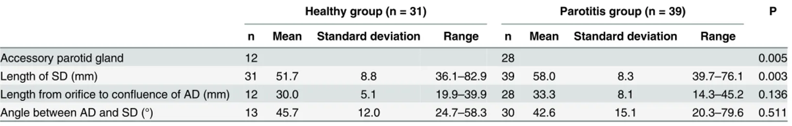
The incidence of APG and the length of SD in patients with parotitis were significantly higher and longer than those in healthy subjects (P = 0.005 and 0.003, respectively). There was no significant difference in the length from the orifice to the confluence of the AD (P = 0.136) or the angle between the AD and SD (P = 0.511) between the healthy and parotitis groups. Table 1shows the results in detail as means, standard deviations, and ranges.
Discussion
The exact pathogenesis of parotitis is still not completely understood. Reduced salivary flow is likely to be the most important factor, since it causes the repeated appearance of ascending infection in the parotid gland. This leads to further destruction of the acini and an increase in the mucous substances of the saliva, which aggravates the disease [1–4]. The obstructive causes of swelling in the parotid gland are examined by sialendoscopy, and include sialoliths (20.3–
52.2%), stenosis of the duct, foreign bodies, and fibrous embolus (32.6–56.3%). However, obstructive causes are only reported to be found in 53.5–89.3% of cases [8,9]. This is quite simi-lar to our clinical experience of parotitis. It is often hard to explain the cause of symptoms because nearly half of patients with parotitis do not have any sialoliths or stenosis in their ducts. It is known that there are complex rheological forms of salivary flow such as flow at the duct entrance, secondary flow, spiral flow, flow separation, and recirculation at the turnings and confluences of the ducts [10]. If we regard saliva flow in the ductal system as a micro flow field, changes of fluid mechanics in the ductal system of the parotid might explain the reduced salivary flow observed in patients without obstruction.
The APG is an anatomical variant with an incidence of 21–56% [5,6]. However, Horsburgh et al. found that the presence of an APG is more common in patients with parotitis, with an inci-dence of 59% [7]. Therefore, we focused on the role of the APG in the etiology of parotitis. In our study, we found the incidence of APG in patients with parotitis to be 71.8%, which was signifi-cantly higher than the incidence of APG in healthy subjects. Thus, we speculate that the high inci-dence of APG correlates to the pathogenesis of parotitis. Perhaps the existence of an APG increases the risk of parotitis. It may be possible that the extra gland and confluence contribute to the development of sialoliths or mucus embolus, which in turn could lead to reduced salivary flow.
The component of saliva produced by the APG might differ from that produced by the parotid gland. Previous studies report that 71–90% of cases of sialolithiasis occur in the sub-mandibular gland, while 5–29% occur in the parotid [11,12]. The parotid is a serous gland and the saliva that it produces is less likely to develop sialoliths due to its low viscosity in compari-son with the saliva produced by the submandibular gland. Previous reports have measured the Table 1. Comparison of Anatomical Parameters of the Parotid Gland Between Two Groups.
Healthy group (n = 31) Parotitis group (n = 39) P
n Mean Standard deviation Range n Mean Standard deviation Range
Accessory parotid gland 12 28 0.005
Length of SD (mm) 31 51.7 8.8 36.1–82.9 39 58.0 8.3 39.7–76.1 0.003
Length from orifice to confluence of AD (mm) 12 30.0 5.1 19.9–39.9 28 33.3 8.1 14.3–45.2 0.136
Angle between AD and SD (°) 13 45.7 12.0 24.7–58.3 30 42.6 15.1 20.3–79.6 0.511
SD, Stenson’s duct; AD, accessory duct
viscosity of saliva produced by each salivary gland. The zero-shear-rate viscosity of saliva in the submandibular gland is 3.1–10 times that of the parotid gland [13,14]. Toh et al. found that 26.7% (8/30) of APGs contain both serous and mucous acini [5]. As it is produced by mixed acinus, the saliva of the APG is more sticky, which is more likely to contribute to parotitis. There is a case report of a female with sialadenitis in the APG. After total parotidectomy was carried out, the patient complained of pain and swelling as before. Finally, it was found that mucoid saliva was being produced by the APG [15]. Furthermore, this kind of saliva might facilitate the development of sialoliths in the confluence of the AD and SD. Cases of sialolithia-sis occurring in the APG have been reported, which highlights the possibility of sialoliths devel-oping in the APG [16–18]. This might suggest a pathophysiological reason as to why the presence of an APG correlates to the pathogenesis of parotitis.
Abnormal morphology of the salivary duct can interfere with the flow of saliva. An anatom-ical malformation has been discovered in the submandibular hilus, known as a pelvis-like for-mation. In this anatomical variation, the bifurcation or trifurcation of the hilus is replaced by a basin-like structure. The accumulation and convergence of saliva in this pelvis-like formation causes obstruction and even lithogenesis [9]. Moreover, in 1991, Katz et al. were the first to describe the observation of smooth muscle strands around the walls of Wharton’s duct, which are thought to have a sphincter-like mechanism. It is considered possible that this anatomical malformation might play a role in the process of lithogenesis [19]. Subsequently, Nahlieli et al. also observed and documented this anatomical malformation in SD, which is located posteri-orly, in the vicinity of the ramification [9]. In our study, there was no difference in APG mor-phology between the two groups, including the location of and angle between the AD and SD. Therefore, we suspect that the confluence itself might lead to disturbance of salivary flow. In the typical case shown inS3 Fig, the patient presented with swelling and pain in the parotid without sialoliths or stenosis as determined by sialendoscopy. As shown inS3 Fig, the sialo-graphic features of the duct were separated by the confluence. It appeared that the dilated upstream section of the duct was blocked in the region of the confluence, although there were no sialoliths or stenosis present.
Changes in salivary flow occur essentially either because of changes in saliva composition or differences in ductal morphology. Computational fluid dynamics analysis of salivary flow in the ductal system would be a useful future direction in etiologic studies of parotitis. At present, computational fluid dynamics analysis is commonly applied in the study of blood flow in the cardiovascular system and air flow in the respiratory tract, but is rarely used in the study of sali-vary flow. Zhu et al. explored the effect of salisali-vary flow on sialolithogenesis using computa-tional fluid dynamics analysis. They demonstrated a low velocity zone and a vortex zone forming around sialoliths, which exacerbated their development [20]. We speculate that if there is a special flow field in the confluence of the AD with SD, this could block salivary flow and led to the increased risk of parotitis. This might explain the reduced salivary flow observed in patients without obstruction.
The APG might be related to the pathogenesis of parotitis. It remains uncertain whether the existence of this anatomical variant increases the risk of parotitis. More histopathological, hydromechanical, and clinical studies of the APG are needed to support this hypothesis.
Supporting Information
S1 Fig. Typical cases from the healthy subjects group.(A) No accessory parotid gland (APD) present. (B) One APD present (arrowhead). (C) One APD with two accessory ducts (ADs) present (arrowhead).
S2 Fig. Typical cases from the parotitis group.(A) No accessory parotid gland (APD) pres-ent. Patient diagnosed with irregular duct segmental sialectasis (arrow). (B) One APD present (arrowhead) in a case with punctiform or globular dilatation (arrows). (C) One APD with two accessory ducts (ADs) present (arrowhead).
(JPG)
S3 Fig. Example of a typical case.The upstream Stenson’s duct (SD) was dilated and the downstream duct was normal, bounded by the confluence of the accessory duct (AD) with SD (arrowhead).
(JPG)
Author Contributions
Conceived and designed the experiments: QT WZ XL. Performed the experiments: WZ FH. Analyzed the data: WZ SG. Contributed reagents/materials/analysis tools: WZ FH SG. Wrote the paper: QT WZ.
References
1. Saunders JR Jr, Hirata RM, Jaques DA. Salivary glands. Surg Clin North Am. 1986; 66: 59–81. PMID:
3003940
2. Chitre VV, Premchandra DJ. Recurrent parotitis. Arch Dis Child. 1997; 77: 359–363. PMID:9389246 3. Watkin GT, Hobsley M. Natural history of patients with recurrent parotitis and punctate sialectasis. Br J
Surg. 1986; 73: 745–748. PMID:3756441
4. Nichols RD. Surgical treatment of chronic suppurative parotitis. A critical review. Laryngoscope. 1977; 87: 2066–2081. PMID:926971
5. Toh H, Kodama J, Fukuda J, Rittman B, Mackenzie I. Incidence and histology of human accessory parotid glands. Anat Rec. 1993; 236: 586–590. PMID:8363063
6. Frommer J. The human accessory parotid gland: its incidence, nature, and significance. Oral Surg Oral Med Oral Pathol. 1997; 43: 671–676.
7. Horsburgh A, Massoud TF. The role of salivary duct morphology in the aetiology of sialadenitis: statisti-cal analysis of sialographic features. Int J Oral Maxillofac Surg. 2013; 42: 124–128. doi:10.1016/j.ijom. 2012.10.006PMID:23137733
8. Koch M, Zenk J, Bozzato A, Bumm K, Iro H. Sialoscopy in cases of unclear swelling of the major sali-vary glands. Otolaryngol Head Neck Surg. 2005; 133: 863–868. PMID:16360504
9. Nahlieli O, Baruchin AM. Long-term experience with endoscopic diagnosis and treatment of salivary gland inflammatory diseases. Laryngoscope. 2000; 110: 988–993. PMID:10852519
10. Sun G, Hu Q, Tang E, Yang X, Huang X. Diagnosis and treatment of accessory parotid-gland tumors. J Oral Maxillofac Surg. 2009; 67: 1520–1523. doi:10.1016/j.joms.2008.12.029PMID:19531428 11. McGurk M, Escudier MP, Brown JE. Modern management of salivary calculi. Br J Surg. 2005; 92: 107–
112. PMID:15573365
12. Bodner L. Salivary gland calculi: diagnostic imaging and surgical management. Compendium. 1993; 14: 572, 574–576. PMID:8358753
13. Holterman S-Gravenmade, Waterman, Mellema, Blom. Flow curves of an adsorbed protein layer at the saliva-air interface. Colloid & Polymer Science. 1990; 268(11): 1036–43.
14. Park MS, Chung JW, Kim YK, Chung SC, Kho HS. Viscosity and wettability of animal mucin solutions and human saliva. Oral Dis. 2007; 13: 181–186. PMID:17305620
15. Ferguson MM, MacDonald DG. Persistent sialadenitis in an accessory parotid gland. Oral Surg Oral Med Oral Pathol. 1978; 45: 696–700. PMID:276787
16. Stenner M, Preuss SF, Huttenbrink KB, Klussmann JP. Accessory parotid gland lesions: case report and review of literature. Eur Arch Otorhinolaryngol. 2008; 265: 1135–1138. doi: 10.1007/s00405-008-0580-5PMID:18193255
18. Bahadir O, Caylan R, Bektas D, Korkmaz O. Sialolithiasis of an accessory parotid gland. Ann Otol Rhi-nol Laryngol. 2004; 113: 52–54. PMID:14763574
19. Katz P. [Endoscopy of the salivary glands]. Ann Radiol (Paris). 1991; 34: 110–113.