Article
The Biochemical Prognostic Factors of Subclinical
Hypothyroidism
Myung Won Lee, Dong Yeob Shin, Kwang Joon Kim, Sena Hwang, Eun Jig Lee
Division of Endocrinology, Department of Internal Medicine, Institute of Endocrine Research, Yonsei University College of Medicine, Seoul, Korea
Background: Patients with subclinical hypothyroidism (SHT) are common in clinical practice. However, the clinical signifi-cance of SHT, including prognosis, has not been established. Further clarifying SHT will be critical in devising a management plan and treatment guidelines for SHT patients. Thus, the aim of this study was to investigate the prognostic factors of SHT. Methods: We reviewed the medical records of Korean patients who visited the endocrinology outpatient clinic of Severance Hospital from January 2008 to September 2012. Newly-diagnosed patients with SHT were selected and reviewed retrospectively. We compared two groups: the SHT maintenance group and the spontaneous improvement group.
Results: The SHT maintenance group and the spontaneous improvement group had initial thyroid-stimulating hormone (TSH) levels that were significantly different (P=0.035). In subanalysis for subjects with TSH levels between 5 to 10 μIU/mL, the spon
-taneous improvement group showed significantly lower antithyroid peroxidase antibody (anti-TPO-Ab) titer than the SHT main-tenance group (P=0.039). Regarding lipid profiles, only triglyceride level, unlike total cholesterol and low density lipoprotein
cholesterol, was related to TSH level, which is correlated with the severity of SHT. Diffuse thyroiditis on ultrasonography only contributed to the severity of SHT, not to the prognosis. High sensitivity C-reactive protein and urine iodine excretion, generally regarded as possible prognostic factors, did not show any significant relation with the prognosis and severity of SHT.
Conclusion: Only initial TSH level was a definite prognostic factor of SHT. TPO-Ab titer was also a helpful prognostic factor for SHT in cases with mildly elevated TSH. Other than TSH and TPO-Ab, we were unable to validate biochemical prognostic factors in this retrospective study for Korean SHT patients.
Keywords: Subclinical hypothyroidism; Thyrotropin; Thyroid peroxidase antibody; Lipids; C-reactive protein; Iodine
INTRODUCTION
In clinical practice, it is not uncommon for patients to present with high serum thyroid-stimulating hormone (TSH) levels but normal free thyroxine (fT4) levels [1]. Such subclinical hypo-thyroidism (SHT) is known to occur in 3% to 8% of the total population, although each study reported various results
ac-cording to their definition of normal TSH level and geograph-ic/ethnic differences of their subjects [2-6]. Unlike overt hypo-thyroidism (OHT) with elevated TSH levels and decreased fT4 levels, SHT is generally asymptomatic, with the diagnosis made solely from laboratory results. Diagnosis of SHT has been recently growing due to a popularized screening test for thyroid function. As a result, concerns about the natural course
Received: 25 April 2013, Accepted: 13 October 2013 Corresponding author: Eun Jig Lee
Division of Endocrinology, Department of Internal Medicine, Yonsei University College of Medicine, 50 Yonsei-ro, Seodaemun-gu, Seoul 120-752, Korea Tel: +82-2-2228-5003, Fax: +82-2-393-6884, E-mail: ejlee423@yuhs.ac
Copyright © 2014 Korean Endocrine Society
and prognosis of SHT and long-term consequences of a persis-tent subclinical hypothyroid state are on the rise. However, the clinical significance of SHT has not been fully characterized. A comprehensive understanding of the clinical significance of SHT will aid in the establishment of a management plan and treatment guidelines for SHT patients.
In this study, we report our experience with 327 SHT pa-tients and aim to define the prognostic factors of SHT.
METHODS
Subjects
We reviewed the medical records of Korean patients who vis-ited the endocrinology outpatient clinic of Severance Hospital from January 2008 to September 2012. Among them, patients who showed subclinical hypothyroid state, defined in this study as TSH >5 μIU/mL and fT4 between 0.73 and 1.95 ng/
dL, were selected. A retrospective study was conducted and data were gathered on age, sex, fT4, TSH, thyroid peroxidase antibody (TPO-Ab), lipid profile, high sensitivity C-reactive protein (hsCRP), urine iodine, as well as ultrasonography (US) findings at diagnosis. According to the independent and sub-jective decision making of five endocrinologists, patients were either administered levothyroxine or no medication based on the following criteria: subject’s clinical presentation, chance of pregnancy in woman of childbearing age, and initial labora-tory findings. In the retrospective analysis, we found that fe-male subjects with lower fT4, higher TSH level, higher anti-TPO Ab titer, lower urinary iodine were more frequently pre-scribed levothyroxine (except for TSH, all others were statisti-cally different between two groups).
During follow-up, thyroid function of patients who were kept under observation had shifted to euthyroid, subclinical hypothyroid, or overt hypothyroid state. The remaining sub-jects were given levothyroxine supplements immediately after the diagnosis and they all had developed a euthyroid state by the follow-up. Regular check-up to determine fT4, TSH, TPO-Ab, lipid profile, and hsCRP status were conducted (the check-up did not include urine iodine and US evaluation). To deter-mine the prognostic factors of SHT, we compared two groups; one group maintained a subclinical hypothyroid state (the SHT maintenance group) and another group improved to a euthyroid state spontaneously (the spontaneous improvement group). To evaluate the benefit of treatment in the SHT, we analyzed the data for the levothyroxine supplement group. Patients with a history of taking antithyroid drugs or thyroid hormone
replace-ment, pregnant women, and those with a previous thyroid dis-ease history or those having other comorbidities that could af-fect lipid profile and hsCRP, such as diagnosed diabetes melli-tus or hyperlipidemia, were excluded.
Thyroid function test and thyroid autoantibodies
Serum fT4 levels were measured using the AMERLEX-MAB* FT4 kits (Trinity Biotech PLC, Wicklow, Ireland), and serum TSH levels were measured by IRMA (TSH-CTK-3, SORIN Biomedica, Saluggia, Italy). The reference ranges were 0.73 to 1.95 ng/dL for fT4 and 0.4 to 5.0 μIU/mL for TSH. Serum TPO-Ab was detected by B.R.A.H.M.S TPO-Ab RIA (B.R.A.H.M.S. AG, Hennigsdorf, Germany), with 60 U/ mL set as the upper normal limit.
Biochemistry
Serum total cholesterol, triglyceride, and high density lipopro-tein cholesterol (HDL-C) were measured with a Hitachi mod-ular 7600 (Hitachi, Tokyo, Japan) automated clinical chemis-try analyzer. The reference values used were 100 to 220 mg/ dL for total cholesterol, 44 to 150 mg/dL for triglyceride, and 40 to 400 mg/dL for HDL-C. Serum low density lipoprotein cholesterol (C) was calculated using the formula: LDL-C=total cholesterol–HDL-C–(triglyceride/5.0). Serum hsCRP
was analyzed using the same automated chemistry analyzer, and the reference values applied were 0 to 3.0 mg/L.
Determination of urinary iodine excretion
Fasting spot urine samples were collected from subjects and analyzed by potentiometric methods using the Metrohm pH/ ion meter model 692. Urinary iodine excretion was expressed as μmoL iodine/g creatinine. The normal urinary iodine excre -tion range given was 8.6 to 41.3 μmol/g of creatinine.
US evaluation of the thyroid gland
conducted fine needle aspiration and included only the lesions confirmed as focal thyroiditis with lymphocytic infiltration.
Statistical analysis
Results are expressed as the mean value with standard devia-tion, the number of subjects with the percentage (%) or medi-an (minimum-maximum). All data were medi-analyzed using IBM SPSS version 21.0 (IBM Co., Armonk, NY, USA). Between two groups (either the SHT maintenance and the spontaneous improvement groups, the observation and levothyroxine sup-plement groups, or the DT+ and - groups), mean values were compared using t test and sex ratio using Pearson chi-square test. Between initial and follow-up parameters in each of the three groups, mean values were compared using paired t test.
RESULTS
Baseline characteristics of the SCH maintenance,
spontaneous improvement, and levothyroxine supplement group
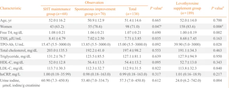
Table 1 shows the baseline clinical and biochemical character-istics of three groups. The data from 68 patients of the SHT maintenance group (maintained subclinical hypothyroid state
without medication), 70 of the spontaneous improvement group (improved to euthyroid state without medication), and 189 of the levothyroxine supplement group (which attained a euthyroid state with medication) were analyzed. The mean fol-low-up period was 10.1 months for the SHT maintenance group, 9.6 months for the spontaneous improvement group, and 13.1 months for the levothyroxine supplement group. Women showed a better prognosis in the course of SHT (P=0.047). In addition, initial TSH level was significantly
dif-ferent (8.41±4.79 μIU/mL in the SHT maintenance group and
7.02±2.50 μIU/mL in the spontaneous improvement group;
P=0.035). Except TSH level and sex ratio, there were no
sta-tistically significant differences in age, fT4, TPO-Ab, lipid pro-file, hsCRP, and urine iodine. We performed further analysis on the subpopulation, according to TSH levels (Table 2). For sub-jects with TSH between 5 to 10 μIU/mL, those in the spontane -ous improvement group showed significantly lower TPO-Ab titer than the SHT maintenance group (P=0.039).
Baseline characteristics of the levothyroxine supplement group were compared with those of the observed population (the SHT maintenance and spontaneous improvement groups) (Table 1). SHT patients with lower fT4, higher TPO-Ab titer, and lower urine iodine were more likely to be considered for
Table 1. Baseline Characteristics of the Subclinical Hypothyroidism Maintenance, Spontaneous Improvement, and Levothyroxine Supplement Group
Characteristic
Observation
P valuea
Levothyroxine supplement group
(n=189)
P valueb SHT maintenance
group (n=68)
Spontaneous improvement group (n=70)
Total (n=138)
Age, yr 52.0±16.2 50.9±12.9 51.4±14.6 0.665 52.0±14.0 0.700
Women 43 (63.2) 55 (78.6) 98 (71.0) 0.047c 158 (83.6) 0.006d
Free T4, ng/dL 1.08±0.21 1.06±0.21 1.07±0.21 0.690 1.00±0.19 0.002
TSH, μIU/mL 8.41±4.79 7.02±2.50 7.71±3.85 0.035 8.68±7.45 0.163 TPO-Ab, U/mL 15.47 (5.5–3000.0) 13.85 (5.5–3000.0) 15.00 (5.5–3000.0) 0.092 39.90 (5.0–3000) 0.028 Total cholesterol, mg/dL 203.0±135.3 192.2±41.0 197.4±98.2 0.553 191.1±34.3 0.463
Triglyceride, mg/dL 131.2±76.7 123.5±85.5 127.1±81.1 0.639 127.9±94.9 0.950
HDL-C, mg/dL 52.0±12.8 56.4±13.3 54.4±13.2 0.095 52.7±13.0 0.343
LDL-C, mg/dL 113.7±30.3 112.3±32.7 112.9±31.5 0.822 113.8±32.3 0.840
hsCRP, mg/L 1.00 (0.18–35.99) 0.98 (0.18–163.0) 0.99 (0.18–163.0) 0.317 1.01 (0.16–18.9) 0.217 Urine iodine,
μmoL iodine/g creatinine60.90 (7.3–450.8) 53.40 (7.0–334.7) 57.3 (7.0–450.8) 0.612 24.0 (6.2–742.0) 0.004
Data are mean±standard deviation, number (%), or median (minimum-maximum).
SHT, subclinical hypothyroidism; TSH, thyroid-stimulating hormone; TPO-Ab, anti-thyroid peroxidase antibody; HDL-C, high den-sity lipoprotein cholesterol; LDL-C, low denden-sity lipoprotein cholesterol; hsCRP, high-sensitivity C-reactive protein.
aP values for the comparison of the mean values between SHT maintenance and Spontaneous improvement group by t test; bP values
for the comparison of the sex ratio by Pearson’s chi-square test; cP values for the comparison of the mean values between Observation
supplement therapy by the doctor. Additionally, the levothy-roxine supplement group had a higher proportion of women than the observation group.
Comparison between initial and follow-up parameters in the SHT maintenance and spontaneous improvement group
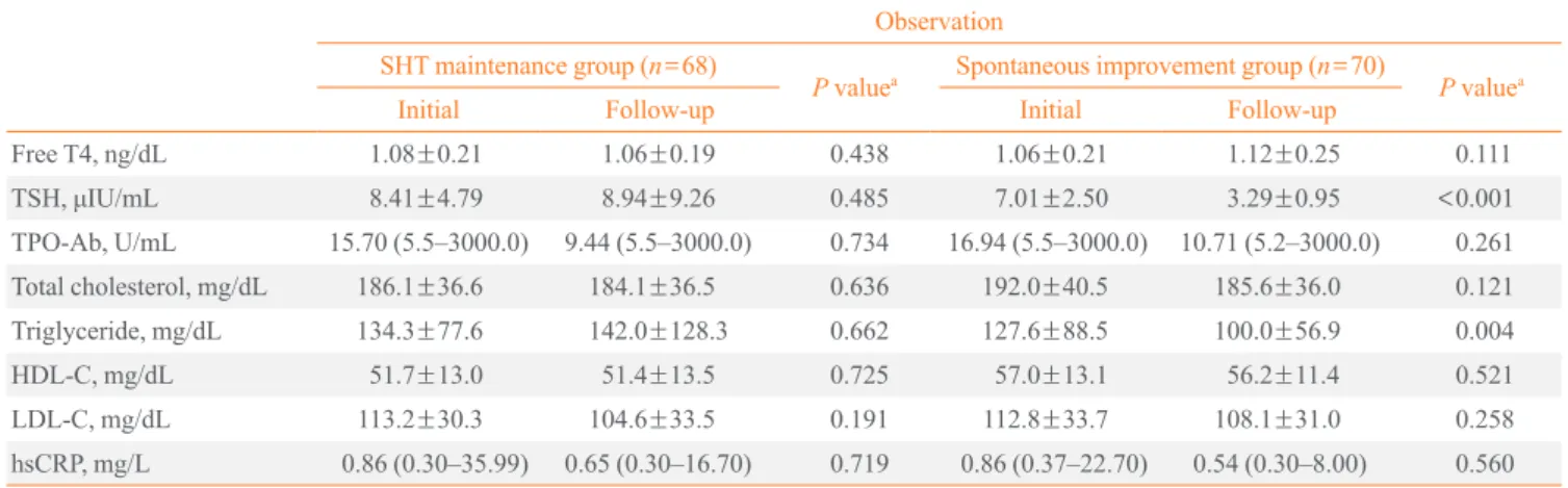
Table 3 shows the changes in parameters during follow-up (about 10 months) in the SHT maintenance and spontaneous improvement group. As expected, none of the measured pa-rameters showed any changes during follow-up in the SHT maintenance group. In the spontaneous improvement group with TSH levels that had returned to normal, there were no sig-nificant improvements in TPO-Ab and hsCRP. TPO-Ab titer showed only a decreasing trend. Analysis of lipid profiles showed triglyceride levels decreasing significantly during fol-low-up (127.6±88.5 mg/dL vs. 100.0±56.9 mg/dL; P=0.004).
Comparison between initial and follow-up parameters in the levothyroxine supplement group
Table 4 shows the changes in parameters during follow-up in the levothyroxine supplement group. We compared the param-eters at the time their thyroid function reached euthyroid state to those at diagnosis. TPO-Ab titer and triglyceride level sig-nificantly decreased during follow-up (for TPO-Ab, median value, 40.06 U/mL vs. 33.97 U/mL, P=0.009; for triglyceride,
mean value, 128.2±97.4 μIU/dL vs. 105.3±52.5 μIU/dL, P=
0.002, respectively). hsCRP titer did not show significant changes.
US finding in SHT
The existence of DT on US at diagnosis was not significantly
Table 3. Comparison between the Parameters at Initial Presentation and Follow-up of the SHT Maintenance and Spontaneous Im-provement Group
Observation
SHT maintenance group (n=68)
P valuea Spontaneous improvement group (n=70) P valuea
Initial Follow-up Initial Follow-up
Free T4, ng/dL 1.08±0.21 1.06±0.19 0.438 1.06±0.21 1.12±0.25 0.111
TSH, μIU/mL 8.41±4.79 8.94±9.26 0.485 7.01±2.50 3.29±0.95 <0.001 TPO-Ab, U/mL 15.70 (5.5–3000.0) 9.44 (5.5–3000.0) 0.734 16.94 (5.5–3000.0) 10.71 (5.2–3000.0) 0.261 Total cholesterol, mg/dL 186.1±36.6 184.1±36.5 0.636 192.0±40.5 185.6±36.0 0.121
Triglyceride, mg/dL 134.3±77.6 142.0±128.3 0.662 127.6±88.5 100.0±56.9 0.004
HDL-C, mg/dL 51.7±13.0 51.4±13.5 0.725 57.0±13.1 56.2±11.4 0.521 LDL-C, mg/dL 113.2±30.3 104.6±33.5 0.191 112.8±33.7 108.1±31.0 0.258
hsCRP, mg/L 0.86 (0.30–35.99) 0.65 (0.30–16.70) 0.719 0.86 (0.37–22.70) 0.54 (0.30–8.00) 0.560
Data are mean±standard deviation or median (minimum-maximum).
SHT, subclinical hypothyroidism; TSH, thyroid-stimulating hormone; TPO-Ab, anti-thyroid peroxidase antibody; HDL-C, high density lipoprotein cholesterol; LDL-C, low density lipoprotein cholesterol; hsCRP, high-sensitivity C-reactive protein.
aP values for the comparison of the mean values between initial and follow-up parameters by using paired t test. Table 2. Baseline Characteristics of SHT Maintenance and
Spontaneous Improvement Group in the Subjects with TSH
between 5 to 10 μIU/mL
Characteristic
Observation
P valuea SHT maintenance
group (n=54)
Spontaneous improvement group (n=65)
Free T4, ng/dL 1.09±0.20 1.07±0.22 0.574
TSH, μIU/mL 6.70±1.26 6.50±1.24 0.374 TPO-Ab, U/mL 15.60 (5.5–3000.0) 11.82 (5.5–3000.0) 0.039 Total cholesterol,
mg/dL
206.8±152.7 192.7±42.3 0.506 Triglyceride, mg/dL 133.5±79.0 123.1±88.4 0.569 HDL-C, mg/dL 51.9±13.0 57.5±13.2 0.053 LDL-C, mg/dL 110.8±29.6 111.9±34.0 0.867 hsCRP, mg/L 1.02 (0.18–19.63) 0.98 (0.18–163.0) 0.232
Urine iodine, μmoL
iodine/g creatinine
55.40 (7.3–450.8) 49.50 (7.0–334.7) 0.695
Data are mean±standard deviation or median (minimum-maximum). SHT, subclinical hypothyroidism; TSH, thyroid-stimulating hormone; TPO-Ab, anti-thyroid peroxidase antibody; HDL-C, high density li-poprotein cholesterol; LDL-C, low density lili-poprotein cholesterol; hsCRP, high-sensitivity C-reactive protein.
Table 5. The Presentation of SHT according to US Finding
Us finding
P valuea Diffuse thyroiditis
(+) (n=150)
Diffuse thyroiditis (-) (n=141)
Age, yr 50.3±12.8 53.4±15.0 0.052 Women, n (%) 120 (80.0) 112 (79.4) 0.904b
Free T4, ng/dL 1.02±0.20 1.04±0.21 0.478
TSH, μIU/mL 8.51±5.92 7.35±2.67 0.033
TPO-Ab, U/mL 220.74 (5.0–3000.0) 11.55 (5.5–3000.0)<0.001 Total cholesterol,
mg/dL
200.3±91.5 187.1±35.8 0.164
Triglyceride, mg/dL
139.8±101.4 109.5±70.5 0.011
HDL-C, mg/dL 52.7±12.5 54.3±14.0 0.384
LDL-C, mg/dL 114.1±29.7 111.6±33.0 0.569 hsCRP, mg/L 0.99 (0.18–22.70) 1.01 (0.16–163.00) 0.112 Urine iodine,
μmol iodine/g
creatinine
28.70 (6.4–742.0) 37.70 (6.4–334.7) 0.770
Data are mean±standard deviation, number (%) or median (mini-mum-maximum).
SHT, subclinical hypothyroidism; US, ultrasohographic; TSH, thy-roid-stimulating hormone; TPO-Ab, anti-thyroid peroxidase antibody; HDL-C, high density lipoprotein cholesterol; LDL-C, low density li-poprotein cholesterol; hsCRP, high-sensitivity C-reactive protein.
aP values for the comparison of the mean values of two groups by
us-ing t test; bP values for the comparison of the Sex ratio of two groups by
using Pearson’s chi-square test.
different between the SHT maintenance group and spontane-ous improvement group (28.3% of the SHT maintenance group and 21.7% of the spontaneous improvement group; P=0.144).
However, when we looked into the clinical and laboratory characteristics at diagnosis according to US finding (Table 5), the existence of DT was associated with higher TSH, TPO-Ab, and triglyceride (P=0.035, P<0.01, and P=0.017,
respective-ly) at that time.
DISCUSSION
Some SHT patients spontaneously recover thyroid function without specific treatment, others maintain a subclinical hypo-thyroid state without improvement, while others progress to an overt hypothyroid state requiring thyroid hormone supple-ment. Therefore, a general understanding of the natural course of SHT, prognostic factors, clinical consequences of a persis-tent subclinical hypothyroid state, and the effect of levothy-roxine supplement, is necessary for clinicians in planning SHT management strategies, including determination of the neces-sity and appropriate time of treatment.
Until now, various studies have regarded TSH level and TPO-Ab titer as the most reliable clinical parameters in pre-dicting the progression of SHT or euthyroidism to OHT. In a
1995 cohort study conducted over the course of 20 years in the United Kingdom [7], the odds ratios (ORs) (with 95% confi-dence intervals [CIs]) of developing hypothyroidism in euthy-roid subjects with raised serum TSH alone (TSH >2 μIU/mL)
were OR, 8; 95% CI, 3 to 20 for women; and OR, 44; 95% CI, 19 to 104 for men. In cases with positive antithyroid antibod-ies alone, the odds ratios were OR 8 (95% CI, 5 to 15) for women and OR 25 (95% CI, 10 to 63) for men [7]. In subjects with both elevated serum TSH and positive antithyroid anti-bodies, the ORs were 38 (95% CI, 22 to 65) for women and OR 173 (95% CI, 81 to 370) for men. In another 4-year cohort study conducted in the United States [8], one-third of subjects with SHT developed OHT over the course of the study. The initial TSH level of all subjects who became overtly hypothy-roid was above 20 μIU/mL, and 80% of all of subjects, regard -less of initial TSH level, also showed high TPO-Ab titer. An-other prospective long-term study of 82 female patients with SHT also showed that higher initial TSH concentrations and positive TPO-Ab allowed initial risk stratification for the de-Table 4. Comparison between the Parameters at Initial
Presen-tation and Follow-up of Levothyroxine Supplement Group
Parameter
Levothyroxine supplement group
(n=189) P valuea Initial Follow-up
Free T4, ng/dL 1.00±0.19 1.26±0.24 <0.001
TSH, μIU/mL 8.68±7.45 2.47±1.19 <0.001 TPO-Ab, U/mL 40.06 (5.0–3000.0) 33.97 (5.0–3000.0) 0.009 Total cholesterol,
mg/dL
191.6±34.3 187.4±33.5 0.135
Triglyceride, mg/dL
128.2±97.4 105.3±52.5 0.002
HDL-C, mg/dL 52.9±13.4 53.0±11.6 0.917 LDL-C, mg/dL 114.3±32.7 113.5±30.0 0.785 hsCRP, mg/L 1.07 (0.30–15.44) 0.98 (0.30–12.30) 0.369
Data are mean±standard deviation or median(minimum-maximum).
SHT, subclinical hypothyroidism; TSH, thyroid-stimulating hormone; TPO-Ab, anti-thyroid peroxidase antibody; HDL-C, high density li-poprotein cholesterol; LDL-C, low density lili-poprotein cholesterol; hsCRP, high-sensitivity C-reactive protein.
aP values for the comparison of the mean values between initial and
velopment of overt thyroid failure within 10 years [9]. Other prospective studies conducted since 2000 also have proved that both elevated TSH level and the presence of TPO-Ab are poor prognostic factors of SHT.
In this study, to determine the prognostic factors of SHT, we compared two groups, the SHT maintenance group and the spontaneous improvement group. Comparison of numerous parameters including age, sex, fT4, TSH, TPO-Ab, lipid pro-file, hsCRP, urine iodine, and the presence of DT on US at ini-tial presentation found that women had a more favorable prog-nosis than men and, among biochemical parameters consid-ered for prognostic factors in many studies, only initial TSH level was significantly different. It is well-known that women have higher prevalence of both SHT and OHT than men. However, sexual difference of disease prognosis has not been established. Subjects with higher TSH level seemed to main-tain a subclinical hypothyroid state, while subjects with lower TSH level showed a tendency to spontaneously improve. Our result is consistent with a study of 107 elderly SHT patients in Spain, which reported that TSH concentration was the most powerful predictor for the outcome of SHT [10]. TPO-Ab, known as the other apparent major predictor of OHT and SHT in several other studies, was not shown to be a significant pre-dictor in our pool of SHT patients. However, when we per-formed further subanalysis for subjects with TSH between 5 to 10 μIU/mL, those in the spontaneous improvement group showed significantly lower TPO-Ab titer than the SHT main-tenance group. Therefore, we can conclude that TPO-Ab titer is also a useful predictor for SHT. Although TSH level is the most powerful prognostic factor in the overall SHT popula-tion, TPO-Ab titer can be used as a predictor in SHT with mildly elevated TSH (between 5 to 10 μIU/mL). This can be explained, at least partially, by the fact that TPO-Ab titer con-tributes to prognosis earlier than TSH in the pathogenesis of SHT. TPO-Ab titer can be regarded as an earlier prognostic factor that is especially helpful in mild SHT. However, TSH level is the strongest prognostic factor, regardless of TPO-Ab titer, in advanced SHT.
We also investigated the changes in parameters during fol-low-up in each group. As for TPO-Ab changes, in the levothy-roxine treatment group, TPO-Ab titer was decreased signifi-cantly. In contrast, in the spontaneous improvement group, there was no significant improvement of TPO-Ab titer, which exhibited a decreasing trend. Given that most of the subjects in the spontaneous improvement group were those who had relatively low TPO-Ab titer (only seven subjects of the
spon-taneous improvement group had TPO-Ab titer >60 U/mL)
and that the subjects in the levothyroxine treatment group had higher TPO-Ab titer, we cannot generalize our results across all SHT subjects. However, our results at least demonstrate a benefit of levothyroxine supplement in SHT for the purpose of decreasing TPO-Ab, thus decreasing autoimmunity and the parenchymal pathologic process.
Regarding lipid profiles, currently there is no conclusive evidence regarding the effect of SHT on lipid profile. In an observational cohort study in the United States, lipid levels in-cluding total cholesterol, LDL-C and triglyceride increased in a graded fashion as thyroid function declined [11]. On the oth-er hand, in anothoth-er randomized placebo-controlled study in SHT where patients were randomly assigned to levothyroxine therapy or placebo and re-evaluated after 6 months of euthy-roidism, triglyceride levels remained similar regardless of TSH levels, although total cholesterol and LDL-C levels were lowered by levothyroxine therapy and did correlate with base-line TSH levels [12]. The study concluded that only LDL-C levels are increased specifically and reversibly in association with SHT. Another study in Switzerland reported found that SHT subjects showed borderline elevated LDL-C and similar total cholesterol and triglyceride concentrations compared to controls matched for age, sex, and body mass index [13]. In our study, in both the spontaneous improvement group and the levothyroxine treatment group, triglyceride level decreased significantly during follow-up. However, most of these pa-tients did not have hypertriglyceridemia (triglyceride >150
mg/dL) at diagnosis, so the change should be interpreted as the decrease of mean triglyceride level, not the improvement of hypertriglyceridemia. Unlike most previous studies stating total cholesterol and LDL-C as lipid profiles correlating with changes in thyroid function, an apparent correlation between triglyceride level and subclinical hypothyroid state was shown in this study. Therefore, we suggest that triglyceride level should not be ignored in evaluating SHT at least in Korean pa-tients.
estab-lished as a potential marker for coronary heart disease, hsCRP elevation in SHT may imply cardiologic problems associated with SHT, although this has not yet been confirmed. However, in this study, hsCRP did not show any significant differences at diagnosis between the SHT maintenance group and the spontaneous improvement group or any significant change during follow-up in all three groups.
Recently, dietary iodine excess has been found to be associ-ated with decreased thyroid function. Iodine is the most nota-ble environmental factor in thyroid dysfunction and also is an indicator of vulnerability to other possible harmful environ-mental factors [18]. However, in this study, higher iodine con-centration in the body, as quantified by urinary iodine excre-tion, was not associated with poor prognosis of SHT. A 1-year observational study of SHT in Denmark reported a significant positive correlation between TSH and urinary iodine excretion in monthly measurements [19]. Furthermore, high urinary io-dine excretion predicted high TSH and TPO-Ab titer during the following month. In another study of adults in an iodine-rich area in Japan [20], hypothyroidism was more prevalent in subjects who were negative for antithyroid antibodies who had high urinary iodine excretion compared with those with nor-mal urinary iodine excretion. They reported significantly higher TSH and lower fT4 levels in SHT patients with high urinary iodine excretion and concluded that the prevalence of hypothyroidism in iodine sufficient areas may be associated with the amount of iodine ingested. Based on our study, how-ever, restriction of excessive iodine intake, which is a possible prognostic factor, if so, maybe only modifiable prognostic fac-tor, has only weak evidence supporting its use in SHT man-agement at present, at least in Korean subjects. As serial mea-surements of urinary iodine excretion after initial laboratory tests were not conducted; however, we cannot rule out the possibility that the altered iodine status during follow-up may influence the clinical course of our subclinical hypothyroid subjects.
Decreased or irregular heterogeneous echogenicity on US is a characteristic finding in diffuse thyroid disease, including both OHT and SHT [21-24]. However, the value of DT on US as a predictor of SHT has not been established until recently. In a 3-year follow-up study of 117 cases of mild SHT in Bra-zil, the likelihood of a progression toward OHT and improve-ment to euthyroidism, respectively, were similar between pa-tients with positive TPO-Ab and/or US alteration and papa-tients with negative TPO-Ab but with positive US alteration, thus suggesting that US findings may be useful in determining the
prognosis of mild SHT [25]. In a retrospective study of Kore-an SHT patients, which aimed to see the difference of response to levothyroxine replacement according to autoantibody status and US finding, patients who initially showed DT on US, re-gardless of thyroid autoantibody level, showed poor response after levothyroxine replacement [26]. This report suggested the possibility that the DT pattern on thyroid US can serve as a prognostic factor when combined with other known parame-ters. In our study, the existence of DT on US at diagnosis was not significantly different between the SHT maintenance group and the spontaneous improvement group. Therefore, this observation indicates that DT on US only reflects the se-verity of the hypothyroid state at the time of US evaluation, which is indicated by higher TSH and TPO-Ab. It could not predict the prognosis of SHT.
This study has several limitations. First, because this is a retrospective study, the impacts and powers of the results are weakened. Second, the study population was not representa-tive of the entire Korean population because the pool of pa-tients was derived from a single tertiary health care center. For the most part, the care of SHT patients can be handled by local physicians at primary health care clinics. Therefore, the num-ber of eligible participants in our study was small compared with the relatively many OHT patients. The third limitation is that the follow-up period was not long enough (less than 13.0 months). Without apparent progression to OHT, patients typi-cally return to the local clinic for follow-up. Fourth, because we lacked additive data, such as body mass index and pre-menopause/postmenopause state, we were unable to interpret our data using these epidemiologic factors, which may have affected the prognosis of SHT. Lastly, as mentioned above, as urinary iodine excretion was not measured serially, altered io-dine status during follow-up may have influenced the clinical course of our SHT subjects.
a large-scaled, prospective trial will be needed to determine the definite prognostic factors of SHT.
CONFLICTS OF INTEREST
No potential conflict of interest relevant to this article was re-ported.
REFERENCES
1. Fowler PB, Swale J, Andrews H, Ikram H, Banim SO.
Grades of hypothyroidism. Br Med J 1973;2:178.
2. Hollowell JG, Staehling NW, Flanders WD, Hannon WH,
Gunter EW, Spencer CA, Braverman LE. Serum TSH, T(4), and thyroid antibodies in the United States population (1988 to 1994): National Health and Nutrition Examination Survey (NHANES III). J Clin Endocrinol Metab 2002;87: 489-99.
3. Riniker M, Tieche M, Lupi GA, Grob P, Studer H, Burgi H.
Prevalence of various degrees of hypothyroidism among patients of a general medical department. Clin Endocrinol (Oxf) 1981;14:69-74.
4. Herrmann J. Prevalence of hypothyroidism in the elderly
in Germany. A pilot study. J Endocrinol Invest 1981;4:327-30.
5. Bilous RW, Tunbridge WM. The epidemiology of
hypothy-roidism: an update. Baillieres Clin Endocrinol Metab 1988; 2:531-40.
6. Kostoglou-Athanassiou I, Ntalles K. Hypothyroidism: new
aspects of an old disease. Hippokratia 2010;14:82-7.
7. Vanderpump MP, Tunbridge WM, French JM, Appleton D,
Bates D, Clark F, Grimley Evans J, Hasan DM, Rodgers H, Tunbridge F, Young ET. The incidence of thyroid disorders in the community: a twenty-year follow-up of the Whick-ham Survey. Clin Endocrinol (Oxf) 1995;43:55-68.
8. Rosenthal MJ, Hunt WC, Garry PJ, Goodwin JS. Thyroid
failure in the elderly. Microsomal antibodies as discrimi-nant for therapy. JAMA 1987;258:209-13.
9. Huber G, Staub JJ, Meier C, Mitrache C, Guglielmetti M,
Huber P, Braverman LE. Prospective study of the sponta-neous course of subclinical hypothyroidism: prognostic value of thyrotropin, thyroid reserve, and thyroid antibod-ies. J Clin Endocrinol Metab 2002;87:3221-6.
10. Diez JJ, Iglesias P. Spontaneous subclinical
hypothyroid-ism in patients older than 55 years: an analysis of natural course and risk factors for the development of overt
thy-roid failure. J Clin Endocrinol Metab 2004;89:4890-7.
11. Canaris GJ, Manowitz NR, Mayor G, Ridgway EC. The
Colorado thyroid disease prevalence study. Arch Intern Med 2000;160:526-34.
12. Caraccio N, Ferrannini E, Monzani F. Lipoprotein profile
in subclinical hypothyroidism: response to levothyroxine replacement, a randomized placebo-controlled study. J Clin Endocrinol Metab 2002;87:1533-8.
13. Althaus BU, Staub JJ, Ryff-De Leche A, Oberhansli A,
Stahelin HB. LDL/HDL-changes in subclinical hypothy-roidism: possible risk factors for coronary heart disease. Clin Endocrinol (Oxf) 1988;28:157-63.
14. Sharma R, Sharma TK, Kaushik GG, Sharma S, Vardey
SK, Sinha M. Subclinical hypothyroidism and its associa-tion with cardiovascular risk factors. Clin Lab 2011;57: 719-24.
15. Tuzcu A, Bahceci M, Gokalp D, Tuzun Y, Gunes K.
Sub-clinical hypothyroidism may be associated with elevated high-sensitive c-reactive protein (low grade inflammation) and fasting hyperinsulinemia. Endocr J 2005;52:89-94.
16. Toruner F, Altinova AE, Karakoc A, Yetkin I, Ayvaz G,
Ca-kir N, Arslan M. Risk factors for cardiovascular disease in patients with subclinical hypothyroidism. Adv Ther 2008; 25:430-7.
17. Park YJ, Lee EJ, Lee YJ, Choi SH, Park JH, Lee SB, Lim S,
Lee WW, Jang HC, Cho BY, Woo JI, Kim KW. Subclinical hypothyroidism (SCH) is not associated with metabolic derangement, cognitive impairment, depression or poor quality of life (QoL) in elderly subjects. Arch Gerontol Geriatr 2010;50:e68-73.
18. Chung HK. Environmental factors and thyroid
dysfunc-tion. Endocrinol Metab 2012;27:191-3.
19. Karmisholt J, Laurberg P. Serum TSH and serum thyroid
peroxidase antibody fluctuate in parallel and high urinary iodine excretion predicts subsequent thyroid failure in a 1-year study of patients with untreated subclinical hypo-thyroidism. Eur J Endocrinol 2008;158:209-15.
20. Konno N, Makita H, Yuri K, Iizuka N, Kawasaki K.
Asso-ciation between dietary iodine intake and prevalence of subclinical hypothyroidism in the coastal regions of Japan. J Clin Endocrinol Metab 1994;78:393-7.
21. Loy M, Cianchetti ME, Cardia F, Melis A, Boi F, Mariotti
22. Rago T, Chiovato L, Grasso L, Pinchera A, Vitti P. Thyroid
ultrasonography as a tool for detecting thyroid autoim-mune diseases and predicting thyroid dsfunction in appar-ently healthy subjects. J Endocrinol Invest 2001;24:763-9.
23. Schiemann U, Avenhaus W, Konturek JW, Gellner R,
Hengst K, Gross M. Relationship of clinical features and laboratory parameters to thyroid echogenicity measured by standardized grey scale ultrasonography in patients with Hashimoto’s thyroiditis. Med Sci Monit 2003;9:MT13-7.
24. Vejbjerg P, Knudsen N, Perrild H, Laurberg P, Pedersen
IB, Rasmussen LB, Ovesen L, Jorgensen T. The associa-tion between hypoechogenicity or irregular echo pattern at thyroid ultrasonography and thyroid function in the gener-al population. Eur J Endocrinol 2006;155:547-52.
25. Rosario PW, Bessa B, Valadao MM, Purisch S. Natural
history of mild subclinical hypothyroidism: prognostic value of ultrasound. Thyroid 2009;19:9-12.
26. Shin DY, Kim EK, Lee EJ. Role of ultrasonography in