Transglutaminase Type I Serving as RNPs Down-Regulate
Astakine-Mediated Hematopoiesis
Yun-Tsan Chang1, Cheng-Yung Lin2, Che-Yiang Tsai1, Vinu S. Siva1, Chia-Ying Chu1,3, Huai-Jen Tsai2,3, Yen-Ling Song1,3*
1Institute of Zoology, National Taiwan University, Taipei, Taiwan, ROC,2Institute of Molecular and Cellular Biology, National Taiwan University, Taipei, Taiwan, ROC, 3Department of Life Science, National Taiwan University, Taipei, Taiwan, ROC
Abstract
Astakine is an important cytokine that is involved in crustacean hematopoiesis. Interestingly, the protein levels of astakine increased dramatically in plasma of LPS-injected shrimp while mRNA levels remained unchanged. Here, we investigated the involvement of astakine 39-untranslated region (UTR) in its protein expression. The 39-UTR of astakine down-regulated the expression of reporter protein but the mRNA stability of reporter gene was unaffected. We identified the functional regulatory elements of astakine 39-UTR, where 39-UTR242–483acted as repressor. The electrophoresis mobility shift assay
(EMSA), RNA pull-down assay and LC/MS/MS were performed to identify the protein association. We noted that crustin Pm4 and shrimp transglutaminase I (STG I) were associated to astakine 39-UTR242–483, while two other proteins have yet to be
revealed. Depletion of hemocytic crustin Pm4 and STG I significantly increased the protein level of astakine while astakine mRNA level remained unaffected. Lipopolysaccharide (LPS) stimulated the secretion of crustin Pm4 and STG I from hemocytes to plasma and increased the astakine level to stimulate the hemocytes proliferation. Altogether, we identified the shrimp crustin Pm4 and STG I as novel RNA binding proteins that play an important role in down-regulating astakine expression at post-transcriptional level and are crucial for the maintenance of hematopoiesis.
Citation:Chang Y-T, Lin C-Y, Tsai C-Y, Siva VS, Chu C-Y, et al. (2013) The New Face of the Old Molecules: Crustin Pm4 and Transglutaminase Type I Serving as RNPs Down-Regulate Astakine-Mediated Hematopoiesis. PLoS ONE 8(8): e72793. doi:10.1371/journal.pone.0072793
Editor:Prasun K. Datta, Temple University, United States of America ReceivedMay 9, 2013;AcceptedJuly 12, 2013;PublishedAugust 27, 2013
Copyright:ß2013 Chang et al. This is an open-access article distributed under the terms of the Creative Commons Attribution License, which permits unrestricted use, distribution, and reproduction in any medium, provided the original author and source are credited.
Funding:This study was supported financially by the National Science Council (Grant no. NSC 100-2313-B-002 -041 -MY2) to Yen-Ling Song. The funders had no role in study design, data collection and analysis, decision to publish, or preparation of the manuscript.
Competing Interests:The authors have declared that no competing interests exist.
* E-mail: song@ntu.edu.tw
Introduction
Crustaceans have open circulatory system in which mainte-nance of homeostasis and innate immune response are closely related, and where hemocytes play important roles against pathogens [1,2]. During infection or massive hemocytes loss, hematopoietic tissues produce hemocytes to maintain homeostasis. Some immunostimulants, such as LPS and laminarin, cause massive depletion of hemocyte. After LPS injection, the circulating hemocyte percentage is considerably decreased to 40% within 3 hours but is restored to 100% in 3–24 hours [3]. Meanwhile, the cells significantly proliferate in hematopoietic tissue of tiger shrimp
Penaeus monodonafter LPS injection [4]. These similar observations have been observed using laminarin stimulation [5]. Thus, it is considered that when shrimp is infected by a pathogen, the involvement of hematopoiesis and hemocyte regulation is crucial response to the pathogen and maintenance of homeostasis.
Astakine is an important cytokine involved in the hematopoiesis of crustaceans. There are two astakine molecules, astakine 1 and 2, cloned from crayfish with a sequence difference of 13 extra amino acids insert in astakine 2. As for tiger shrimp, one astakine molecule but two transcripts with various lengths of 39-UTR have been cloned and reported [6,7]. Shrimp astakine is more similar to crayfish astakine 2 in amino acid sequence (53% identity), but less
similar to crayfish astakine 1 (38% identity). However, shrimp astakine shows functional analogy with crayfish astakine 1 in stimulating hemocyte proliferation in the hematopoietic tissue [6,7]. As for crayfish astakine 2, it fails to stimulate hemocyte proliferation, instead, it stimulates crayfish hemocyte to further differentiate and mature into granulocytes [8]. Serving as hematopoietic growth factors, astakine 1 may be involved in the early developmental stage and astakine 2 in the late stage in crayfish. In a recent study, the expression of crayfish astakine 2 is up-regulated by melatonin in brain during the dark period of the circadian rhythm [9]. However, the intracellular regulatory mechanism of astakine is still unknown.
existing mRNAs allows for more rapid changes in protein levels during nutrient deprivation and stress, development and differen-tiation, nervous system function, aging, and diseases [11–15]. Regulation through the 39-UTR of mRNA is one of the critical mechanisms for post-transcriptional control. Moreover, the average length of 39-UTR has increased during evolution suggesting that its utilization may contribute to organism complexity [16,17]. In invertebrates, the mean length of functional 39-UTR is around 300 bp and the extended 39-UTR length provides potential for transcript-specific regulation [17].
It has been shown that various RNA-binding proteins that interact with 39-UTR to form ribonucleoprotein (RNP) complexes perform a key role as translational regulator [18]. Besides, diverse RNA-binding protein and association of RNPs to specific recognition elements of mRNAs are part of a pervasive mechanism for multi-dimensional regulation of their post-transcriptional fate [19]. Strikingly, not only the ‘classical’ conventional RNPs but many enzymes with well-established cellular functions can act as nonconventional RNPs, and participate potentially in regulating RNA stability and gene expression [20,21]. Still there are many conventional and nonconventional RNPs yet to be revealed, and their role in controlling gene expression is important in many aspects, such as invertebrate hematopoiesis. In the present study, for the first time, we identified two nonconventional proteins associated with the regulatory elements in the 39UTR of astakine and revealed their specific role in regulating the expression of astakine via its 39UTR242–483.
Materials and Methods
RNA secondary structure prediction
The sequence from shrimp long form astakine 39-UTR (GenBank accession no. EU980444) was submitted to RNA fold web server [22] (http://rna.tbi.univie.ac.at/cgi-bin/RNAfold.cgi) for secondary structure prediction.
Cell culture
Sf21cell line fromSpodoptera frugiperdawas cultured at 26uC in TNM-FH insect cell medium (Grace’s insect cell culture medium (Invitrogen).
Shrimp hemocytes were cultured using primary culture method described by Li et al. [23]. Shrimp hemocytes were drawn from the abdominal segment with a 1 ml syringe containing 0.5 ml anticoagulation solution (0.1 M sodium citrate, 0.4 M sucrose, 0.01 M Tris-HCl, pH 7.6, 780615 mOsm/kg). Hemocytes were collected by centrifugation, and were then gently suspended with 1 ml Leibovitz L-15 culture medium (Gibco, USA). Hemocytes were counted and distributed into 24-well culture plates (Corning Life Sciences, USA) with 36105 cells/well. The total volume in each well was adjusted to 500ml/well using the culture medium, and the culture plates were then placed in a 26uC incubator for 2 h before the next treatment.
Construction, transfection and activity assay of luciferase by astakine 39-UTR
Tiger shrimp astakine 39-UTR was divided into eight segments of different length and named as 39-UTR1–965, 39-UTR1–483, 39-UTR484–965, 39-UTR1–241, 39-UTR242–483, 39-UTRde242– 483 (astakine 39-UTR lacking 39-UTR242–483), 39-UTR242– 362 and 39-UTR363–483. All the eight segments were then separately incorporated into pGL3 firefly luciferase reporter plasmid, which has an OpIE2 promoter (pGL3-OpIE2), and the astakine 39UTR segments were inserted behind the firefly
luciferase reporter gene. The phRG Renilla luciferase plasmid with TK promoter (phRG-TK) was served as internal control. The pGL3-OpIE2 vector (800 ng/well) and phRG-TK vector (200 ng/well) were co-transfected into Sf21 cells by using CellfectinTM Transfection Reagent (Invitrogen, CA, USA).
After 48 hrs post transfection, the luciferase assay was performed using Dual-Glo luciferase assay system (Promega) with phRG Renilla luciferase gene vector as internal control to normalize the transfection efficiency. The firefly and Renilla
luciferase activities were measured by the Dual-Glo luciferase assay system according to the manufacturer’s instructions and the chemiluminescence was read by a Luoroskan Ascent FL (Labsystems) reader. The co-expressed Renilla luminescence was used to normalize the firefly luminescence.
Competition experiment
The astakine 39-UTR242–483 was subcloned into pEGFP reporter vector with a CMV promoter, and astakine 39-UTR242– 483 was inserted behind the reporter gene. This competitor plasmid was named as pEGFP-CMV-Ast 39-UTR242,483.
The pGL3-OpIE2-Ast 39-UTR242,483vector (400 ng/well)
was co-transfected with various amounts of pEGFP-CMV-Ast 39 -UTR242,483competitor vector (0 ng/well, 400 ng/well and
1000 ng/well) intoSf21 cells by using CellfectinTM Transfection Reagent (Invitrogen, CA, USA). The pEGFP reporter vector without astakine 39-UTR242–483 was used as a concentration control vector and the phRGRenillaluciferase reporter vector with TK promoter was used as an internal control vector. The procedures for plasmid transfection and luciferase activity assay were performed as above.
RNA-electrophoretic mobility shift assay (RNA-EMSA) in shrimp hemocyte protein extraction
The astakine 39-UTR242–483 was subcloned into pCS2+ vector. The constructed pCS2+-Ast 39-UTR242–483 plasmids were used as template, and SP6 RNA polymerase (Roche) was used for in vitro transcription to synthesize Ast 39-UTR242–483 RNA according to the manufacturer’s instructions. The synthe-sized Ast 39-UTR242–483 RNA was biotinylated at 39end using PierceH RNA 39 End Biotinylation Kit (Pierce, Rockford, IL, USA) according to the manufacturer’s instruction.
Live black tiger shrimps (P. monodon) were purchased from local vendors in Taiwan. The shrimps were then acclimated for one week before the experimental use. To extract hemocyte protein, freshly prepared hemocytes were homogenized in lysis buffer (10 mM HEPES, 1.5 mM MgCl2, 10 mM KCl, 0.5 mM DTT, treated with 16protease inhibitor cocktail (Roche), pH 7.9 and incubated at 4uC for 10 minutes. The cells were centrifuged at 10,000 rpm at 4uC for 15 minutes and the clear supernatant was stored at280uC.
RNA binding protein: biotin pull-down assay
For biotin pull-down assay, the biotin-labeled Ast 39-UTR242– 483 transcripts were incubated with 30mg of total shrimp hemocyte lysate for 30 min at 25uC and then complexes were isolated with DynabeadsHM-280 Streptavidin (Invitrogen). After washing with lysis buffer, the pull-down RNA-binding proteins were analyzed by SDS-PAGE.
Identification of proteins interacting with Ast 39UTR242–483
To identify the RNA-binding proteins, in-gel digestion was performed. In brief, the protein band in 12.5% SDS-PAGE was manually excised from the gel and sliced into pieces. The gel pieces were incubated for 1 hour with 50 mM DTE in 25 mM ammonium bicarbonate, pH 8.5, at 37uC, and subsequently alkylated for 1 hour with 100 mM iodoacetamide in 25 mM ammonium bicarbonate at 25uC. The pieces were then washed with 50% acetonitrile in 25 mM ammonium bicarbonate, dehydrated with 100% acetonitrile, dried, and rehydrated for 16 h in 10mL of 25 mM ammonium bicarbonate containing 0.1mg trypsin (sequencing grade, Promega, USA) at 37uC. Following tryptic digestion, peptides were extracted with 50% acetonitrile containing 5% trifluoro acetic acid with moderate sonication. The extracted peptides were evaporated under vacuum. Thereafter, the digested peptides were desalted firstly by using C18 Zip-Tip and sent for LC/MS/MS analysis. The search for matched peptides was done by Mascot algorithm (www. matrixscience.com).
RNAi againstP. monodonSTG1 and Crustin Pm4 in primary hemocyte
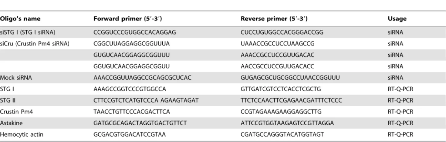
Specific siRNA sequences directed against P. monodon STG I and crustin Pm4 mRNA (GenBank accession no. AY074924.1 and FJ686015.1) were designed and ordered from Sigma. The antisense strand sequences of both siRNAs are: siSTG1: 59 -CUCCUGUGGCCACGGGACCGG-39; siCru: 59 -UAAACC-GCCUCCUAAGCCG- 39, 59 -AAACCGCCUCCGUUGAC-AC-39and 59-AACCGCCUCCGUUGACACC-39(Table 1).
The siRNAs were transfected into shrimp primary hemocytes culture, respectively, using CellfectinTM Transfection Reagent (Invitrogen, CA, USA). To deplete the target genes, hemocytes were transfected with 200ml siRNA transfection mixture contain-ing 4ml CellfectinTM, 30 pmol siRNA, and 196ml serum-free L-15 medium. In the untreated (UT) well, transfection mixture was
replaced with 200ml of serum-free L-15 medium. For mock transfections, hemocytes were transfected with a mixture of 4ml CellfectinTMand 196ml serum-free L-15 medium. At 5 hours post transfection, the transfection mixture was removed and 300mL L-15 medium with 16% FBS was added into the wells and all hemocytes were placed in a 26uC humid incubator before the subsequent treatments.
Hemocytes cDNA and protein preparation
Hemocytes were harvested at 24 hours post transfection and total RNA was extracted using TRIZOLH reagent (Invitrogen) followed by DNase I (Invitrogen) treatment. The cDNA was synthesized using the SuperScriptTM III First-Strand Synthesis System for RT-PCR according to the manufacturer’s instruction (Invitrogen).
Hemocyte proteins were extracted from siRNA knock-down hemocyte cultures and mock-transfection hemocyte cultures with TRIZOLH reagent (Invitrogen). Proteins were dissolved in cell lysis buffer (7 M urea and 2 M thiourea) and quantified using the Bradford method.
Real time PCR
The cDNAs were used as template along with STG I, STG II, crustin Pm4 and astakine gene primers (Table 1) and 26KAPATM SYBRH qPCR Master Mix (KAPA Biosystems). Real time PCR was performed in an ABI 7500 Q-PCR system using the standard program. The P. monodon b-tubulin gene served as an internal control. The relative expression ratio was represented by the equation: (each gene expression level)/(b-tubulin expression level). The data were analyzed using analysis of variance (ANOVA) and Duncan’s multiple range test (Duncan’s MRT) to determine differences between groups. The specificity of real-time PCR products was confirmed by melting curve analysis.
LPS Injection
The shrimps were injected at the second abdominal segment with lipopolysaccharide (LPS, E. coli 055:B5; Sigma-Aldrich). Hemolymph from each of the four shrimps was first withdrawn with anticoagulant as untreated sample and then injected with LPS (1mg/g shrimp) dissolved in MCHBSS (Modified Complete Hank’s Balanced Salt Solution) (10 mM CaCl2, 3 mM MgCl2, 5 mM MgSO4, 24 mg mL21HBSS (Sigma); 780615 mOsm kg21). Hemolymph was again withdrawn from each of the four
Table 1.Sequences of oligonucleotides used in this study.
Oligo’s name Forward primer (59-39) Reverse primer (59-39) Usage
siSTG I (STG I siRNA) CCGGUCCCGUGGCCACAGGAG CUCCUGUGGCCACGGGACCGG siRNA siCru (Crustin Pm4 siRNA) CGGCUUAGGAGGCGGUUUA UAAACCGCCUCCUAAGCCG siRNA
GUGUCAACGGAGGCGGUUU AAACCGCCUCCGUUGACAC siRNA
GGUGUCAACGGAGGCGGUU AACCGCCUCCGUUGACACC siRNA
Mock siRNA AAACCGGUUAGGCCGCAGCGCUCAC GUGAGCGCUGCGGCCUAACCGGUUU siRNA
STG I AAAGCCGGTCCCGTGGCCA GTTGATCGTCCTCACCTCGCTG RT-Q-PCR
STG II CTTCCGTCTCATGTCCCA AGAAGTAGAT TTCTCCAACTTCGAGAACGATTTCTCCC RT-Q-PCR
Crustin Pm4 TAACCTGTTCCCACGACTTCA CCGTAGAAAGAAGGAGGCTTG RT-Q-PCR
Astakine GATGCGCAGACTAGGTGACTGTTCT ATTCCGTGGTAAGAGTCCGTTAGGA RT-Q-PCR
Hemocytic actin GCGACGTGGACATCCGTAA CGATGCCAGGGTACATGGTAGT RT-Q-PCR
shrimps at 3 hours post-injection using a syringe with anticoag-ulant.
Western blotting
The New Zealand white rabbits were given intra-spleen injection with rAst [7], crustin Pm4 peptide (GSGTYGGGG-SYGGGGSYGGC) and STG I peptide (VATGGFFKSD), respectively, five times with 2-week intervals and the antiserum was separated from the blood of the rabbit (Genomics, Taiwan).
The hemolymph and the hemocyte or plasma protein were analyzed by electrophoresis in 15% sodium dodecyl sulfate polyacrylamide gel electrophoresis (SDS-PAGE). The gel was transferred to a PVDF membrane for Western blot, where primary antibody such as STG I antibody (1:1000 dilute), crustin Pm4 antibody (1:1000 dilute), rAst antibody (1:10000 dilute) and GAPDH antibody (1:5000 dilute; GeneTex) were used. Goat anti-rabbit IgG conjugated alkaline phosphatase antibody (1:1000 dilute; Abcam; USA) was used as secondary antibody. TBS containing 4-Nitro blue tetrazolium chloride and 5-Bromo-4-chloro-3-indolyl-phosphate (NBT/BCIP) stock solution (Roche) were used for the color development in the dark.
The membrane was scanned and quantified by MataMorphH version 7.0 software. The relative expression ratio was defined as the expression level of STG I, crustin Pm4 or astakine to GAPDH. The data were analyzed using analysis of variance (ANOVA) and Duncan’s multiple range test (Duncan’s MRT) to determine differences between groups.
Results
Characterization of the regulatory element in astakine 39 -UTR
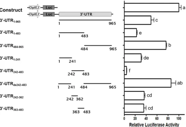
To examine whether shrimp astakine 39-UTR can regulate upstream gene expression, the full-length astakine 39-UTR was subcloned into pGL3-OpIE2 vector. We observed that the full length of astakine 39-UTR down-regulated firefly luciferase activity to about 50% relative to pGL3-OpIE2 vector without whole astakine 39-UTR (Fig 1, lane 1 and 2). To identify the key regulation segment of astakine 39-UTR, various regions of astakine 39-UTR were amplified and subcloned into pGL3-OpIE2 vector for the luciferase reporter assay inSf21 cells. The main regulatory region of astakine 39-UTR was located in 39-UTR242–483, which down-regulated the firefly luciferase activity to about 5% relative to pGL3-OpIE2 vector without 39-UTR (Fig 1, lane 6). To examine the function of the astakine 39-UTR242–483, we subcloned 39-UTRde242–483 (astakine 39-UTR lacking 39 -UTR242–483) into pGL3-OpIE2 vector, and measured the luciferase activity in Sf21 cells. Based on the results shown in Fig. 1, the 39-UTRde242–483 effected the down-regulation of reporter activity and even increased the luciferase activity to higher than that of the full-length astakine 39-UTR. When we divided the 39-UTR242–483 into 39-UTR242–362 and 39 -UTR363–483, and assayed the regulatory function separately, the down-regulation activity was restored to 35%. These data indicate that the key regulatory region of astakine 39-UTR is located at 39-UTR242–483. Interestingly, other regions such as 39 -UTR484–965 may act as an enhance element, which can increase the reporter activity to around 30% as compared to full-length astakine 39-UTR construct.
mRNA stability assay
In order to understand whether astakine 39-UTR regulated the upstream mRNA stability, pGL3-OpIE2 vectors, with or without full-length astakine 39-UTR, were transfected into Sf21 cell and
their RNA stabilities were measured. RNA was extracted at 0, 30, 60 and 120 min after adding actinomycin D and their relative luciferase mRNA levels were measured using RT-qPCR. The two fitting curves almost overlapped, and the estimated half-life (t1/2) of RNA for the cells transfected with pGL3-OpIE2 vector containing or not containing full-length astakine 39-UTR were 60 min and 63 min, respectively (Fig. 2). This result showed that astakine 39 -UTR would not affect the mRNA stability.
Prediction of astakine 39-UTR RNA structures
To investigate the secondary structure of astakine 39-UTR, the sequence of long form astakine 39-UTR was submitted for RNA structure prediction. The astakine 39-UTR sequence is highly structured (Fig. 3). In the region of 39-UTR242–483 where it exhibits the regulatory activity, we found two secondary loop structures. We hypothesized that the predicted secondary structure could be recognized by specific RNA-binding proteins (RBP) and might be involved in the regulation of astakine protein expression.
The variation of luciferase activity after co-transfection with competitor plasmid contains astakine 39-UTR242–483
To confirm this prediction, we performed competition assay using competitor plasmid pEGFP-CMV-Ast 39-UTR242,483.
Various ratios of two plasmids, pGL3-OpIE2-Ast 39 -UTR242,483 and pEGFP-CMV-Ast 39-UTR242,483, were
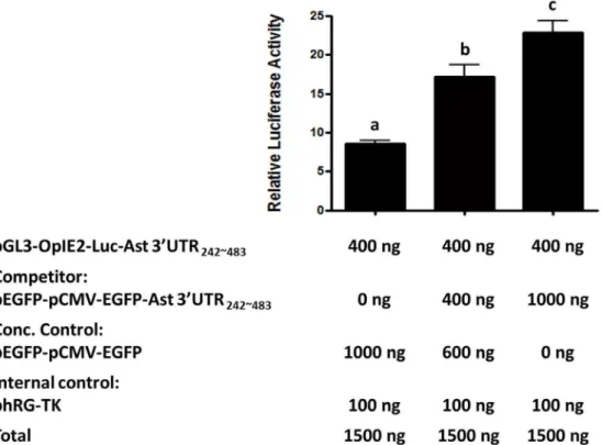
co-transfected into Sf21 cells then the relative luciferase activity was measured. The results revealed that the luciferase activity for reporter was increased with respect to the increase in competitor plasmid, suggesting the relief of repression through 39-UTR242– 483 (Fig. 4). With the high amount of 39-UTR242–483 of competitor mRNA, fewer regulatory factors can bind to the 39 -UTR242–483 of firefly luciferase mRNA. Hence, the firefly luciferase activity was restored.
Multiple protein complexes bind to astakine 39-UTR242– 483
To characterize the RNA binding proteins at astakine 39 -UTR242,483, we performed RNA-EMSA. The astakine 39
-UTR242–483 transcript was synthesized in vitro and incubated with cytoplasmic protein extracts prepared from shrimp hemocyte. Four protein complexes, C1–C4, with various molecular weights were detected by RNA-EMSA (Fig. 5). The specificity of the interaction between astakine 39-UTR242–483 transcripts and binding protein complexes was confirmed by adding the unlabeled probe as competitor. The amount of binding protein complexes to biotin-labeled astakine 39-UTR242–483 transcripts was reduced after the addition of the unlabeled astakine 39-UTR242–483 transcripts.
To identify specific RNA-binding proteins in C1–C4, biotin-labeled Ast 39-UTR242–483 transcripts were used to pull down the RNA-binding proteins from shrimp hemocytes and were analyzed by LC/MS/MS. The molecular weights of the four detected proteins were determined as 25 kDa, 35 kDa, 70 kDa or 100 kDa. After LC/MS/MS analysis, two proteins were identified as crustin Pm4 (25 kDa) and STG I (100 kDa) and the other two proteins were unidentified (Table 2). Crustin Pm4 and STG I represented as candidates for further investigation.
Depletion of STG I and crustin Pm4 does not affect the astakine mRNA
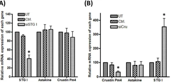
significantly decreased STG I mRNA level to 40% but did not affect the astakine (Fig. 6A) and STG II mRNA level.
To deplete the crustin Pm4 in shrimp, siCru was used. Compared to untreated group (UT) and mock transfected group (Ctrl.), siCru significantly decreased crustin Pm4 mRNA expres-sion to 40% and did not affect the astakine mRNA expresexpres-sion. Interestingly, knock-down of hemocytic crustin Pm4 mRNA induced STG I mRNA expression to about 3.5-fold (Fig. 6B).
Depletion of STG I and crustin Pm4 increased the protein level of astakine
The knock-down of STG I and crustin Pm4 was assayed at protein level by Western blot using anti-STG I and anti-crustin Pm4 antibody. The examination of STG I and crustin Pm4 protein expression demonstrated a clear reduction in protein after transfection with siSTG I and siCru simultaneously (Fig. 7A). Western blot for astakine showed that the level of astakine protein was increased to 1.6-fold after STG I and crustin Pm4 double knock-down. However, there was no significant change of astakine protein level after STG I or crustin Pm4 knock-down (Fig. 7).
Figure 1. Characterization of regulatory element in astakine 39-UTR.Luciferase activity assay in Sf21 cells transfected with reporter
constructs. Eight fragments containing various regions of astakine 39-UTR were constructed into pGL3-OpIE2, a firefly luciferase reporter vector with the OpIE2 promoter. Relative luciferase activity for each construct was measured and normalized to that of pGL3-OpIE2 empty vector without 39-UTR (n = 6).
doi:10.1371/journal.pone.0072793.g001
Figure 2. Astakine 39-UTR effect on mRNA stability.The
pGL3-OpIE2 vector with or without full-length astakine 39-UTR was transfected into Sf21 cell and their RNA stabilities were measured. The estimated half-life (t1/2) of luciferase RNA with or without astakine 39-UTR was 60 min and 63 min, respectively.
doi:10.1371/journal.pone.0072793.g002
Figure 3. Prediction of astakine 39-UTR RNA. The secondary
structure of full-length astakine 39-UTR was predicted by RNA fold. In the region of 39-UTR242–483where the putative regulatory sequence is located, two major secondary loop structures were found.
These data showed that crustin Pm4 and STG I would act as functional redundancy in astakine expression at the post-transcriptional level.
LPS stimulation released STG I and crustin Pm4 from hemocyte in vivo
In order to understand the regulatory mechanism during LPS injection, Western blotting assay was employed to compare the protein level expression of regulatory proteins crustin Pm4 and STG I in hemolymph, hemocyte and plasma of both LPS-injected
Figure 4. The down-regulation of reporter activity by astakine 39-UTR242–483inSf21 cells co-transfected with vector expressing
astakine 39-UTR as the competitor.Luciferase activity assay was conducted inSf21 cells which was co-transfected with plasmid pGL3-OpIE2-Ast
39-UTR242,483and competitor plasmid pEGFP-CMV-Ast 39-UTR242,483. Luciferase activity is expressed relative to pGL3-OpIE2 vector without 39-UTR and data represent mean6S.D. (n = 6). Different letters represent statistically significant differences as compared to each plasmid-transfectedSf21 cell according to the Duncan’s multiple range test (p,0.05).
doi:10.1371/journal.pone.0072793.g004
Figure 5. Multiple protein complexes are associated with Ast 39-UTR242–483.RNA EMSA analysis of Ast 39-UTR242–483RNA incubated with
and untreated shrimp. The result showed that LPS injection increased the amount of astakine in the plasma (Fig. 8A) and both STG I and crustin Pm4 were considerably decreased in hemocyte after LPS injection (Fig. 8). However, STG I protein was increased in the hemolymph and plasma (Fig. 8). The data revealed that two astakine translational repressor proteins were released from hemocytes to plasma after LPS injection.
Discussion
Astakine, an endocrinal cytokine, is an important humoral factor that regulates crustacean hematopoiesis [6,24]. Two forms of shrimp astakine transcripts have been reported, with various lengths of 39-UTR for each form. The additional 671 bp in the 39 -UTR of long astakine transcript suggests that the inserted fragment of 39UTR may play an important role in regulating the expression of astakine. In this present study, we provided evidence of how astakine is regulated at post-transcriptional level in the immune response via its inserted 39-UTR.
Because no stable shrimp cell line is available at present, we used the insect Sf21 cell line to perform the reporter assay for astakine 39-UTR in this study. We have established the reporter constructs with OpIE2 promoter that can be expressed inSf21cells [25]. Therefore, the reporter assay in Sf21 cell should recapture the 39UTR activities of shrimp astakine. Our data suggest that the full length 39-UTR of the long astakine transcript can down-regulate the expression of reporter gene to around 50% at protein
level compared to the control. To further characterize the function of astakine 39UTR, various deletion mutants of astakine 39UTR were constructed in the reporter system to find the localization of the repressor as well as enhancer of the transcript. Our results revealed that certain regions function as repressors or enhancers, which can fine-tune the expression of astakine. The main negative regulatory element was in 39-UTR242–483nt, which down-regulated the reporter gene expression to around 92% at protein level. 39-UTR484–965nt acted as an enhancer to restore the expression level of the reporter gene. This was clearly determined by the specific construct of 39-UTR484–965nt, and also by the deletion of repressor region 39-UTR242–483nt. Moreover, the repressor region was further analyzed by reporter assay for the 39 -UTR242–362nt and 39-UTR363–483nt constructs. Our results showed that the repressor activity was relieved in both constructs compared to 39-UTR242–483, which may be due to disruption of its secondary structure. Altogether, we identified the main repressor region in astakine 39-UTR, the 39-UTR242–483. Surprisingly, this region was localized inside the inserted 671 bp sequence which is only found in the long-form astakine transcripts. Apart from this repressor region, we believe that other regions of 39UTR are also involved in regulating the expression of astakine through multiple mechanisms. In this study, we are interested in further dissecting the regulatory mechanism of 39-UTR242–483 because this region down-regulates almost 90% of astakine protein expression.
Table 2.Proteins identified by LC/MS/MS.
No. Gene name Matched Gene ID Mass (kDa) Score
1 Transglutaminase [Penaeusmonodon] (STG I) gi|33694274 84.66 823
2 Unknown protein - ,70
-3 Unknown protein - ,35
-4 Crustin Pm4 antimicrobial peptide [Penaeusmonodon] gi|229459067 24.229 154
doi:10.1371/journal.pone.0072793.t002
Figure 6. Depletion of STG I and crustin Pm4 does not affect the mRNA level of astakine.(A) Knock-down of hemocytic STG I mRNA after
siSTG I transfection. siSTG I significantly decreased STG I mRNA level (n = 8), while no effect was found on the mRNA of astakine. (B) Knock-down of hemocytic crustin Pm4 mRNA expression after siCru transfection. siCru significantly decreased the level of crustin Pm4 mRNA but did not affect astakine mRNA (n = 8). Knock-down of hemocytic crustin Pm4 mRNA increased the level of STG I mRNA. Data represent mean6SD. Different letters represent statistically significant differences, Duncan’s multiple range test (p,0.05).
The translational control mechanisms are modulated via the interaction of RNA binding proteins (RBPs) [17,26–29] or microRNA[30] at 39-UTR of mRNA. The stability and the rate of the mRNA translation are strictly regulated by some specific RBPs. For example, the half-life and translational rate of the
transcripts which contain AU-rich element at the 39-UTR is controlled by specialized RBPs [27,28,31] or the mRNA decay is promoted by ARE-RBPs such as AU-binding factor 1 [31–33]. Some RBPs like Hu proteins and NF90 are involved in increasing the mRNA stability and also modulating the translation [34,35]. However, our result showed the half-life or stability of RNA was not affected by astakine 39-UTR (Fig. 2). Meanwhile, we found neither AU-rich element nor microRNA binding sites within astakine 39-UTR from microRNA database. From the 39-UTR RNA structure prediction, we found several secondary loop structures that could be recognized by RNA binding proteins. Reportedly, some RBPs like TIA-1 and TIAR or FUBP3 have been used to suppress or enhance the translation, respectively [36– 38]. Hence, a competition assay was conducted to confirm the involvement of astakine 39-UTR-RBP. Our results suggested that some proteins might have been involved in regulating the expression of astakine, because the competition between two constructs, one of which was 39-UTR242–483, significantly affected the repression of luciferase gene expression.
To further corroborate this evidence, RNA-EMSA assay was performed to verify the RNA-protein interaction. Results showed four protein complexes with different molecular sizes were associated with 39-UTR242–483. To disclose the protein infor-mation, RNA-pull down analysis was employed, and it revealed the participation of four proteins, which were specifically bound to 39-UTR242–483. Two of these four proteins, the shrimp transglutaminase I (STG I) and crustin Pm4, had been identified in the shrimp. However, two other proteins were not recognized via LC/MS/MS analysis because the shrimp genome database has not been completed yet. Therefore, the N-terminal sequencing and cloning are necessary to get further information regarding these unknown proteins.
Crustins are among the most important antimicrobial peptides (AMPs) found in decapod crustaceans. Crustin Pm4 is classified as a member of crustin family, but the molecular weight is larger than other crustin Pm isoforms, and its antimicrobial activity is still unknown. Although the antimicrobial peptide or other peptides can nonspecifically bind to DNA or RNA [39], we found only one antimicrobial peptide, crustin Pm4, from our RNA pull down assay. Recent studies showed that antimicrobial peptides have multiple functions and participate not only in antimicrobial function but also in other physiological functions. For example, shrimp penaeidin reportedly behaves as a cytokine for attracting penaeidin-positive granulocytes to the wound tissue, thus, it functions as an autocrine to repair the damaged tissue [23,40]. LL37, an AMP, allegedly interacts with dsRNA to enhance the TLR3 response against poly(I:C) and viral dsRNAs [39]. As far as STGs are concerned, TGs are a family known for their roles in blood coagulation. There are two types of TGs, STG I and STG II, have been cloned and identified from shrimpP. monodonand STG II plays the roles in blood coagulation. However, STG I does not exhibit coagulation activity in our previous studies [41,42]. STG I protein has been found to be abundant in hemocytes and high levels of STG I mRNA expression have been detected in hematopoietic tissue based on our previous study [41]. Recently, crayfishP. leniusculus TG was preventing the differentiation and migration of hematopoietic stem cells where astakine decreased the TG activity [43]. The amino acid identity ofP. leniusculusTG closed toP. monodonSTG II, but the function of STG I was not fully revealed yet. Hence, it is not surprising that STG I, as a non-conventional RNP, has one of the notable functions to control the intricate mechanisms. In this study, we provided evidence that crustin Pm4 and STG I are two known proteins with novel Figure 7. Co-depletion of STG I and crustin Pm4 increases the
protein level of astakine. (A) siSTG I and siCru co-transfected
hemocytes demonstrate a decrease in protein levels of STG I and crustin Pm4. (B) The proteins from siRNA transfected primary cultured hemocyte and medium were collected and extracted for Western blot of astakine expression. (C) The relative expression of astakine protein was quantified by MataMorphHv7.0 software using GAPDH as internal control (n = 7). Astakine protein expression increased after siSTG I and siCru co-transfection. Data represent mean6SD. Symbol ‘*’ represent statistically significant difference, Duncan’s multiple range test (p,0.05).
function as nonconventional RNPs participating in astakine repression by interacting with its 39UTR242–483.
Knock-down of crustin Pm4 and STG I was associated with an increase in astakine protein expression, but no effect on astakine mRNA expression. The result supported our hypothesis that astakine regulation was in translational level, not in transcriptional level. At the same time, there were no differences found in the astakine protein expression with respect to knock-down of crustin Pm4 and STG I individually. Therefore, both crustin Pm4 and STG I were important for the repression of astakine. With the
crustin Pm4 knock-down the mRNA expression of STG I was significantly increased, but the upregulation of crustin Pm4 mRNA expression did not happen with the STG I knock-down. It was speculated that when crustin Pm4 was down-regulated, STG I would be highly expressed in order to repress astakine protein expression. Hence, crustin Pm4 was a major protein that collaborated with STG I as RNP rather than an enzyme that participated in astakine repression because we could not find the TG catalytic sites on crustin Pm4, and STG I enzyme activity was very low [41]. In addition to crustin Pm4 and STG I, two
Figure 8. LPS injection induces STG I and crustin Pm4 released from hemocytesin vivo.(A) Western blot of astakine in plasma after LPS
injection. LPS injection induces increased amount of astakine in the plasma. (B) Western blots of STG I and crustin Pm4 in hemolymph, hemocyte and plasma after LPS injection. The relative expression of STG I (C) and crustin Pm4 (D) protein was quantified by MataMorphHv7.0 software (n = 4). STG I and crustin Pm4 were considerably decreased in hemocyte and STG I protein increased in hemolymph and plasma after LPS injection. 40mg of proteins from extracted protein were loaded on SDS-PAGE. Data represent mean6SD. Symbol ‘*’ represent statistically significant difference, Duncan’s multiple range test (p,0.05).
unknown RNA binding proteins were not identified yet. The function and relationship of these two proteins with crustin Pm4 and STG I in astakine regulation system should be further investigated. Expression of shrimp astakine is shown to be down-regulated by binding of hemocytic proteins, crustin Pm4 and STG I to astakine 39-UTR at post-transcriptional level. Interestingly, the expression of crayfish astakine 2 is up-regulated by melatonin, which affects the core clock of crayfish brain, during the dark period of circadian rhythm [9]. Whether expression of the intracellular crustin Pm4 and STGI proteins will be affected during the dark period of circadian rhythm is worthy to further study.
LPS causes the hematopoiesis phenomenon in shrimps such as cell proliferation in hematopoietic tissue [4], down regulation of total hemocyte count (THC), and normalization of hemocyte count after few hours [3]. In addition, LPS injection can also induce the increased amount of astakine protein in plasma [7]. In this study, we confirmed that the increased amount of astakine protein in plasma after LPS injection was because of the regulatory proteins. Once the regulatory proteins, crustin Pm4 and STG I, are secreted from hemocyte to plasma, the translation of astakine mRNA is not repressed, and the increasingly secreted astakine influences the hematopoietic tissue for the production of hemo-cytes to maintain homeostasis. This mechanism and physiological function of crustin Pm4 and STG I secretions from hemocytes to plasma after LPS injection also need further investigation.
In conclusion, we found that crustin Pm4 and STG I interacted with astakine transcript at 39-UTR242,483 and functioned as a
nonconventional RNP to down-regulate the astakine protein expression. Furthermore, the depletion of crustin Pm4 and STG I resulted in increasing the astakine protein expression but did not affect its mRNA expression. These results revealed that crustin Pm4 and STG I regulate astakine protein expression through a mechanism at post-transcriptional level which provides new aspect for gene regulation in crustacean immune response.
Acknowledgments
We appreciate the assistance of the Tungkang Biotechnology Research Center and the Fisheries Research Institute of the Council of Agriculture for generously offering the tiger shrimp for experimental use, and the Technology Commons, College of Life Science, NTU for providing us the technical assistance needed.
We are also thankful to Dr. Ching-Yu Li for his valuable suggestions about AMPs and Dr. San-Tai Shen for protein identification and also Dr. Apolinario V. Yambot (College of Fisheries-Freshwater Aquaculture Center, Central Luzon State University, Philippines) for his critical review of the manuscript.
Author Contributions
Conceived and designed the experiments: YTC YLS CYC. Performed the experiments: YTC CYL CYT. Analyzed the data: YTC CYT CYC YLS. Contributed reagents/materials/analysis tools: CYL HJT. Wrote the paper: YTC VSS.
References
1. Aguirre-Guzman G, Sanchez-Martinez JG, Campa-Cordova AI, Lima-Gonza´-lez A, Ascencio F (2009) Penaeid shrimp immune system. Thai Journal of Veterinary Medicine 39: 205–215.
2. Van de Braak C, Botterblom M, Taverne N, Van Muiswinkel W, Rombout J, et al. (2002) The roles of haemocytes and the lymphoid organ in the clearance of injectedVibriobacteria inPenaeus monodonshrimp. Fish & shellfish immunology 13: 293–309.
3. Lorenzon S, De Guarrini S, Smith V, Ferrero E (1999) Effects of LPS injection on circulating haemocytes in crustaceansin vivo. Fish & shellfish immunology 9: 31–50.
4. Van de Braak C, Botterblom M, Liu W, Taverne N, Van der Knaap W, et al. (2002) The role of the haematopoietic tissue in haemocyte production and maturation in the black tiger shrimp (Penaeus monodon). Fish & shellfish immunology 12: 253–272.
5. So¨derha¨ll I, Bangyeekhun E, Mayo S, So¨derha¨ll K (2003) Hemocyte production and maturation in an invertebrate animal; proliferation and gene expression in hematopoietic stem cells ofPacifastacus leniusculus. Developmental & Comparative Immunology 27: 661–672.
6. Hsiao CY, Song YL (2010) A long form of shrimp astakine transcript: Molecular cloning, characterization and functional elucidation in promoting hematopoiesis. Fish & shellfish immunology 28: 77–86.
7. So¨derha¨ll I, Kim YA, Jiravanichpaisal P, Lee SY, So¨derha¨ll K (2005) An ancient role for a prokineticin domain in invertebrate hematopoiesis. The Journal of Immunology 174: 6153–6160.
8. Lin X, Novotny M, So¨derha¨ll K, So¨derha¨ll I (2010) Ancient cytokines, the role of astakines as hematopoietic growth factors. Journal of Biological Chemistry 285: 28577–28586.
9. Watthanasurorot A, Saelee N, Phongdara A, Roytrakul S, Jiravanichpaisal P, et al. (2013) Astakine 2–the dark knight linking melatonin to circadian regulation in crustaceans. PLoS genetics 9: e1003361.
10. Grech G, von Lindern M (2012) The Role of Translation Initiation Regulation in Haematopoiesis. Comparative and Functional Genomics 2012.
11. Mazan-Mamczarz K, Lal A, Martindale JL, Kawai T, Gorospe M (2006) Translational repression by RNA-binding protein TIAR. Science Signalling 26: 2716.
12. Sonenberg N, Hinnebusch AG (2009) Regulation of translation initiation in eukaryotes: mechanisms and biological targets. Cell 136: 731–745.
13. Mangone M, Manoharan AP, Thierry-Mieg D, Thierry-Mieg J, Han T, et al. (2010) The landscape ofC. elegans39UTRs. Science 329: 432–435. 14. Spriggs KA, Bushell M, Willis AE (2010) Translational regulation of gene
expression during conditions of cell stress. Molecular cell 40: 228–237. 15. Bandziulis RJ, Swanson MS, Dreyfuss G (1989) RNA-binding proteins as
developmental regulators. Genes & development 3: 431–437.
16. Wickens M, Bernstein DS, Kimble J, Parker R (2002) A PUF family portrait: 39
UTR regulation as a way of life. TRENDS in Genetics 18: 150–157.
17. Mazumder B, Seshadri V, Fox PL (2003) Translational control by the 39-UTR: the ends specify the means. Trends in biochemical sciences 28: 91–98. 18. Szostak E, Gebauer F (2013) Translational control by 39-UTR-binding proteins.
Briefings in Functional Genomics 12: 58–65.
19. Hogan DJ, Riordan DP, Gerber AP, Herschlag D, Brown PO (2008) Diverse RNA-binding proteins interact with functionally related sets of RNAs, suggesting an extensive regulatory system. PLoS biology 6: e255.
20. Scherrer T, Mittal N, Janga SC, Gerber AP (2010) A screen for RNA-binding proteins in yeast indicates dual functions for many enzymes. PLoS One 5: e15499.
21. Cie la J (2006) Metabolic enzymes that bind RNA: yet another level of cellular regulatory network? Acta Biochim Pol. 53: 11–32.
22. Gruber AR, Lorenz R, Bernhart SH, Neubo¨ck R, Hofacker IL (2008) The vienna RNA websuite. Nucleic acids research 36: W70–W74.
23. Li CY, Yan HY, Song YL (2010) Tiger shrimp (Penaeus monodon) penaeidin possesses cytokine features to promote integrin-mediated granulocyte and semi-granulocyte adhesion. Fish & shellfish immunology 28: 1–9.
24. Lin X, So¨derha¨ll I (2011) Crustacean hematopoiesis and the astakine cytokines. Blood 117: 6417–6424.
25. Chuang K-H, Ho S-H, Song Y-L (2007) Cloning and expression analysis of heat shock cognate 70 gene promoter in tiger shrimp (Penaeus monodon). Gene 405: 10– 18.
26. Gebauer F, Hentze MW (2004) Molecular mechanisms of translational control. Nature Reviews Molecular Cell Biology 5: 827–835.
27. Bevilacqua A, Ceriani MC, Capaccioli S, Nicolin A (2003) Post-transcriptional regulation of gene expression by degradation of messenger RNAs. Journal of cellular physiology 195: 356–372.
28. Wilusz CJ, Wilusz J (2004) Bringing the role of mRNA decay in the control of gene expression into focus. TRENDS in Genetics 20: 491–497.
29. Zhang T, Kruys V, Huez G, Gueydan C (2002) AU-rich element-mediated translational control: complexity and multiple activities of trans-activating factors. Biochemical society transactions 30: 952–958.
30. Moor CH, Meijer H, Lissenden S (2005) Mechanisms of translational control by the 39UTR in development and differentiation. Elsevier. 49–58.
31. Chen CYA, Shyu AB (1995) AU-rich elements: characterization and importance in mRNA degradation. Trends in biochemical sciences 20: 465–470. 32. Loflin P, Chen CYA, Shyu AB (1999) Unraveling a cytoplasmic role for hnRNP
D in the in vivo mRNA destabilization directed by the AU-rich element. Genes & development 13: 1884–1897.
33. Zhang W, Wagner B, Ehrenman K, Schaefer A, DeMaria C, et al. (1993) Purification, characterization, and cDNA cloning of an AU-rich element RNA-binding protein, AUF1. Molecular and Cellular Biology 13: 7652–7665. 34. Brennan C, Steitz J (2001) HuR and mRNA stability. Cellular and Molecular
35. Kuwano Y, Kim HH, Abdelmohsen K, Pullmann R, Martindale JL, et al. (2008) MKP-1 mRNA stabilization and translational control by RNA-binding proteins HuR and NF90. Molecular and cellular biology 28: 4562–4575.
36. Gau BH, Chen TM, Shih YHJ, Sun HS (2011) FUBP3 interacts with FGF9 39
microsatellite and positively regulates FGF9 translation. Nucleic acids research 39: 3582–3593.
37. Gueydan C, Droogmans L, Chalon P, Huez G, Caput D, et al. (1999) Identification of TIAR as a protein binding to the translational regulatory AU-rich element of tumor necrosis factoramRNA. Journal of Biological Chemistry 274: 2322–2326.
38. Piecyk M, Wax S, Beck ARP, Kedersha N, Gupta M, et al. (2000) TIA-1 is a translational silencer that selectively regulates the expression of TNF-a. The EMBO journal 19: 4154–4163.
39. Lai Y, Adhikarakunnathu S, Bhardwaj K, Ranjith-Kumar C, Wen Y, et al. (2011) LL37 and Cationic Peptides Enhance TLR3 Signaling by Viral Double-stranded RNAs. PloS one 6: e26632.
40. Li CY, Song YL (2010) Proline-rich domain of penaeidin molecule exhibits autocrine feature by attracting penaeidin-positive granulocytes toward the wound-induced inflammatory site. Fish & shellfish immunology 29: 1044–1052. 41. Huang CC, Sritunyalucksana K, So¨derha¨ll K, Song YL (2004) Molecular cloning and characterization of tiger shrimp (Penaeus monodon) transglutaminase. Developmental & Comparative Immunology 28: 279–294.
42. Chen MY, Hu KY, Huang CC, Song YL (2005) More than one type of transglutaminase in invertebrates? A second type of transglutaminase is involved in shrimp coagulation. Developmental & Comparative Immunology 29: 1003– 1016.