___________________________
Corresponding author: Ana Milosevic-Djeric, Department of Gynecology and Obstetrics,
Hospital Uzice, Milosa Obrenovica 17, 31 000, tel +38131 561255, Uzice, Serbia, mail: ana.mdjeric@gmail.com.
UDC 575 DOI: 10.2298/GENSR1501119M Original scientific paper
INACTIVATION OF Stigmatella aurantiaca csgA GENE IMPARES RIPPLING FORMATION
Ana MILOSEVIC-DJERIC1, Susanne MÜLLER2, and Hans URLICH SCHAIRER3
1 Department of Gynaecology and Obstetrics, Hospital Centre Uzice, Uzice, Serbia 2 Univeristy of Iowa, Iowa City, USA
3Zentrum fur Molekulare Biologie, ZMBH, Heidelberg, Germany
Milosevic-Djeric A., S. Müller, and H. Urlich Schairer (2015): Inactivation of Stigmatella aurantiaca csgA gene impares rippling formation- Genetika, Vol 47, No. 1, 119-130.
Stigmatella aurantiaca fruiting body development depends on cell-cell interactions. One type of the signaling molecule stigmolone isolated from S. aurantiaca cells acts to help cells to stay together in the aggregation phase. Another gene product involved in intercellular signaling in S. aurantiaca is the csgA homolog of Myxococcus xanthus.
In close relative M. xanthus C signal the product of the csgA gene is required for rippling, aggregation and sporulation. Isolation of homologous gene in S. aurantiaca implicates a probable role of CsgA in intercellular communication. Inactivation of the gene by insertion mutagenesis caused alterations in S. aurantiaca fruiting. The motility behavior of the cells during development was changed as well as their ability to stay more closely together in the early stages of development. Inactivation of the csgA gene completely abolished rippling of the cells. This indicates the crucial role of the CsgA protein in regulating this rhythmic behavior.
Key words: cell-cell signaling, development, fruiting body formation, myxobacteria, Stigmatella aurantiaca
INTRODUCTION
reassembling fruiting bodies consisting of a stalk bearing several sporangiols. Inside the sporangioles rod shaped vegetative cells differentiate into spherical resistant myxospores. Spores are generally more resistant than vegetative cells and can survive long starvation period (SHIMKETS
and BRUN, 2000). Some cells remain outside the fruiting bodies and after some time they undergo lysis that reflect programmed cell death (NARIYA and INOUYE, 2008; BOYTON et al.., 2013). Formation of multicellular structures as well as differentiation of cells during development is regulated by extracellular signaling molecules (KROOS, 2007; KAISER et al., 2010). A fine coordination between cells depends on highly coordinated changes in gene expression during development (KROOS and INOUYE,2008). The most studied myxobacteria is Myxococcus xanthus
which represents model organism to understand the molecular mechanisms underlying fruiting body formation (HUNTLEYet al., 2011). On the other hand Stigmatella is much more interesting than Myxococcus because it forms more complex fruiting bodies but it is also so much more
demanding and much more harder to handle. This is the main reason for the small number of known information about this intriguing bacterium. Stigmolone is only one isolated chemical signal that acts in concentrations of about 1 nM to accelerate aggregation of Stigmatella cells at the beginning of development (PLAGAet al., 1998). Its activity depends on cell density and therefore it may have a role in “quorum sensing”. Additionally, cells have to be in direct contact or on the solid surface to secrete stigmolone. The stigmolone biosynthetic pathway as well as the putative pheromone receptor and signaling molecular mechanisms are still unknown (PLAGAet al., 1998). In contrast to S. aurantiaca in Myxococcus several signaling pathways were identified up to now. One of them C signal is necessary for rippling, aggregation, sporulation and expression of many developmental genes. All mutants unable to synthesized C signal carry mutation in the csgA gene. Transcription of the csgA gene is induced in response to starvation (CRAWFORD and SHIMKETS,
2000). The csgA gene encodes 25 kDa protein that become proteoliticaly cleaved by the PopC protease to generate p17, the actual C-signal to 17 kDa protein (LOBEDANZ and SØGAARD -ANDERSEN, 2003; SØGAARD-ANDERSEN 2008; ROLBETZKI et al., 2008). PopC accumulates in vegetative cells and is secreted when cells are starving (ROLBETZKI et al., 2008). PopC secretion is prohibited by interaction with PopD and PopD is degraded in a RelA-dependent manner (KONOVALOVAet al., 2012).
According to the close phylogenetic relationship between S. aurantiaca and M.xanthus
it was speculated that the CsgA protein might plays a role in communication between S. aurantiaca cells during development. To investigate this possibility an S. aurantiaca csgA insertion-mutant was constructed. Inactivation of the gene abolished rippling and caused alternation in cells migration pattern during aggregation.
MATERIALS AND METHODS Bacterial strains and plasmids
The strains and plasmids used in this study are listed in Table.1. S. aurantiaca wild-type DW4/3-1 cells were grown at 32°C in tryptone medium (1% Bacto Tryptone, 0.2%MgSO4) supplemented with streptomycin sulfate (120 µg/ml)or with kanamycin sulfate (50 µg/ml) or oxytetracycline (7.5 µg/ml). Fruiting body formation and sporulation assays were done as previously described (MÜLLERet al., 2006).
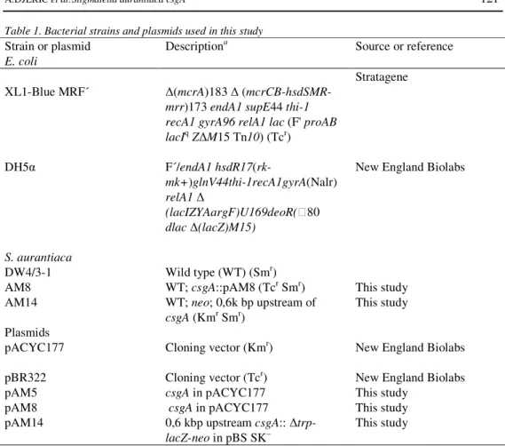
Table 1. Bacterial strains and plasmids used in this study
Strain or plasmid Descriptiona
Source or reference E. coli
XL1-Blue MRF´ (mcrA)183 (
mcrCB-hsdSMR-mrr)173 endA1 supE44 thi-1 recA1 gyrA96 relA1 lac (F' proAB lacIqZ M15 Tn10) (Tcr)
Stratagene
DH5 F´/endA1 hsdR17(rk-
mk+)glnV44thi-1recA1gyrA(Nalr) relA1
(lacIZYAargF)U169deoR(80 dlac (lacZ)M15)
New England Biolabs
S. aurantiaca
DW4/3-1 Wild type (WT) (Smr)
AM8 WT; csgA::pAM8 (Tcr Smr) This study
AM14 WT; neo; 0,6k bp upstream of
csgA (Kmr Smr)
This study
Plasmids
pACYC177 Cloning vector (Kmr) New England Biolabs
pBR322 Cloning vector (Tcr) New England Biolabs
pAM5 csgA in pACYC177 This study
pAM8 csgA in pACYC177 This study
pAM14 0,6 kbp upstream csgA::
trp-lacZ-neo in pBS SK–
This study
a Apr, ampicillin resistance; Cmr, chloramphenicol resistance; Kmr, kanamycin resistance; Smr, streptomycin resistance; Tcr, tetracycline resistance; WT, wild type
DNA manipulations
Standard genetic techniques for in vitro DNA manipulations andcloning were used. The method described by Neumann and co-workers was used for the isolation of total DNA from S. aurantiaca (MÜLLERet al., 2006). Southern blot hybridization and detection wereperformed using the Biotin-Detection-System (RocheDiagnostics). Sequencing of the DNA was performed with ABI Prism tm 377 Sequence System (Perkin-Elmer Corporation) in the sequence facilities of the ZMBH.
RT-PCR
thermal cycle (30 sec at 94°C, 1 min at 54°C and 1 min at 68°C). This was followed by a final extension for 7 min at 68°C. After electrophoresis on an agarose gel, the cDNA was stained with the ethidiumbromide for 30 min, and analysed by UV illumination. Pimer pair pair used was csgA7CTGGATGTGCTCATCAAC/csgA8CTGGAAGTCGAGGAACATG.
Construction of plasmid pAM8 and mutant AM8
Plasmid pAM8 was generated by digesting pAM5, plasmid that contains the csgA locus, (MILOSEVIC, 2003), with the SacI and SphI in order to remove the internal part (340 bp) of the csgA gene. After restriction 5’-overhanging ends were filled using T4 DNA polymerase and ligate to tetracycline resistance gene amplified by PCR from pBR322 with the primer pair TcfwXba and TcrvXba. Plasmid pAM8 was integrated into the genome of S. aurantiaca wild type by electroporation. Oxytetracycline resistance clones were selected and double recombination event was confirmed by Southern analysis.
Construction of plasmid pAM14 and mutant AM14
Plasmid pAM14 was constructed by ligation of 0,9 kbp fragment harbouring 0,6 kbp of the csgA promoter, upstream and 0,3 kbp downstream of putative ATG start codon amplified by PCR (primer pair NotI csgA 21 and csgA 20 XbaI), to plasmid pSM62 (kindly provided by S. Müller). Plasmid pSM62 (derivative of pBSSK-) contains the promoterless trpAlacZ gene fused to a neo cassette for selection of recombinants after transformation. The plasmid pAM14 sequence was reconfirmed by restrictional analysis and sequencing.
In order to produce a csgA merodiploid mutant strain, a plasmid pAM14 was integrated into the S. aurantiaca wild type csgA locus bya single recombination event. Kanamycin-resistant clones were selected and single recombination event was confirmed by Southern blot (data not shown) resulting in AM14. ß-Galactosidase activity in strain AM14 was determined under vegetative anddevelopmental conditions as described previously. Fifty micrograms oftotal protein in 0.1 ml MOPS (morpholinepropanesulfonic acid)buffer (50 mM MOPS, pH 7.5, 10 mM MgCl2, 10 mM dithiothreitol,1 mM phenylmethylsulfonyl fluoride) was mixed with 0.3 ml Naphosphate buffer (10 mM Na phosphate, pH 7, 0.1 M NaCl methylumbelliferyl, 1 mMMgCl2) containing the fluorescent substrate 4-MUG (4- ß-D-galactopyramoside). After incubation at 37°Cfor 30 min the reaction was stopped with 3 ml of 0.1M glycine buffer (pH 10.3). The substrate was cleaved by ß-galactosidase releasing the fluorescent 4- methylumbelliferon (4-MU) that was measured at an excitation wavelength of 360 nm and an emission wavelength of 450 nm using a Shimadzu RF 5000 fluorescence spectrophotometer.
RESULTS
Inactivation of S. aurantiaca csgA gene and physiological function of CsgA
aggregation centers in outside ring (Fig.2). With progression of the development from 8 to 12 h the first differences between the two strains could be observed. During the indicated time period mutant cells migrated to the outer part of the spot forming a circle with high cell density. Unlike mutant cells, wild type cells concentrated mostly in the inner part of the spot and the edge of the spot was transparent as at the beginning (Fig.2). From 12 to 20 h mutant cells continued to accumulate in an outer circle. They formed aggregation centers very close to each other in the outer ring. Importantly specific rippling trails were not observed with the mutant cells during this time period. Rippling is highly organized cells movements in concentric traveling waves that precede aggregation. Wild type cells formed many aggregation centers between 12 and 20 h from which fruiting bodies will arise in later stages. Aggregation of the wild type cells was also visible in the inner part of the spot. This is not the case during aggregation of the mutant cells. The rippling was well visible when analyzing development of wild type cells. After 20 to 26 h mutant as well as wild type cells formed fruiting bodies. No changes in the appearance of the fruiting bodies were observed after 48 h. The mutant fruiting bodies were located in the outer ring of the circle whereas the wild type ones were also present in the inner part of the circle. Inactivation of the csgA gene resulted in alternation in cell migration as mutant cells completely lost ability for rippling but they were still able to develop wild type fruits.
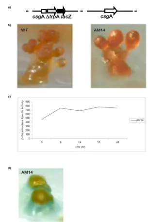
Fig.1a Construction of the csgA insertion mutant.
Fig. 3. Expression of csgA detected by RT-PCR. The RT-PCR reactions were carried out using the same amounts of total RNA. Lane 1: 100 bp DNA Ladder (New England BioLabs); lane 2: control provided by kit (Promega); lane 3: vegetative cells; lane 4: indol induced sporulation after 10 min; lane 5: indol induced sporulation 30 min; lane 6: indol induced sporulation 60 min; lane 7: indol induced sporulation 120 min, lane 8: cells after 8h of development; lane 9: cells after 20h of development, lane 10: cells after 30h of development.
Expression of csgA gene in S. aurantiaca
Transcription of the csgA gene in S. aurantiaca during vegetative growth, development, or artificially induced sporulation was determined by qualitative RT-PCR. Total RNA isolated from cells under different conditions was transcribed with AMV reverse transcriptase and cDNA was amplified with primer pair csgA7/csgA8 as described in the method part. The transcription of csgA was detected during vegetative growth as well as during development (Fig.3). The csgA expression was detected after 8, 20, 30 h of development. Additionally expression was also detected during artificially induced sporulation with indol after 10, 30, 60 and 120 min.
To study the level of the csgA expression a promoter region of csgA was fused to trpA- lacZ reporter gene. Plasmid pAM14 containing promotor csgA- trpA- lacZ-neo fusion was integrated into the genome by a single recombination event leading to a merodiploid mutant AM14 with a wild type csgA allele and a csgA promotor- trpA- lacZ-neo fusion allele in tandem (Fig.4 a). Mutant AM14 displayed the same behaviorduring development and formed wild type fruiting bodies enclosing viable spores (Fig.4 b). This led to the conclusion that the upstream region of 0,6 kbp used to construct the merodiploid mutant is sufficient for expression of the csgA gene.
Fig. 4a Construction of the merodiploid strain AM1.
Fig. 4b Side-view of representative fruiting bodies formed by AM14 and wild type after 48h of starvation on water agar.
Fig. 4c Determination of the b-galactosidase activity of strain AM14 during fruiting body formation (50 mg).
Fig. 4d Fruiting body of the merodiploid strain AM14 on starvation agar containing X-gal.
.
DISCUSSION
intensively studied in M. xanthus. C signal is necessary for regulation of the cells movement behavior that results in the formation of rippling waves and aggregation of the cells. Whereas rippling is completely abolished in the csgA mutant, aggregation is impaired under certain conditions. Unlike wild type cells, which aggregate into compact translucent mounds after 12 h of starvation on the agar surface, csgA mutants aggregate only after 18 h into larger, less compact mounds and ridges.
The C signal acts about 6 h after the beginning of development. A low concentration of the C signal is required for rippling and aggregation. A higher concentration induces sporulation and C signal dependent gene expression including csgA itself. Therefore the model of the C signalling pathway indicates two branches. One branch leads to the regulation of the movement responds of cells and the other branch controls sporulation and expression of the late developmental genes.
The function of the S. aurantiaca CsgA protein in intercellular communication was elucidated by inactivation of the gene. The detectable phenotype that was observed as a consequence of csgA inactivation by insertion mutagenesis suggests an involvement of CsgA in intercellular communication of S. aurantiaca.
Inactivation of csgA in S. aurantiaca impaired rippling, mutant cells showed somehow altered migration and aggregation patterns during development, whereas the shape of the fruiting body showed no obvious differences as compared to the wild type. Additionally, mutant cells differentiated into viable myxospores enclosed in the fruiting bodies.
The expression of the csgA gene in S. aurantiaca was determined by qualitative RT-PCR. The csgA mRNA was detected in vegetative cells and also during development. Analysis with promoterless trpA-lacZ gene as reporter gene indicatedthat the csgA was expressed at a low level vegetatively with slight increase during development in mutant AM14. Also determination of the in situ beta galactosidase activity as well as RT PCR indicate that the csgA gene is expressed during development in S. aurantiaca.
This result shows that CsgA protein is required for formation of rippling wave pattern during S. aurantiaca development. Rippling is observed only in myxobacteria and it is organized cells movement in concentric equidistant ridges (BERLEMAN and KIRBY, 2009; ZHANGet al., 2011). Myxococcus xanthus cells self-organize into periodic bands of traveling waves, termed ripples, during multicellular fruiting body development and predation on other bacteria (ZHANG et al., 2012b). In M. xanthus rippling dependents on CsgA (SAGER and KAISER, 1994). C-signal acts on top of a signaling pathway regulating developmental gene expression and motility (GRONEWOLD and KAISER, 2007). The C-signal suppresses cell reversals in a cell-cell contact dependent manner. As a result cells stream into aggregates forming fruiting bodies that contain dormant spores. The cell surface C-factor activates a receptor on neighboring cells causing the suppression of cell reversals through methylation of FrzCD, a receptor of the Frz chemosensory pathway (ZHANGet al., 2012a).
assay performed with the wild type cells. Therefore one possibility might be that stigmolone is a substrate for CsgA in S aurantiaca. In that case the reduction of the keto group in stigmolone by CsgA might lead to a real signal molecule, stigmolol, which is exchanged between the cells during development. To investigate this possibility it would be necessary to purify the putative stigmolol from wild type cells. The csgA mutant cells would not have stigmolol but the mutant phenotype could be rescued by adding this substance. M. xanthus does not respond to stigmolone by accelerating fruiting body formation in a bioassay (PLAGAet al., 1998). As mention before in S. aurantiaca forms complex fruiting bodies that consist of a branched stalk bearing several sporangioles, whereas M. xanthus forms only mounds filled with spores. Thus, complexity of S. aurantiaca fruiting bodies implicates requirements for a more subtile communication network in S. aurantiaca. It is also possible that there is a way to bypass the csgA mutational block in S. aurantiaca since the pressure to survive under nutrient limitation is high. It might be that mutant cells adopt some suppressor mutations that allow them to bypass the csgA block. It is known that overproduction of SocE in M. xanthus csgA mutant cells bypasses the csgA mutation and cells are able to aggregate and to produce fruiting bodies in the absence of C signaling (CRAWFORD and
SHIMKETS, 2000). However, this study revealed connection between cell motility behavior and csgA gene and open possibility for further investigation of signaling molecular mechanism in S. aurantiaca.To fully understand the CsgA dependent modulation of cell behavior it will be also necessary to analyse the effect of the C-signal in the context of other cell-cell interactions in S. aurantiaca during the complex developmental process.
ACKNOWLEDGMENTS
Thanks Richard Herrmann, Wulf Plaga, Diana Hofman for support and helpful discussions. This research was supported by the Deutsche Forschungsgemeinschaft.
Received September15th, 2014 Accepted January 28th, 2015
REFERENCES
BERLEMAN, J. E., J. R. KIRBY (2009): Deciphering the hunting strategy of a bacterial wolfpack. FEMS Microbiol Rev. 33: 942–957.
BOYNTON, T.O., J.L. MCMURRY, L.J. SHIMKETS (2013): Characterization of Myxococcus xanthus MazF and implications for a new point of regulation. Mol Microbiol. 6:1267-76.
CLAESSEN, D., D. E. ROZEN, O. P. KUIPERS, L. SØGAARD-ANDERSEN,& G. P.VAN WEZEL (2014): Bacterial solutions to multicellularity: A tale of biofilms, filaments and fruiting bodies. Nature Reviews Microbiology. 12(2): 115-124.
CRAWFORD, E. W. JR., L. J. SHIMKETS (2000): The stringent response in Myxococcus xanthus is regulated by SocE and the CsgA C-signaling protein. Genes. Dev.14(4): 483-92.
GRONEWOLD, T.M.A., D. KAISER (2007): Mutations of the act promoter in Myxococcus xanthus. J Bacteriol. 189: 1836– 1844.
HUNTLEY, S., N. HAMANN, S. WEGENER-FELDBRUGGE, A. TREUNER-LANGE, M. KUHE, R. REINHARDT, S. KLAGE, R. MULLER,
C. RONNING, W. NEIRMAN, W., L. SOGAARD-ANDERSEN (2011): Comparative genomic analysis of fruiting body formation in Myxococcales. Mol Biol Evol. 28(2): 1083-1097.
KONOVALOVA, A., S. LÖBACH, L. SØGAARD-ANDERSEN (2012): A RelA-dependent two-tiered regulated proteolysis cascade controls synthesis of a contact-dependent intercellular signal in Myxococcus xanthus.Mol Microbiol. 84(2):260-75.
KROSS, L. (2007): The Bacillus and Myxococcus developmental networks and their transcriptional regulators. Annu. Rev. Genet. 41:13-39.
KROOS, L., S. INOUYE (2008): Transcriptional regulatory mechanisms during Myxococcus xanthus development. In Myxobacteria, multicellularity and differentiation (ed. Whitworth D.E., ASM Press, Washington, D.C.), pp. 149–168.
LOBEDANZ, S., L. SOGAARD-ANDERSEN (2003): Identification of the C-signal, a contact-dependent morphogen coordinating multiple developmental responses in Myxococcus xanthus. Genes. Dev. 17: 2151-2161.
MILOSEVIC, A. (2003): Ph.D. thesis. Ruprecht-Karls-Universität, Heidelberg, Germany.
MÜLLER S, H. SHEN, D. HOFMANN, H.U. SCHAIRER, J.R. KIRBY (2006): Integration into the phage attachment site, attB, impairs multicellular differentiation in Stigmatella aurantiaca.J Bacteriol.188(5):1701-9.
NARIYA, H., M. INOUYE (2008): MazF, an mRNA interferase, mediates programmed cell death during multicellularMyxococcus development. Cell.132:55–66.
PLAGA, W., I. STAMM, H. U. SCHAIRER (1998): Intercellular signalling in Stigmatella aurantiaca: purification and characterization of stigmolone, a myxobacterial pheromone. Proc. Natl. Acad. Sci. USA. 95: 11263-11267.
ROLBETZKI, A, M. AMMON, V. JAKOVLJEVIC, A. KONOYALOVA, L. SOGAARD- ANDERSEN (2008): Regulated secretion of a protease activates intercellular signaling during fruiting body formation in M. xanthus. Dev. Cell. 15: 627-634.
SAGER, B., D. KAISER (1994): Intercellular C-signaling and the traveling waves of Myxococcus.Gens Dev.8 (23):2793-804.
SHIMKETS, L.J., Y.V., BRUN (2000). Procariotic development stages to enhance survival. In: Procariotic development (ed. Y.V. Brun and L.J. Shimkets, American Society for Microbiology, Washington D.C.): 1-7.
SHIMKETS, L., M. DWORKIN, H. REICHENBACH (2006): The myxobacteria. In: The prokaryotes (ed. M. Dworkin, S. Falkow, E. Rosenberg, K. H. Schleifer and E. Stackenbrandt, Springer, New York): 31-115.
SØGAARD-ANDERSEN, L. (2008): Contact-dependent signaling in Myxococcus xanthus: The function of the C-signal in fruiting body morphogenesis. (ed. D.E. Whitworth, ASM Press, Washington, D.C. ) : 77–91.
STAMM, I., A. LECLERQUE, W. PLAGA (1999): Purification of cold-schok-like proteins from Stigmatella aurantiaca-molecular cloning and characteriyation of the scgA gene.Arch.Microbiol.172 (3) :175-81.
ZHANG, H., S. ANGUS, M. TRAN, C. XIE, C., O.A. IGOSHIN (2011): Quantifying aggregation dynamics during Myxococcus xanthus development. J. Bacteriol. 193: 5164–5170.
ZHANG, Y., A. DUCRET, J. SHAEVITZ, T. MIGNOT (2012a): From individual cell motility to collective behaviors: insights from a prokaryote, Myxococcus xanthus. FEMS Microbiol Rev. 36(1):149-64
INAKTIVACIJA Stigmatella aurantiaca csgA GENA ONEMOGU AVA RITMI KO KRETANJE
Ana MILOŠEVI ERI 1, Susanne MÜLLER2 i Hans Urlich SCHAIRER3
1Ginekologija i akušerstvo, Zdravstveni Centar Užice, Užice, Srbija 2
Universitet Iowa, Iowa, SAD
3Centar za molekularnu biologiju, ZMBH, Heidelberg, Nema ka
Izvod
Stigmatella aurantiaca je myxobacteria poznata po formiranju više eliskih struktura kao odgovor na nepovoljne uslove životne sredine. U okviru više eliskih formacija koje podse aju na strukturu drveta vegetativne elije se diferenciraju u višestruko otporne spore koje preživljavaju negativne uslove spoljašnje sredine. S obzirom na neobi an životni ciklus ove bakterije, ona predstavlja dobar model sistem za prou avanje gena i proteina uklju enih u me u elijsku komunikaciju. Druga myxobacteria koja je poznatija i bolje prou ena je Myxococcus xanthus. Kod M. xanthus-a product csgA gena je neophodan za me u elijsku komunikaciju. U ovom radu je prikazana uloga csgA gena kao i CsgA proteina u interakciji me u elijama Sigmatelle u toku razvoja više eliskih struktura i formiranja visoko rezistentnih myxospora.