Original Article
A Study on the Photobleaching Effect of 5-ALA Conjugated Gold Nanoparticles
in a CT26 Tumor Model During Photodynamic Therapy
Nafiseh Sobhani1, Ameneh Sazgarnia1*, Omid Rajabi2, Samaneh Soudmand1, Nadia Naghavi3
Abstract
Introduction
During the process photodynamic therapy (PDT), bleaching of photosensitizer induced by irradiation and generation of reactive oxygen species (ROS) can provide some information concerning the efficiency of treatment. Since gold nanoparticles (GNPs) have been highlighted as efficient drug delivery systems, in this study, by utilizing GNPs conjugated with 5 aminolevulinic acid (5-ALA-GNPs), the photobleaching of ALA-induced protoporphyrin IX (PpIX) was estimated on a colon carcinoma tumor model.
Materials and Methods
CT26 tumor models were prepared by subcutaneous injection of 5×10⁵ cells into the right flank of Balb/c inbred mice. To estimate the time required to reach maximum concentration of PpIX in the tumors, the fluorescence signal of PpIX was monitored and PDT was performed by intratumoral injection of 5-ALA-GNPs, 5-ALA-GNPs, and 5-ALA in separated groups for 15 min with a cycle of 5 min irradiation and 1 min darkness. The photobleaching rate was calculated from recorded fluorescence signals at the darkness intervals.
Results
The maximum fluorescence of PpIX was recorded 3 and 5 hr after injection of 5-ALA and 5-ALA-GNPs, respectively. Despite the low PpIX accumulation in tumors receiving conjugate, the photobleaching rate of PpIX was determined to be higher than 5-ALA. The reduction of the fluorescence signal due to 5-ALA-GNPs clearance was higher than that of 5-ALA.
Conclusion
Administration of 5-ALA-GNPs, intensification of ROS generating and the subsequent elevation of photobleaching results in higher treatment efficiency. Also, more rapid clearance of PpIX has an important implication in clinical application of 5-ALA-GNPs that decreases the undesirable effects on healthy tissues.
Keywords:5 -ALA-Conjugated Gold Nanoparticles, CT26 Tumor Model, Photobleaching, Photodynamic
amount of time after injection of PS for efficient accumulation in the targeted tissue, and delivery of suitable light dosage with an appropriate wave length for optical tissue destruction.
5-aminolevulinic acid is a precursor of PS Protoporphyrin IX (PpIX) that is widely used in PDT. The PpIX induced by 5-ALA is cleared from the body more rapidly than other PSs such as Photophyrin [1, 2]. Furthermore, 5-ALA can be locally administered which reduces the undesired optical side effects [1]. Although the hydrophilic nature of 5-ALA limits its penetration through tissues and cell membranes, improvement in drug delivery through the cell membrane has the potential to enhance the efficacy of PDT. Recent studies have shown that the use of nano-sized particles can be a promising approach in drug delivery [3].
The binding of nanoparticles to
photosensitized molecules can elevate the reactive oxygen species (ROS) formation. These nonaparticles can accumulate passively in target tissues through the rich permeable vasculature around the tumor [3].
One of the modalities of dosimetry for predicting the efficacy of PDT is estimation of the photobleaching rate of PS. Photobleaching is attributed to the reduction of light absorption in PS after irradiation.
using 5-ALA conjugated to GNPs.
2. Materials and Methods
The CT26 cell line, derived from colon carcinoma of Balb/c mice was first provided by the Pasteur Institute (Tehran, IRAN) and was co-cultured monolayerly in RPMI1640 medium with 10% FBS from the Gibco Company in addition to antibiotics. After incubation at 37 °C with 5% CO2, the cells were trypsinized with trypsin EDTA which was purchased from the Biogen Company. In the next step and after counting, 500000 tumoral cells were injected into the inbred balb/c hairless 4-6 week old mice. When the tumor diameter of the induced tumors reached 8-12 mm, three of them were randomly selected, then underwent pathologic studies, and entered this clinical trial.
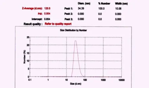
For preparing the conjugate, 5-ALA which was purchased from the Sigma Company and gold nanoparticles with a maximum distribution size of around 34 nm were used [5]. After providing the suspension of gold nanoparticles in de-ionised water, the distribution curves were examined by the Malvern Instruments Analyzing System (Figure 1).
Figure 1. Size distribution curve of GNPs.
The fluorescence spectrum was recorded using
the AvaSpec-2048TEC dual-channel
spectrometer, equipped with a cooled Charge Coupled Device (CCD) in the wavelength of
200-1100 nm (resolution=1 nm) and a
counting sensitivity of 2000 per mJ of incoming radiation.
In this system, the CCD temperature was cooled down to 30 °C below the ambient temperature and the collected data were analysed with the AvaSoft7 software. The light transmission was performed by a two-branch fiber optic bundle (FCR-UV, 2 IND) consisting of a central optical probe with a core diameter of 600 μm for transmission of light emission from the tissue to the spectrometer. This excitation fiber was surrounded by 12 receiving fibers with a 200
μm diameter which transferred light from the
laser to the tissue (energy dissipation coefficient in fiber=0.1 dB/m and in SMA connector of probe=0.5 dB). A single-branched probe was applied for leading light of the Helium-Neon laser (made by the
signal of PpIX was recorded from five points of each tumor and the autofluorescence signal was subtracted.
The study was performed in two parts. First, after 5-ALA and nanoconjugate injection, the time duration of maximum PpIX concentration in the tumor was measured; second, the photobleaching level of PS was assessed. The tumors were randomly divided into five groups. Three groups entered the study in the first step whereas the other two entered in the second step. For the first three groups, 5-ALA, gold nanoparticles and nanoconjugates were administered, respectively; each group with a dosage equivalent to 40 mg/kg of 5-ALA. In order to apply the excitation wavelength, the light of a 405 nm wavelength laser diode (Roithner Laser Technik, Austria, RLDE405-12, : 405 nm, output power: 12 mW, divergence: 0.6 mrad, output aperture: 5 mm) which was fed by a direct voltage source Matrix model: Mps-3003L-3 was used. Based on the maximum rate of the emission peak, the
of the tumoral tissue was measured and then subtracted from the light spectrum recorded after drug administration.
Finally, in the first group, the reduction of the fluorescence signal level was attributed to photobleaching, and in the second group this reduction was defined by the protoporphyrin removal from the tumoral tissues.
2.1. The Photobleaching Evaluation and Statistical Analysis
For the assessment of the fluorescence signal level in 635 nm and the normalized data according to the maximum fluorescence signal level of each tumor during different times after
the administration of PS, the PpIX
fluorescence signal level was compared before and after PDT [7], the photobleaching factor [8], and the time coefficients t10%, t37%, and t90% [9]. The bleaching factor is defined as the ratio of the fluorescence signal level before irradiation to the fluorescence signal level after irradiation.
Time coefficients are obtained by subtracting the base fluorescence signal level from the maximum value multiplied by the assigned percentage. In this study, because of the nonlinear changes in the signal level and for
more accurate computation of time
coefficients, based on the best statistical functions that fit the data with a correlation coefficient close to one, the irradiation time that is required for the signal level in order to drop to 10%, 37%, and 90% of its initial value was evaluated. The data were analyzed using Repeated Measures Test.
3. Results
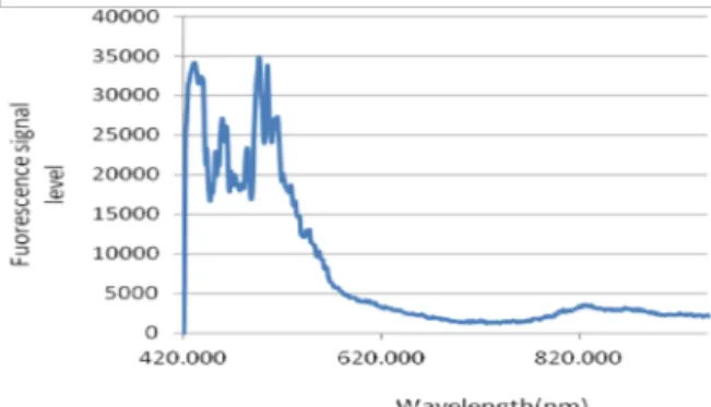
According to the results of the first part and since GNPs do not have fluorescence
Figure 2. Fluorescence spectrum of nanoparticles after 40 mg/kg GNPs administration.
To determine the maximum presence time of PpIX in tissue which is the optimum time for starting PDT, the fluorescence emission was recorded before and 0.5, 1, 2, 3, 3.5, 4, and 5 hr after injection. Data were normalized based on the maximum signal level of each group. As seen in Figures 2 and 3, the maximum accumulation of PpIX induced by 5-ALA in a tumor was recorded 3 hr after injection and showed a significant difference compared to the time points before that (p<0.008).
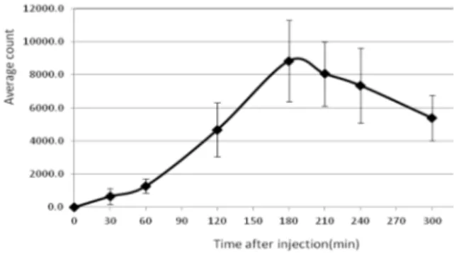
Figure 4. Average count of the fluorescence signal level of PpIX in animals at each time point after excitation by a 405 nm laser diode and injection of 40 mg/kg 5-ALA to the five tumor models. The base signal level obtained from the tumor surface of each animal before injection is subtracted from all the other data.
Furthermore, in order to obtain the time of maximum PpIX presence after administration of the nanoconjugate, the fluorescence signal was recorded before and 0.5, 1, 2, 3, 4, 5, 6, 7, and 8 hr after the injection. Data was normalized based on the maximum signal level of each group. As shown in Figures 4 and 5, the maximum accumulation of PpIX induced by the nanoconjugate in the tumor was recorded at 5 hr after injection which indicated significant differences compared to the different previous times (p<0.02).
In the second part for the purpose of evaluating the photobleaching rate of drugs, in the time of maximum PpIX presence in tissue, PDT was performed during three consecutive, 5 min irradiation followed by 1 min of darkness intervals to collect the fluorescence signal of the tumor. During PDT, the fluorescence signal was reduced due to the photobleaching of PpIX. Comparing the fluorescence signal level of 5-ALA and the nanoconjugate after irradiation, showed a higher photobleaching percentage for the nanoconjugate in comparison with 5-ALA.
Figure 5. Percentage of PpIX induced fluorescence signal normalized based on the maximum signal level at each time point for each animal after excitation by a 405 nm laser diode and administration of 40 mg/kg nanoconjugate to the tumor models. Data are presented as meanstandard error.
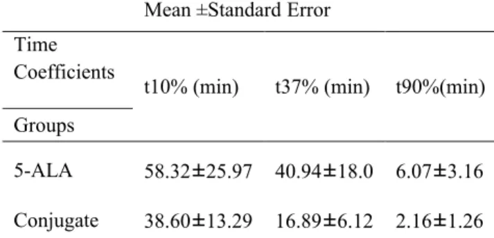
Table 1: Time coefficients t10%, t37%, and t90% of PpIX photobleaching in each animal group after 40 mg/kg 5-ALA administration isolated or in a conjugate based on the recorded fluorescence during three 5 min PDT intervals.
4. Discussion
Based on the findings of this research, 3 and 5 hr after the administration of 5-ALA and nanoconjugate, the peak PpIX was recorded in the tumor tissue, respectively. However, the recorded signal level for 5-ALA was higher than the nanoconjugate during both time points. Although in several in vitro studies the effect of gold nanoparticles on the higher entrance of 5-ALA into cells has been demonstrated, it seems that in the present in vivo study, 5-ALA conjugated to GNPs has not acted as an efficient delivery factor in PDT.
These findings were achieved in spite of Khaing Oo et al. study which reported that the use of 5-ALA conjugated to GNPs had enhanced the uptake rate of 5-ALA by fibrosarcoma cells in comparison with free 5-ALA and reported twice the ROS production with the conjugate [10].
saturation, tissue scattering, and its dependency on wavelength are related to the PpIX photobleaching rate and consequently the treatment efficiency [6].
Atif et al. studied the effect of the local mTHPC concentration on the dynamics of photobleaching [7]. They found out that at a fixed laser fluence-rate, photobleaching as a function of the delivered optic dose proceeded more rapidly at higher drug concentrations. Naghavi and his colleagues in 2011 studied the PpIX concentration during PDT in a colon carcinoma tumor model in Balb/c mice (the tumor model which was administered in our study) by fluorescence spectroscopy and showed a decrease in the PpIX concentration of the tumor during PDT. Their results were
consistent with the findings of the
mathematical simulation tissue response and the photobleaching phenomenon [11].
So far, no study has been conducted to compare photobleaching of a nano-conjugate and 5-ALA. According to many studies such
as the one conducted by Johnson,
photobleaching of a photosensitizer is in direct correlation with the PDT efficiency [12]. In this study, it is shown that the efficiency of PDT with the use of a designed nano-conjugate would be higher than 5-ALA. In
many studies about the fluorescence
spectrometry of photosensitizers such as PpIX, increasing the exposure time has led to an enhancement in the photobleaching rate [13]. In the current study, the nanoconjugate has also followed the mentioned process, similar to 5-ALA.
In the present study, the photobleaching of 5-ALA conjugated with gold nanoparticles during PDT was studied. Comparison of the tissue fluorescence spectrum before and after Mean ±Standard Error
t90%(min) t37% (min) t10% (min) Time Coefficients Groups
6.07±3.16 40.94±18.0
58.32±25.97 5-ALA
2.16±1.26 16.89±6.12
illumination showed that reduction in the fluorescence signal of the nanoconjugate due to photobleaching was higher than the same value for 5-ALA. The time coefficients t10%, t37%, and t90% were calculated in 5-ALA and in the nanoconjugate which were in agreement with the previous results indicating a higher tissue bleaching rate for the nanoconjugate. Measuring the bleaching factor also confirmed
a higher photobleaching rate for the
nanoconjugate.
According to the previous studies and with the aim of justifying the role of GNPs in increasing the treatment efficiency, it can be said that GNPs on their own cannot increase the production rate of PpIX but can lead to the formation of singlet oxygen molecules (O2). As the surface plasmon resonance (SPR) effect of GNPs,enhances the photocurrent between GNPs and PpIX after light irradiation, it
increases the energy level of the
photosensitizer and PpIX with the high transferred energy and in the presence of oxygen molecules results in an elevated ROS production rate and consequently higher treatment efficiency.
5. Conclusion
The results of this study showed that the administration of a nanoconjugate did not increase the PpIX formation level in a tumor in comparison with 5-ALA injection.
As in other studies, the amount of bleaching is used as a dosimetric agent in PDT and the results indicate a higher level of bleaching with the nanoconjugate compared to that of 5-ALA. Therefore, it can be concluded that the application of a conjugate can increase the efficiency of PDT. This can be due to the elevated ROS formation in the presence of GNPs which is in direct correlation with the PDT efficiency.
Acknowledgements
This work has been extracted from an MSc thesis of Medical Physics and was funded by the Research Deputy of Mashhad University of Medical Sciences. The authors would also like to thank Mr. Ali Shad who has kindly cooperated in this research.
References
1. Dolmans DE, Fukumura D, Jain RK. Photodynamic therapy for cancer. Nat Rev Cancer. 2003 May; 3(5):380-387.
2. Marcus SL. Photodynamic therapy of human cancer. Proceedings of the IEEE. 1992; 80(6):869-89.
3. Cai W, Gao T, Hong H, Sun J. Applications of gold nanoparticles in cancer nanotechnology. Nanotechnol Sci Appl. 2008;1(1):17-32.
4. Bonnett R, Martı́nez G. Photobleaching of sensitisers used in photodynamic therapy. Tetrahedron. 2001; 57(47):9513-47.
5. Ghahremani FH, Sazgarnia A, Bahreyni-Toosi MH, Rajabi O, Aledavood A. Efficacy of microwave hyperthermia and chemotherapy in the presence of gold nanoparticles: an in vitro study on osteosarcoma. Int J Hyperthermia. 2011; 27(6):625-36.