carbons) with the lone electron pair of the hydroxyl radical. Moreover, quantum chemical calculations were carried out on selected GSH/OHNcomplexes and on appropriate GSH conformers to describe the energy profile of the recognition process. The relative enthalpy and the free energy changes of the radical recognition of the strongest complexes varied from242.4 to227.8 kJ/mol and from 221.3 to 9.8 kJ/mol, respectively. These complexes, containing two or more intermolecular interactions, would be the starting configurations for the hydrogen atom migration to quench the hydroxyl radicalvia different reaction channels.
Citation:Fiser B, Jo´ja´rt B, Csizmadia IG, Viskolcz B (2013) Glutathione – Hydroxyl Radical Interaction: A Theoretical Study on Radical Recognition Process. PLoS ONE 8(9): e73652. doi:10.1371/journal.pone.0073652
Editor:Claudio M. Soares, Instituto de Tecnologica Quı´mica e Biolo´gica, UNL, Portugal
ReceivedMay 28, 2013;AcceptedJuly 30, 2013;PublishedSeptember 9, 2013
Copyright:ß2013 Fiser et al. This is an open-access article distributed under the terms of the Creative Commons Attribution License, which permits unrestricted use, distribution, and reproduction in any medium, provided the original author and source are credited.
Funding:This work was supported by the following grants: ‘‘Supercomputer as a national virtual laboratory’’ (project ID: TA´MOP-4.2.2.C-11/1/KONV-2012-0010) and ‘‘New functional material and their biological and environmental answers’’ (project ID: TA´MOP-4.2.2.A-11/1/KONV-2012-0047). The publication fee was supported by ‘‘University of Szeged Talent Publishing Support’’. The funders had no role in study design, data collection and analysis, decision to publish, or preparation of the manuscript.
Competing Interests:The authors have declared that no competing interests exist.
* E-mail: [email protected]
Introduction
Molecular recognition – the interaction between a larger host and smaller guest molecules – is one of the most important biochemical processes [1]. This complex mechanism can take place during intra- and intercellular communication, the induction of the immune system and the response to external stimulietc. [2]. The cell itself evolves its own defensive mechanism against external actions, which can damage cells or cellular organelles, and this mechanism has been investigated for a long time. Free radical initiated oxidation is one of these external actions and one of the most important antioxidants [3], used as a defense mechanism, is a small tripeptide, glutathione (c -L-glutamyl-L-cysteinyl-glycine, GSH,Figure 1).
GSH is a nonprotein free thiol present in high concentrations in the living organisms [4] and it is essential in a number of biochemical processes [5–7]. GSH exhibits antioxidant, radical scavenging activity by its electron donating ability [8,9]. This enables GSH to neutralize free radicals, especially reactive oxygen species (ROS) such as the superoxide, hydroperoxyl, hydroxyl (OHN) radicals, having electron acceptor ability. The hydroxyl
radical is one of the most reactive of the ROS [10] and has a key role in the oxidative stress related events, such as lipid peroxidation [11] and DNA oxidation [12,13]. The rate constants of the reactions between GSH and radicals, as well as the GSH
and hydroxyl radical reaction were studied previously by experimental methods, e.g. by laser photolysis, absorption spectroscopy and pulse radiolysis [9,14]. The theoretical calcula-tions were focused on the calculation of the potential energy surface [3] and the rate constants of elementary steps [15]. Furthermore, conformational analyses of GSH were carried out by NMR spectroscopy [16–18] and molecular dynamics (MD) [18– 21] methods, which showed that GSH is flexible and does not adopt a strongly preferred conformation. Recently Machuqueiro
et al. performed constant-pH MD simulations for GSH and GSSG (oxidized form of GSH) [20]. They concluded that the conforma-tional flexibility of GSH is pH-dependent and it has reduced flexibility at higher pH (pH.10) values.
The large flexibility of GSH can be the weak point of the classical potential energy surface calculations, because the most reliable initial conformations are difficult to find. Moreover, the radical scavenging mechanism depends on the interactions formed between GSH and radicals, and the steric properties of collisions and attractive interactions can strongly influence the overall kinetics and the mechanism of this bimolecular reaction. To overcome this limitation, structures for furtherab initiocalculations could be determined by non-reactive molecular dynamics simulations. Therefore, we set a long lasting, comparative MD simulation for GSH and GSH/OHNsystems. The MD trajectories
and OHN. Moreover, the non-reactive MD trajectories combined
withab initiocalculations allow us to describe a detailed free radical recognition process.
Materials and Methods
The glutathione anion is found to be dominant at physiological pH, where the c-L-glutamic acid predominantly exists in its zwitterionic form, while the carboxyl group of the glycine residue prefers to be deprotonated [21]. For these reasons, to obtain the most reliable theoretical model for GSH in water, its anionic form was considered and is hereinafter referred to as GSH.
Five independent molecular dynamics (MD) simulations (56240 ns) were performed for the GSH and GSH/OHNsystems,
respectively. GSH was solvated with TIP3P [22] water molecules and one Na+
ion was also placed in the box in order to ensure the electro-neutrality of the system. The simulation box was cubic (373 A˚3), where the minimum distance between any atom of the GSH and the wall of the box was 12 A˚ . The simulations were conducted with the Desmond v. 30110 [23] software using the CHARMM22 [24] force field. The short range van der Waals and electrostatic cut-off values were set to 9.0 A˚ and the long-range electrostatic interaction was calculated via the Particle Mesh Ewald [25] method.
The missing bond parameters and charges for the OHNradical
were calculated with the Force Field Toolkit Plugin [26] implemented in Visual Molecular Dynamics (VMD) [27].
The simulation protocol was as follows: 1) steepest descent minimization (with and without solute restraints); 2) NVT MD (T = 10 K,Dt = 12 ps) with the Berendsen thermostat [28]tT= 0.1 ps and restrained solute heavy atoms; 3) NPT MD (T = 10 K,
Dt = 12 ps, p = 1 bar) with Berendsen thermo- and barostat (tT= 0.1 ps,tp= 50 ps, separate coupling for solute and solvent) and no restraints; 4) NPT MD (T = 310 K,Dt = 12 ps, p = 1 bar) with Berendsen thermo- and barostat (tT= 0.1 ps, tp= 50 ps, separate coupling for solute and solvent) and restrained solute heavy atoms; 5) NPT MD (T = 310 K,Dt = 24 ps, p = 1 bar) with Berendsen thermo- and barostat (tT= 0.1 ps,tp= 2.0 ps, separate coupling for solute and solvent) and no restraints. 6) NPT MD (T = 310 K, p = 1 bar) with Berendsen thermo- and barostat [28] (tT= 0.1 ps,tp= 2.0 ps, separate coupling for solute and solvent) and no restraints.
The structures were saved every 9.8 ps, which resulted in 25 000 frames for each simulation. The protocol was repeated 5 times with different random velocities and a total of 1.2ms of simulations were obtained for each GSH and GSH/OHNsystem, respectively.
The metadynamics [29] method was used to reconstruct the free energy surface of interaction between GSH and OH radical. A 24 ns biased molecular dynamics simulation was performed with two collective variables: the distance between the oxygen of OHNand Gly.OT1 atom (d (OT1 – OHN) and distance between
the oxygen of OHNand Cys.SG atom (d (SG – OHN). During the
calculation, the height of the Gaussian was set to 0.03 kcal/mol, the width was set to 0.05 A˚ , and barriers were applied at 6 A˚ in order to prevent large changes in the direction of the variables.
Intra- and intermolecular interactions were identified by the geometric analysis using the following criteria: d(A666D),3.5 A˚ and a(A666H-D) .100.0u. The structural analysis was
per-formed with the ptraj module of the AmberTools 1.5 program package and Visual Molecular Dynamics [27] was used to prepare the 3D structures in the figures.
To describe these interactions more in detail, electron density analyses (Atoms in Molecules, AIM) [30,31] were carried out on some geometrically selected structures at the B3LYP/6-31G (d) level of theory. The AIM analysis was carried out with the AIM2000 program [32]. Previously, similar refinements were successfully used to improve the analysis of intramolecular interactions in the case of human galactokinase enzyme [33].
Geometry optimization were conducted on properly selected GSH/OHNcomplexes and GSH conformers with the
BHandH-LYP density functional combined with the 6–31G(d) split valence basis set. To mimic the bulk water, the solvation model ‘‘D’’ (SMD) [34] was used. Normal mode analysis was also carried out at the same level of theory in order to confirm that the structures obtained are minima on the respective potential energy surface. The quantum chemical calculations were carried out using the Gaussian 09 program package [35].
Results and Discussion
Interactions between water and radicals have been investigated earlier by classical MD methods [36], wherein the authors used the same van der Waals parameters for the radicals (OHN, HO
2N) as
for the water molecules. The obtained free energy profiles through the water slab were in good agreement with experimental studies; Figure 1. The chemical structure of GSH, with the atomic naming scheme (indicated with green).The three amino acid residues, c-glutamic acid, cysteine and glycine are indicated in blue, gold and black, respectively. The non-regularc-peptide bond between the glutamic acid and the cysteine residues is also labelled.
the GSH was indeed very flexible during the simulations, which is in good agreement with previous studies [18–21]. The compact-ness of the GSH structure during the simulations was measured by the radius of gyration of heavy atoms (Rgyr). The Rgyrvalues for
most of the GSH structures varied from 3.0 to 4.5 A˚ both with and without hydroxyl radical. The percentage of the distribution of the structures on the RMSD – Rgyr surface was also calculated
(Figure 2). In order to describe the flexibility of GSH from another structural point of view we used the same parameters as Machuqueiro et al. [20]. All possible distances were measured between the Gly.OT1/OT2 and GGL.OT1/OT2/N and the minimum value among these 6 distances was used as the head-tail distance (HT). The other parameter was the minimum distance between CYS.SG and the above mentioned atoms (CYS-HT). Thereafter, using a 0.25 A˚ bin width, the distribution of the structures on this surface was determined.
Similar distributions were obtained for the simulations with and without OHN, which can be seen on the difference surfaces (right
panel,Figure 2). The largest deviation obtained was only about 60.4% and60.1% on the RMSD – Rgyrand HT – CYS-HT
surfaces, respectively. In case of the HT – CYS-HT surface, the same distribution was obtained also in our case comparing the results of Machuqueiroet al.: the HT values varied between 2 and 12 A˚ , while the CYS-HT values were between 2 and 8 A˚.
This indicates that the presence of OHNhas no large impact on
the dynamic nature of GSH in a non-reactive, classical MD simulation.
The most populated regions of the GSH and GSH/OHN
systems (12.9% and 13.1% of the structures, respectively) are located in the same [1.75 A˚ #RMSD,2.00 A˚ ; 3.75 A˚#Rgyr
,4.00 A˚ ] range of the RMSD – Rgyrsurface. The averages of the
RMSD and the Rgyr values in this most populated range were
calculated and the structures which had the smallest deviation from the averages were selected for comparison as representative structures. The RMSD between the two representative GSH structures is 1.34 A˚ , which means that the representative structures fit to each other very well (Figure 2). The largest deviation was obtained for the glycine (GLY) residue and the peptide bond between the cysteine (CYS) and GLY residues. In the represen-tative structures we did not obtain any intramolecular hydrogen bonds, except the interaction between the protonated amine and the deprotonated caboxyl group in the c-glutamic acid (GGL) residue. Furthermore, the representative structures of GSH are in good agreement with the conformations obtained previously by NMR and MD studies [17–21].
Intramolecular hydrogen bond analyses were carried out as well for GSH in both systems for each snapshot from the MD simulations. The intramolecular hydrogen bonds were formed in
attack points of GSH where theg(r)values show high maxima for OHN. During theg(r)curve analysis and in the further discussion all
CA atoms were considered, although in the case of Gly the terminal carboxyl group is responsible for the enrichment around this region. Nevertheless, we were interested in these regions as well, because the a-carbon atoms are highly vulnerable attack points in the proteins [39]. Hydrogen atom abstraction by radicals from these positions can easily happen if the a-carbons are accessible, because the process is favorable from the thermody-namics point of view [40]. In the case of the c-glutamic acid residue, the OHN enrichment is conspicuous near the a-carbon
(GGL.CA), the carboxyl carbon (GGL.C) and both carboxyl oxygens (GGL.OT1/GGL.OT2). The b-carbon (CYS.CB) and the sulfur (CYS.SG) in the cysteine residue are potential radical attractors, as shown by the corresponding g(r) values. The a -carbon (GLY.CA), the carboxyl -carbon (GLY.C) and carboxyl oxygens (GLY.OT1 and GLY.OT2) in the glycine residue also have radical control ability. The distribution functions of the GLY.OT1/2 show the same characteristics as the corresponding
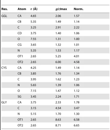
g(r)s of the GGL residue (see Figure 3). To determine the probability of finding the OHNmolecule near GSH, the maxima of
the g(r) curves (g(r)max) were analyzed and these values were collected inTable 1with the corresponding distances.
Based on theseg(r)max values, the most probable OHNattack
points can be selected. The highestg(r)max values for the OHN
belong to the carboxyl oxygens of GLY (8.63 and 8.71). These were followed by the carboxyl oxygens of GGL (5.25 and 6.00), while the lowest value (1.31) was attributed to the carbonyl oxygen of GGL (GGL.O). Compared to the probability at the carbonyl oxygen, we obtained more than 1.5 times higher probability values around the a-carbons (GGL.CA and GLY.CA), the carboxyl carbons, oxygens (GGL.C, GLY.C and GGL.OT1,2, GLY.OT1,2) and the sulfur (CYS.SG), altogether around 9 atomic positions. Seven of these cases are situated closer than the maximum distance criteria of the hydrogen bonding interaction (,3.5 A˚ ). The high probability of finding of OHN around the
carboxyl carbons (GLY.C and GGL.C) is caused by the strong H-bonds between OHN and negatively charged carboxyl oxygen
atoms. Therefore we will omit these from the further analysis, since there are no direct interactions between OHN and these
carbon atoms.
To obtain a more detailed description about the interactions between the hydroxyl radical and the different functional groups of GSH, interaction pattern analyses were carried out. The interactions were determined between the OHNand those heavy
OT1, OT2). All in all, 2305 structures were found where at least one interaction was established between the OHNradical and the
corresponding parts of GSH. The most frequently occurring interaction (,78%) was present between the OHNand the anionic carboxyl groups of GSH. Due to the fact that the -SH group is responsible for the antioxidant activity of GSH, we determined the structures in which the OHN is interacting with the sulfhydryl
group (,4%) as well.
In around 36% (838) of the structures, the OHNinteracts with at
least two heavy atoms of GSH. The distribution of OHNaround
GSH in such structures was depicted as a volumetric map (see
Figure 4).
The volumetric map also demonstrates that the carboxyl oxygens (GGL.OT1,2 and GLY.OT1,2) of GSH are dominant in the interactions, and that the OHNis mostly hydrogen-bonded
with these atoms of GSH. The thiol (CYS.SG) and the protonated amine group (GGL.N) are also important for intermolecular interactions between GSH and OHN. 82% of these structures
contained hydrogen bonds where the OHN is interacting with
carboxyl oxygens. These structures can be divided into two subgroups, depending on which site of GSH (GLY or GGL) takes part in the interaction (GLY,48%, GGL,34%). In 11% of the structures, the hydroxyl radical formed interactions with a carboxyl oxygen and with a hydrogen bonded atom at the same time.
The analysis of the intermolecular interactions revealed 12 cases in which three interactions were established between GSH and
OHN (see Figure 5), and this is the highest number of such
(GSH666OHN) intermolecular interactions. These are the most
stable complexes, because of the high number of interactions between the two molecules. For this reason, AIM analysis was carried out on these structures to describe these interactions and investigate their existence from a molecular electron density point of view (seeFigure 5, second and fourth columns).
Two main types of interactions can be identified in these cases, zwitterionic and anionic, based on the interacting functional groups of GSH (seeFigure 5,I. and II.). The zwitterionic group (2NH3
+
666OHN6662CO22) contains 4 members, where one
of the anchor points is the protonated amine group of GGL (GGL.N) and the other one is a deprotonated carboxyl group. In the anionic group (2CO2–666OHN6662X-H) there are 8
structures where the main interaction between OHNand GSH is
formed with the carboxyl group of the GLY or the GGL residue. Besides this interaction, others were also found in these complexes with the comprising of GGL.CA or GLY.CA and/or CYS.CA/ CB/SG. The AIM analysis resulted in bond critical points (BCPs) indicating the existence and the strength of the interactions. The BCPs varied from 0.002 a.u. to 0.058 a.u. (higher electron densities at the BCPs represent stronger interactions). The weakest interactions correspond to geometrically not investigated ones (GLY[–N-HN]666OHN), which appeared in two cases (I/4,
zwitterionic group and II/7, anionic group). The AIM analysis showed that the strongest interactions (H-bonds) were between the carboxyl oxygens of the glycine and the hydroxyl radical Figure 2. The percentage distribution of the structures on the RMSD – Rgyr(upper panel) and HT – CYS-HT surfaces was calculated
for the GSH and the GSH/OHNsystems (left panels).Representative structures from the most populated region from the RMSD – R
gyrsurface
are shown as well. The different origin of the representative structures is indicated by colored carbon atoms (green – GSH, brown – GSH/OHN). The differences between surfaces were also calculated (right panel).
Figure 3. The radial distribution functions (g(r)) were calculated between the oxygen atom of water (blue curves) or OHN(black
curves) and all heavy atoms in GSH for the GSH/OHNsystem.The maximumg(r)values and the corresponding distance for OHNare indicated in the lower right corner of each graph.
(GLY.OT1,2666OHN). This finding is in good agreement with
the g(r)max values calculated.
Geometry optimization and frequency calculations were con-ducted on selected GSH/OHNcomplexes and GSH conformers at
the BHandHLYP/6-31G(d) level of theory to describe the energy profile of the recognition process. The representative GSH structure from the molecular dynamics simulation of GSH without OHNwas selected as a reference structure. The relative enthalpy of
other conformers with respect to this structure is in the range of
227.1–5.1 kJ/mol (seeFigure 6).
The lowest enthalpy conformer is the 3rdfrom the zwitterionic group (seeFigure 5, right upper corner). The complex formation between the hydroxyl radical and the GSH is highly exothermic, regardless of the molecular motion of GSH, as well as the attack point of OHN on the glutathione. The enthalpy of complex
formation is between 242.4 and 227.8 kJ/mol. The relative enthalpy of the formed complexes varies from267.2 to224.5 kJ/ mol compared to the reference conformer and the hydroxyl radical. These results show that the radical recognition process of GSH (GSH/OHNcomplex formation) is energetically favorable.
In summarizing our results, we outline a possible general radical recognition process as follows. The g(r) curves show that the probability of finding OHN is 4 times larger around the
GLY.OT1,2 (g(r)max .8.6) compared to CYS.SG (g(r)max = 2.2), which is the common antioxidant part of GSH. Addition-ally, the relative amount of the GLY.OT1,2666OHNH-bonds
compared to the total number of interactions shows us that the terminal GLY may have a double role in the radical recognition: catching and steering. In the first step of the process, the
GLY.OT1,2 most often forms a H-bonded intermolecular complex with OHN, however the GGL.OT1,2 and/or GGL.N
are also capable of this, quasi catching the radical from the bulky solvent phase. In the second step, owing to the high flexibility of GLY, it can further steer the radical in the direction of the CYS. In this step, new interactions are forming, e.g. with the CYS.CB, and the original interactions (GLY.OT1,2666OHN) also remain. In
our previous work [3] we showed that the bond dissociation energies of the hydrogen atoms from CB are one of the highest among all possibilities. Therefore, hydrogen abstraction from this
b-carbon atom is unlikely. The OHN does not stop here, but
continues to move forward on the way to the –SH group. After the radical recognition, the detoxification of OHNby GSH can take
place, the OHNis forwarded to CYS.SG and abstracts a hydrogen
from this group.
The recognition mechanism is based on selected structures from a large ensemble, therefore one may assume that it lacks the statistical relevance. We have to emphasize here that during the calculations we saved the structures at every 9.8 ps to sample diverse conformations. In order to confirm further our results we have several possibilities. We can perform additional calculations with more frequent structure sampling, or we can focus around the region of catching and steering. We decided to use a biased MD simulation by means of metadynamics using two collective variables. Based on this calculation the free energy surface of the recognition mechanism was determined (Figure 7).
As one can see in Figure 7, if we use the above mentioned collective variables, we obtain a deep free energy valley with two minima. The first minimum is located on the surface between 3 A˚
,d (OT1 – OHN),4.2 A˚ and 3.8 A˚,d(SG – OHN),4.8 A˚ and a
local barrier at d(OT1 – OHN) = 3.0 A˚ with a height lower than
1 kcal/mol. Data obtained from unbiased MD calculations indicate that the OH radical can easily access this local maximum because we obtained a sharp peak on the g(r) curve at 2.65 A˚ . The second minimum lies in the 2.4 A˚ ,d(OT1 – OHN),2.8 A˚ and
3.5 A˚ ,d(SG – OHN),4.8 A˚ range. This second minimum valley
confirms our proposed steering mechanism: after catching the OHNby one of the carboxylate oxygen of Gly, and due to its high
flexibility, GLY can steer OHNtoward the thiol group of Cys. In
this process we did not obtain any barrier, indicating that there is no thermodynamic control regarding steering. The lowest free energy point lies at 2.65 A˚ (d(OT1 – OHN)) 3.65 A˚ (d(SG – OHN))
on the surface and two of the selected structures correspond to this point: the 3rdand the 5th structure from the anionic group (see
Figure 5). Table 1.The maximum values of the radial distribution
functions (g(r)) between the oxygen atom of OHNand all heavy atoms in GSH.
Res. Atom r (A˚ ) g(r)max Norm.
GGL CA 4.65 2.06 1.57
CB 5.35 1.49 1.14
C 3.25 2.91 2.22
CD 3.75 1.40 1.06
O 7.55 1.31 1.00
CG 3.65 1.32 1.01
N 5.35 1.53 1.17
OT1 2.65 5.25 4.01
OT2 2.65 6.00 4.58
CYS CA 4.25 1.49 1.14
CB 3.85 1.76 1.34
C 3.95 1.62 1.23
N 5.65 1.39 1.06
O 7.15 1.47 1.12
SG 3.45 2.24 1.71
GLY CA 3.75 2.33 1.78
C 3.15 4.54 3.47
N 5.15 1.70 1.30
OT1 2.65 8.63 6.58
OT2 2.65 8.71 6.65
The normalized values (Norm.) were calculated (Norm. =g(r)max/min[g(r)max]) and are also tabulated.
doi:10.1371/journal.pone.0073652.t001
Figure 4. The volumetric map was created for the radical (OHN)
occurrence around those 838 structures where the OHN
interacts with at least two heavy atoms of the GSH.
Conclusions
Two, in total 1.2ms comparative MD simulations were conducted on GSH and GSH/OHN systems to explore the
molecular recognition process and identifying the OH radical attractor regions of GSH.
The high flexibility of GSH was preserved during the simulation of the GSH/OHNsystem and this is one of the driving forces of the
radical recognition process. Two main steps of the detailed molecular radical recognition process of GSH were assigned, namely catching and steering. In ,78% of all interactions characterized, strong complexes were formed between anionic
Figure 5. The structures that contained the maximum (3) number of interactions between GSH and the OHN based on the
geometrical criteria are depicted (I. – zwitterionic group, II. – anionic group).The interactions based on geometrical criteria are indicated with blue, dashed lines, while those resulted from the AIM analyses are depicted with green points (bond critical points, BCPs).
carboxyl groups and the OH radical. After the catching step, the strong carboxyl-OHNcomplexes could evolve additional
interac-tions with the other parts of GSH, stabilized by both the donor and acceptor features of the OH radical.
The glycine residue dominates the steering role in the recognition step, while the glycine-hydroxyl radical complexes could facilitate further interactions with the thiol group,a- andb -carbons of the cysteine residueviathe OHNlone pair electron. The
glutamic acid residue does not show this property during the MD simulations. Quantum chemical calculations on selected GSH/
OHNcomplexes revealed exothermic heats of formation between
242.4 and227.8 kJ/mol, and these strong complexes become the starting configurations of individual bond rearrangement to complete the radical scavenging mechanism.
Supporting Information
Figure S1 The RMSD of the heavy atoms of the GSH along the 56240 ns long trajectories in the case of GSH and the GSH/OHN
systems. (TIF)
Acknowledgments
The authors thank M. Laba´di and L. Mu¨ller for the administration of the computer clusters used for this work. We also thank Sz. Feje´r, M. Owen and R. Izsa´k for fruitful discussions.
Author Contributions
Conceived and designed the experiments: BF BJ BV. Performed the experiments: BF BJ. Analyzed the data: BF BJ BV. Wrote the paper: BF BJ IGC BV.
References
1. Schneider H-J (1991) Mechanisms of Molecular Recognition: Investigations of Organic Host-Guest Complexes. Angew Chem Int Ed Engl, 30: 1417–1436. 2. Sampson NS, Mrksich M, Bertozzi CR (2001) Surface molecular recognition.
Proc Natl Acad Sci U S A 23: 12870–12871.
3. Fiser B, Szori M, Jo´ja´rt B, Izsa´k R, Csizmadia IG, et al. (2011) Antioxidant potential of glutathione: a theoretical study. J Phys Chem B 115: 11269–77. 4. Schafer FQ, Buettner GR (2001) Redox environment of the cell as viewed
through the redox state of the glutathione disulfide/glutathione couple. Free Radical Bio Med 30: 1191–1212.
5. Meister A, Anderson ME (1983) Glutathione. Annu Rev Biochem 52: 711–760. 6. Arrigo AP (1999) Gene expression and the thiol redox state. Free Radical Bio
Med 27: 936–944.
7. Voehringer DW (1999) BCL-2 and glutathione: alterations in cellular redox state that regulate apoptosis sensitivity. Free Radical Bio Med 27: 945–950. 8. Sjo¨berg L, Eriksen TE, Re´ve´sz L (1982) The reaction of the hydroxyl radical
with glutathione in neutral and alkaline aqueous solution. Radiat Res 89: 255– 263.
9. Mezyk SP (1996) Rate Constant Determination for the Reaction of Hydroxyl and Glutathione Thiyl Radicals with Glutathione in Aqueous Solution. J Phys Chem 100: 8861–8866.
10. Lipinski B (2011) Hydroxyl radical and its scavengers in health and disease. Oxidative Medicine and Cellular Longevity 2011: 1–9.
11. Ghosh S, Chakraborti T, Banerjee A (1996) Role of hydroxyl radical in superoxide induced microsomal lipid peroxidation: Protective effect of anion channel blocker. J Biosciences 21: 35–43.
12. Mate´s JM, Pe´rez-Go´mez C, Nu´n˜ez de Castro I (1999) Antioxidant enzymes and human diseases. Clin Biochem 32: 595–603.
13. Balasubramanian B, Pogozelski WK, Tullius TD (1998) DNA strand breaking by the hydroxyl radical is governed by the accessible surface areas of the
hydrogen atoms of the DNA backbone. Proc Natl Acad Sci U S A 95: 9738– 9743.
14. Pru¨tz WA, Butler J, Land EJ (1994) The glutathione free radical equilibrium, GS.+GS2«GSS.2G, mediating electron transfer to FE(III) -cytochrome c. Biophys Chem 49: 101–111.
15. Galano A, Alvarez-Idaboy JR (2011) Glutathione: mechanism and kinetics of its non-enzymatic defense action against free radicals. RSC Advances 1: 1763– 1771.
16. Fujiwara S, Formicka-Kozlowska G, Kozlowski H (1977) Conformational study of glutathione by NMR. B Chem Soc JPN 50: 3131–3135.
17. York M, Beilharz G, Kuchel PW (1987) Conformation of reduced glutathione in aqueous solution by 1H and 13C nmr. Int J Pept Prot Res 29: 638–646. 18. Zhang R, Wu W (2011) Studies on the structures and interactions of glutathione
in aqueous solution by molecular dynamics simulations and NMR spectroscopy. J Mol Liq 162: 20–25.
19. Lampela O, Juffer AH, Rauk A (2003) Conformational analysis of glutathione in aqueous solution with molecular dynamics. J Phys Chem A 107: 9208–9220. 20. Vila-Vicosa D, Teixeira VH, Santos HAF, Machuqueiro M (2013)
Conforma-tional Study of GSH and GSSG Using Constant-pH Molecular Dynamics Simulations. J Phys Chem B 117: 7507–7517.
21. Yan H, Zhu H, Shen J (2007) Molecular dynamics simulation study on zwitterionic structure to maintain the normal conformations of Glutathione. Sci China Ser B 50: 660–664.
22. Jorgensen WL, Chandrasekhar J, Madura JD, Impey RW, Klein ML (1983) Comparison of simple potential functions for simulating liquid water. J Chem Phys 79: 926–935.
23. Bowers KJ, Sacerdoti FD, Salmon JK, Shan Y, Shaw DE, et al. (2006) Molecular dynamics – Scalable algorithms for molecular dynamics simulations on commodity clusters. In Proceedings of the 2006 ACM/IEEE conference on Supercomputing – SC906; ACM Press: New York, New York, USA, p. 84.
Figure 6. The relative enthalpy of the optimized GSH conformers and the GSH/OHNcomplexes.The reference conformer (red line) was the representative structure obtained from the molecular dynamics simulation of GSH without OHN. The calculations were carried out at the BHandHLYP/6-31G(d) level of theory combined with the SMD implicit (continuum) solvent model.
doi:10.1371/journal.pone.0073652.g006
Figure 7. The free energy surface of the GSH666OHNradical
interaction determined by metadynamics calculation.