D I A B E T E S & M E T A B O L I S M J O U R N A L
his is an Open Access article distributed under the terms of the Creative Commons At-tribution Non-Commercial License (http://creativecommons.org/licenses/by-nc/3.0/) which permits unrestricted non-commercial use, distribution, and reproduction in any medium, provided the original work is properly cited.
Changing Clinical Characteristics according to Insulin
Resistance and Insulin Secretion in Newly Diagnosed
Type 2 Diabetic Patients in Korea
Jang Won Son1,*, Cheol-Young Park2,*, Sungrae Kim1, Han-Kyu Lee3, Yil-Seob Lee3; Insulin Resistance as Primary Pathogenesis
in Newly Diagnosed, Drug Naïve Type 2 Diabetes Patients in Korea (SURPRISE) Study Group
1Division of Endocrinology and Metabolism, Department of Internal Medicine, Bucheon St. Mary’s Hospital, College of Medicine, he Catholic University of
Korea, Bucheon,
2Department of Endocrinology and Metabolism, Kangbuk Samsung Hospital, Sungkyunkwan University School of Medicine, Seoul, 3GlaxoSmithKline, Seoul, Korea
Background: he role of increased insulin resistance in the pathogenesis of type 2 diabetes has been emphasized in Asian popu-lations. hus, we evaluated the proportion of insulin resistance and the insulin secretory capacity in patients with early phase type 2 diabetes in Korea.
Methods: We performed a cross-sectional analysis of 1,314 drug-naive patients with newly diagnosed diabetes from primary care clinics nationwide. he homeostasis model assessment of insulin resistance (HOMA-IR) was used as an index to measure insulin resistance, which was deined as a HOMA-IR ≥2.5. Insulin secretory defects were classiied based on fasting plasma C-peptide levels: severe (<1.1 ng/mL), moderate (1.1 to 1.7 ng/mL) and mild to non-insulin secretory defect (≥1.7 ng/mL).
Results: he mean body mass index (BMI) was 25.2 kg/m2; 77% of patients had BMIs >23.0 kg/m2. Up to 50% of patients had
central obesity based on their waist circumference (≥90 cm in men and 85 cm in women), and 70.6% had metabolic syndrome. Overall, 59.5% of subjects had insulin resistance, and 20.2% demonstrated a moderate to severe insulin secretory defect. Among those with insulin resistance, a high proportion of subjects (79.0%) had a mild or no insulin secretory defect. Only 2.6% of the men and 1.9% of the women had both insulin resistance and a moderate to severe insulin secretory defect.
Conclusion: In this study, patients with early phase type 2 diabetes demonstrated increased insulin resistance, but preserved in-sulin secretion, with a high prevalence of obesity and metabolic syndrome.
Keywords: Diabetes mellitus, type 2; Insulin resistance; Obesity
Corresponding author: Sungrae Kim
Division of Endocrinology and Metabolism, Department of Internal Medicine, Bucheon St. Mary’s Hospital, College of Medicine, he Catholic University of Korea, 327 Sosa-ro, Wonmi-gu, Bucheon 14647, Korea
E-mail: [email protected]
*Jang Won Son and Cheol-Young Park contributed equally to this study as
INTRODUCTION
here are several diferences in the susceptibility to type 2 dia-betes among ethnic groups [1]. In Asia, diadia-betes is distinguished by an explosive increase in its prevalence within a relatively short period of time and a trend toward developing diabetes with a lesser degree of obesity compared with patients in the
West [2]. hese characteristics have been explained by the fact that some Asians are unable to increase insulin secretion even if there is a slight decrease in insulin sensitivity because they have vulnerable β-cells [3]. Several relevant studies have demonstrat-ed that β-cell dysfunction rather than insulin resistance may be the initial basis for diabetes development in Asian populations [4-8].
Rapid socioeconomic change has occurred because of West-ernization; the prevalence of obesity has gradually increased over the last decade. According to data from the Korean Nation-al HeNation-alth and Nutrition Examination Surveys (KNHANES), the overall obesity prevalence in a Korean adult with a body mass index (BMI) >25 kg/m2 was 30.6% and the prevalence of
meta-bolic syndrome was 31.3%, which has increased 0.6% annually since the late 1990s [9,10]. Recent reports demonstrate that up to 40% of diabetics are obese, which is approximately 2-fold greater than the rate reported 20 years ago, indicating that the Korean diabetic patient’s body shape is changing rapidly [11,12]. Obesity and metabolic syndrome are closely associated with in-sulin resistance [13,14]. Based on these epidemiological charac-teristics, insulin resistance may be becoming increasingly im-portant in the pathogenesis of impaired glucose metabolism. Individualized therapy for patients with diabetes has recently been emphasized. It has therefore become more important to identify changes in the pathogenesis of diabetes to ensure ap-propriate treatment, and the characteristics of type 2 diabetics must be investigated from a pathophysiological perspective. One recent study revealed that the proportion of Korean type 2 diabetic patients with insulin resistance was greater than that of patients with insulin secretory defects [11]. However, this study consisted of patients who had a long duration of diabetes and who were already exposed to various antidiabetic drugs, which may have biased the evaluation of insulin resistance and β-cell dysfunction. hus, limited data are available regarding the clin-ical characteristics of early phase diabetes based on insulin se-cretion and insulin resistance in Asian populations.
To address this issue, utilizing a nationwide cross-sectional primary care clinic-based study, we evaluated whether insulin resistance or insulin deiciency is a possible primary patho-genesis in newly diagnosed, drug-naive Korean patients with type 2 diabetes.
METHODS
Study subjects
he study was performed between September 2009 and July 2010. Data were collected using a nationwide cross-sectional primary care clinic-based format from a total of 100 organiza-tions that were randomly selected based on the geographical population distribution. he geographical population distri-bution and sample representation were considered for subject recruitment. First, we evaluated the geographical population
distribution using data from the National Statistical Oice in 2008, and patients were distributed into equal proportions by dividing the total population among four regions. he allow-able error for each region was ±15%. Patients who were older than 18 years of age and who had been diagnosed with type 2 diabetes within the past 3 months were selected. Type 2 diabe-tes was diagnosed based upon the 2009 American Diabediabe-tes Association guidelines [15], and patients with type 2 diabetes who had not been administered oral hypoglycemic agents were selected for the study. Patients with C-peptide levels less than 0.6 ng/mL or who had type 1 diabetes, deined as ketosis at diagnosis, were excluded. However, there might be a mis-classiication bias due to a lack of information about auto-anti-bodies for type 1 diabetes and genetic testing for maturity-on-set diabetes of the young. Written informed consent was ob-tained from all subjects. he study protocol was performed in compliance with the Declaration of Helsinki principles (as re-vised in 2000) and was approved by the local Institutional Re-view Board.
Anthropometric and laboratory assessments
Height (m) and body weight (kg) were measured for all patients, and BMI (kg/m2) was calculated. Waist measurements (cm)
were taken from the bottom of the lower lumbar spine to the middle of the pelvic iliac crest using a tapeline in the upright position. Systolic and diastolic blood pressure were measured with an automatic blood pressure gauge ater 5 minutes of sit-ting to calm the patients. All subjects fasted for more than 8 hours before their blood was collected to measure the fasting plasma glucose (FPG), glycated hemoglobin, total cholesterol, high density lipoprotein cholesterol (HDL-C), triglyceride, low density lipoprotein cholesterol, fasting plasma insulin (FPI), and C-peptide levels. Pancreatic β-cell function and insulin resis-tance were calculated using the homeostasis model assessment (HOMA) index [16]: HOMA–insulin resistance (IR)=[FPI (μIU/mL)×FPG (mmol/L)]/22.5; HOMA-β=20×FPI (μIU/ mL)/[FPG (mmol/L)–3.5]. Patients with a HOMA-IR ≥2.5 were placed into the insulin resistant group, and patients with a HOMA-IR <2.5 were placed into the insulin sensitive group [17]. Patients with fasting serum C-peptide concentrations <1.1 ng/mL (0.37 nmol/L), 1.1 to 1.7 ng/mL (0.37 to 0.56 nmol/L), or more than 1.7 ng/mL (0.57 nmol/L) were classiied as having a severe secretory defect, moderate secretory defect, or mild to no secretory defect, respectively [11,18].
BMIs following the Asia-Paciic region obesity standard as de-termined by the World Health Organization. Patients were classiied as underweight (BMI <18.5 kg/m2), normal weight
(BMI 18.5 to 22.9 kg/m2), overweight (BMI 23.0 to 24.9 kg/m2),
obesity stage I (BMI 25.0 to 29.9 kg/m2), and obesity stage II
(BMI ≥30 kg/m2) [19]. Men and women with waist
circumfer-ences >90 or 85 cm, respectively, were deined as having cen-tral obesity based on the Asia-Paciic region abdominal obesity standard [20]. Metabolic syndrome was deined according to the Modiied Adult Treatment Panel III guidelines. Because all of the subjects had diabetes, the presence of metabolic syn-drome was deined as a subject meeting two or more of the fol-lowing criteria: (1) increased waist circumference (>90 cm for men and >85 cm for women); (2) elevated plasma triglyceride levels (≥1.69 mmol/L); (3) low plasma HDL-C levels (<1.04 mmol/L for men and <1.29 mmol/L for women); and (4) in-creased blood pressure (≥130 mm Hg systolic and/or ≥85 mm Hg diastolic) [21].
Statistical analysis
All data are represented as the mean±standard deviation or as percentages. he patient distribution data were based on the de-gree of obesity and metabolic syndrome components and were expressed as patient numbers and percentages. For study sub-jects with or without insulin resistance as assessed by HOMA-IR, signiicant diferences in continuous and categorical vari-ables were determined using independent t-test and chi-square
tests, respectively. Statistical data analyses were performed uti-lizing SPSS version 11.00 (SPSS Inc., Chicago, IL, USA), and
P<0.05 was considered to be statistically signiicant.
RESULTS
A total of 1,439 subjects participated in this study. Of these par-ticipants, 95 subjects did not comply with the inclusion and ex-clusion criteria; 20 subjects did not provide blood samples; one subject overlapped (did not comply with study criteria and did not provide blood samples); and nine type 1 diabetic patients with C-peptide levels <0.6 ng/mL were excluded from the study. herefore, a total of 1,314 subjects (693 males [52.7%] and 621 females [47.3%]) were evaluated in the current study.
Table 1 shows the clinical and biochemical characteristics of the study subjects. he mean patient age was 55 years, and the mean BMI was 25.3 kg/m2 for men and 25.2 kg/m2 for women.
he waist circumferences for men and women were 89.5 and
85.2 cm, respectively. Hypertension was the most frequent ac-companying disorder (563 subjects, 42.8%), followed by dys-lipidemia (315 subjects, 24.0%), and fatty liver (93 subjects, 7.1%). Overall, the FPG, triglyceride, HDL-C, and total cho-lesterol levels were 9.34, 10.26, 2.60, and 10.92 mmol/L, re-spectively. Male patients had significantly higher FPG (P=
0.0091) and triglyceride (P<0.0001) levels but signiicantly
low-er HDL-C levels (P<0.0001) compared with female patients.
he FPI, C-peptide, and glycosylated hemoglobin (HbA1c) lev-els were 14.0 μIU/mL, 1.02 mmol/L, and 7.6%, respectively, with no significant gender differences. The HOMA-IR and HOMA-β values were 6.18 and 29.4, respectively, and no signif-icant gender diferences were observed.
As demonstrated in Fig. 1, 49.8% of the subjects were obese with BMIs >25.0 kg/m2, and 27.5% of all patients were
over-weight (BMI 23.0 to 24.9 kg/m2), indicating that 77.3% of the
subjects were overweight or obese (BMI ≥23.0 kg/m2). Overall,
Table 1. Baseline characteristics of the study subjects
Variable Male
(n=693)
Female (n=621)
Total (n=1,314)
Age, yr 53.8±11.6 57.4±11.4a 55.5±11.6
Weight, kg 72.4±10.0 61.3±9.1a 67.2±11.1
Body mass index, kg/m2 25.3±3.0 25.2±3.4 25.2±3.2
Waist circumference, cm 89.5±9.3 85.2±9.6a 87.5±9.7
Family history of diabetes 193 (27.9) 167 (26.9) 360 (27.5)
SBP, mm Hg 128.8±13.6 128.5±13.7 128.7±13.7
DBP, mm Hg 81.1±9.7 79.7±9.4b 80.5±9.6
HbA1c, % 7.70±1.83 7.60±1.87 7.65±1.85
Total cholesterol, mmol/L 10.83±2.22 11.03±2.21 10.92±2.21
Triglycerides, mmol/L 11.28±9.43 9.13±5.98a 10.26±8.06
HDL-C, mmol/L 2.49±0.60 2.72±0.64a 2.60±0.63
FPG, mmol/L 9.65±4.78 8.99±4.26b 9.34±4.55
FPI, μIU/mL 13.5±15.4 14.6±19.4 14.0±17.4
Fasting C-peptide, ng/mL 1.01±0.62 1.03±0.74 1.02±0.68
HOMA-IR 5.98±8.25 6.40±10.0 6.18±9.12
HOMA-β 28.1±38.5 30.7±41.7 29.4±40.0
Anti-hypertensive agents 265 (38.2) 272 (43.8)b 537 (40.9)
Lipid-lowering agents 107 (15.4) 119 (19.2) 226 (17.2)
Values are presented as mean±standard deviation or number (%). SBP, systolic blood pressure; DBP, diastolic blood pressure; HbA1c, glycosylated hemoglobin; HDL-C, high density lipoprotein choles-terol; FPG, fasting plasma glucose; FPI, fasting plasma insulin; HOMA-IR, homeostasis model assessment of insulin resistance.
the central obesity prevalence was 49.8%. In addition, 70.6% of the subjects had more than three of the ive metabolic syndrome components; of these subjects, 68.4% were men and 73.3% were women. When classified according to insulin resistance, as shown in Table 2, the mean HbA1c level (7.93% vs. 7.25%,
P<0.0001) was signiicantly higher in insulin resistant patients
compared with insulin sensitive patients. As expected, the
insu-lin resistant subjects also had a high prevalence of metabolic syndrome as well as central and overall obesity.
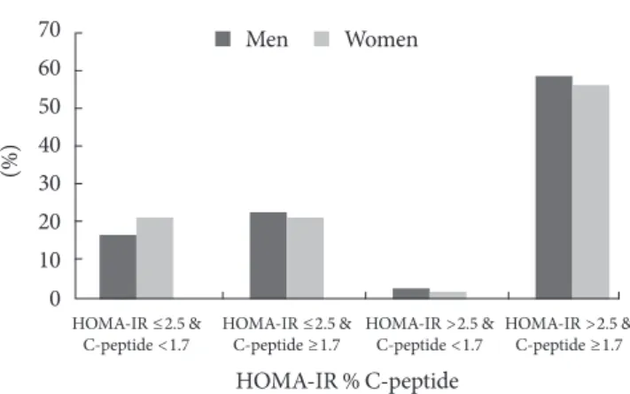
When the subjects were divided according to their insulin re-sistance and insulin secretion phenotypes (Fig. 2), 782 patients (59.5%) exceeded a HOMA-IR of 2.5. In contrast, only 3.3% of patients had severe insulin secretory defects with C-peptide lev-els <1.1 ng/mL, while 17.7% of subjects had moderate insulin secretion defects; most of the subjects (79.0%) had mild or non-secretory defects. Only 2.6% of men and 1.9% of women had both insulin resistance and decreased insulin secretion. Conse-quently, the subjects who were insulin resistant with preserved 50
45
40
35
30
25
20
15
10
5
0
(%)
≤22.9 23.0–24.9 25.0–29.9 30≤
BMI
Men Women
35
30
25
20
15
10
5
0
(%)
1 2 3 4 5
Components of metabolic syndrome
Men Women
Fig. 1. The proportion of study subjects according to body mass index (BMI) and the presence of metabolic syndrome among newly diagnosed patients with type 2 diabetes.
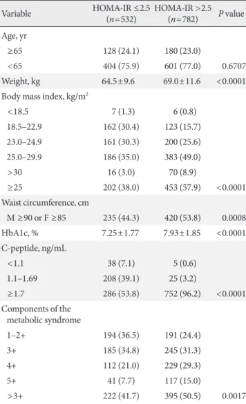
Table 2. he relationship between HOMA-IR, C-peptide lev-els, metabolic syndrome, and obesity
Variable HOMA-IR ≤2.5
(n=532)
HOMA-IR >2.5 (n=782) P value
Age, yr
≥65 128 (24.1) 180 (23.0)
<65 404 (75.9) 601 (77.0) 0.6707
Weight, kg 64.5±9.6 69.0±11.6 <0.0001
Body mass index, kg/m2
<18.5 7 (1.3) 6 (0.8)
18.5–22.9 162 (30.4) 123 (15.7)
23.0–24.9 161 (30.3) 200 (25.6)
25.0–29.9 186 (35.0) 383 (49.0)
>30 16 (3.0) 70 (8.9)
≥25 202 (38.0) 453 (57.9) <0.0001
Waist circumference, cm
M ≥90 or F ≥85 235 (44.3) 420 (53.8) 0.0008
HbA1c, % 7.25±1.77 7.93±1.85 <0.0001
C-peptide, ng/mL
<1.1 38 (7.1) 5 (0.6)
1.1–1.69 208 (39.1) 25 (3.2)
≥1.7 286 (53.8) 752 (96.2) <0.0001
Components of the metabolic syndrome
1–2+ 194 (36.5) 191 (24.4)
3+ 185 (34.8) 245 (31.3)
4+ 112 (21.0) 229 (29.3)
5+ 41 (7.7) 117 (15.0)
>3+ 222 (41.7) 395 (50.5) 0.0017
Values are presented as number (%) or mean±standard deviation. he data were obtained from an independent t-test or chi-square test.
insulin secretion were the most predominant group in this study.
DISCUSSION
Although both insulin deiciency and insulin resistance are in-volved in type 2 diabetes pathogenesis, an investigation of the main pathogenesis involved in the development of type 2 dia-betes is important. In the present study, only 21% of drug-na-ive patients with type 2 diabetes had moderate to severe insu-lin secretory defects, whereas 59.5% exhibited insuinsu-lin resis-tance. Within the insulin resistant group, there was a high pro-portion of subjects with mild secretory defects (C-peptide >1.7 ng/mL). he patient group with both insulin resistance and insulin deiciency was the smallest (2.6% in men and 1.9% in women). hese indings suggest that in Korea, insulin resis-tance is more likely related to the pathophysiology of type 2 diabetes than insulin deiciency.
Recently, obesity has increased because of rapid socioeco-nomic changes, consumption of a high fat diet, and low physi-cal activity, and obesity is an important factor that has been associated with diabetes incidence since 2000 [22]. During this period, the mean BMI of Korean diabetics has gradually increased from 23.8 [23] to 25.6 kg/m2 [12]. Although 20% to
30% of diabetic patients were obese in the 1980s and 1990s [24], recent studies (including ours) have demonstrated that 40% to 50% of diabetics in Korea are obese [11,12]. In addition to general obesity, up to 50% of the subjects in our study have central obesity. Asians demonstrate prominent central obesity at a given BMI, which may explain the diferent association
between BMI and diabetes risk in interethnic groups [25]. his alteration in body fat distribution is another possible explana-tion for increased insulin resistance with less obesity in Asian populations compared with Western populations [26,27]. Fur-thermore, a higher metabolic syndrome frequency of up to 80% was observed among type 2 diabetes patients, and these patients had exacerbated insulin resistance and worse glucose tolerance, which aligns with our results [28]. he prevalence of metabolic syndrome in Korean children and adolescents also doubled between KNHANES 1998 and KNHANES 2007 [29]. Together, these changes support the premise that insulin resis-tance prevalence explosively increased in Korean diabetics af-ter rapid Wesaf-ternization.
Several studies could potentially explain our findings. Be-cause Korean populations are genetically close to Japanese populations, indings in diabetic Japanese Americans indicate how environmental changes can afect pathophysiologic het-erogeneity in type 2 diabetic populations in Korea. Previously, comparing a study of Japanese individuals living in Hiroshima to those in Hawaii, the Hawaiian Japanese subjects demon-strated a higher prevalence of type 2 diabetes and increased in-sulin resistance without diferences in inin-sulin secretory capaci-ty [30]. his inding explained the potential environmental ef-fects on diabetes prevalence after Westernization. Data ob-tained from a 75-g oral glucose tolerance test also demonstrat-ed that the prevalence of isolatdemonstrat-ed impairdemonstrat-ed fasting glucose lev-els increased from 17% to 28.8% between the early 1990s [23] and the mid-2000s [31] in pre-diabetic Korean adults. The main pathogenesis of impaired fasting glucose levels is closely related with increased insulin resistance rather than an insulin secretory defect [32]. herefore, this increasing importance of insulin resistance in pre-diabetes was aligned with the results of our study.
Unexpectedly, the ratio of subjects with insulin secretion disorders in our study was much lower than that of previous studies of Korean subjects. It is diicult to explain this discrep-ancy. Such diferences among studies may have occurred be-cause these previous studies were performed in a single center in an urban area, the subjects had diabetes for a longer period of time, or because the subjects were exposed to insulin secre-tagogues or insulin. However, our observation does not dimin-ish the importance of β-cell dysfunction in the development of type 2 diabetes. here is a strong genetic susceptibility that is represented by early β-cell failure in some Asian populations. Numerous studies have already demonstrated the relative im-70
60
50 40
30
20 10
0
(%)
HOMA-IR ≤2.5 & C-peptide <1.7
HOMA-IR ≤2.5 & C-peptide ≥1.7
HOMA-IR >2.5 & C-peptide <1.7
HOMA-IR >2.5 & C-peptide ≥1.7
HOMA-IR % C-peptide
Men Women
portance of an early phase insulin secretory defect compared with insulin resistance in the development of glucose intoler-ance, independent of the degree of obesity in Korean subjects [33-35]. hus, although C-peptide can be used to assess endog-enous insulin secretion in our study [36], fasting C-peptide levels are not representative of the insulin secretory response or early diabetes progression. Taken together, we assumed that our subjects who were exposed to a Western lifestyle did not produce a sufficient insulin secretion response to overcome rapidly increasing insulin resistance, which resulted in higher diabetes prevalence. hese results must be conirmed by fur-ther investigation.
The current study had some limitations. Although the HOMA-IR is a useful estimate of insulin resistance, certain limitations should be noted. HOMA-IR has merit because it is easily used to conirm type 2 diabetic patient clinical properties in large-scale studies, and reasonable reference intervals for HOMA-IR have recently been established. It was difficult to elucidate causal relationships because of the limitation of the cross-sectional design and because there were no comparative patient groups, such as those with a normal glucose tolerance or pre-diabetes, but we were able to demonstrate the clinical properties of newly diagnosed diabetes patients in a large, na-tional investigation compared with other small cohort studies. In conclusion, the present study demonstrated remarkably increased obesity and metabolic syndrome-associated insulin resistance in early phase diabetic patients in Korean popula-tions. his inding suggests that the main pathogenesis of type 2 diabetes may have shited from insulin deiciency to insulin resistance in the Korean population. Considering that insulin resistance-related components were modiiable, unlike β-cell dysfunction, which may be genetically determined, one must evaluate the degree of insulin resistance and establish individ-ual treatment approaches for insulin resistance or insulin se-cretory defects. Based on our indings, interventions for im-proving insulin resistance would be a more efective treatment for newly diagnosed, drug-naïve Korean type 2 diabetics. In the future, along with multi-institutional prospective studies, more evidence is required to determine whether insulin secre-tion or insulin resistance is important for the type 2 diabetes pathophysiology in Asian populations.
CONFLICTS OF INTEREST
his work was supported by GlaxoSmithKline Korea, Inc.
REFERENCES
1. Zimmet P, Alberti KG, Shaw J. Global and societal implications of the diabetes epidemic. Nature 2001;414:782-7.
2. Yoon KH, Lee JH, Kim JW, Cho JH, Choi YH, Ko SH, Zimmet P, Son HY. Epidemic obesity and type 2 diabetes in Asia. Lan-cet 2006;368:1681-8.
3. Chan JC, Malik V, Jia W, Kadowaki T, Yajnik CS, Yoon KH, Hu FB. Diabetes in Asia: epidemiology, risk factors, and pathophysi-ology. JAMA 2009;301:2129-40.
4. Kim DJ, Lee MS, Kim KW, Lee MK. Insulin secretory dysfunc-tion and insulin resistance in the pathogenesis of Korean type 2 diabetes mellitus. Metabolism 2001;50:590-3.
5. Chen KW, Boyko EJ, Bergstrom RW, Leonetti DL, Newell-Mor-ris L, Wahl PW, Fujimoto WY. Earlier appearance of impaired insulin secretion than of visceral adiposity in the pathogenesis of NIDDM. 5-Year follow-up of initially nondiabetic Japanese-American men. Diabetes Care 1995;18:747-53.
6. Matsumoto K, Miyake S, Yano M, Ueki Y, Yamaguchi Y, Aka-zawa S, Tominaga Y. Glucose tolerance, insulin secretion, and insulin sensitivity in nonobese and obese Japanese subjects. Diabetes Care 1997;20:1562-8.
7. Fukushima M, Suzuki H, Seino Y. Insulin secretion capacity in the development from normal glucose tolerance to type 2 dia-betes. Diabetes Res Clin Pract 2004;66 Suppl 1:S37-43. 8. Rattarasarn C, Soonthornpan S, Leelawattana R, Setasuban W.
Decreased insulin secretion but not insulin sensitivity in normal glucose tolerant hai subjects. Diabetes Care 2006;29:742-3. 9. Kim DM, Ahn CW, Nam SY. Prevalence of obesity in Korea.
Obes Rev 2005;6:117-21.
10. Lim S, Shin H, Song JH, Kwak SH, Kang SM, Won Yoon J, Choi SH, Cho SI, Park KS, Lee HK, Jang HC, Koh KK. Increasing prevalence of metabolic syndrome in Korea: the Korean Na-tional Health and Nutrition Examination Survey for 1998-2007. Diabetes Care 2011;34:1323-8.
11. Kim DJ, Song KE, Park JW, Cho HK, Lee KW, Huh KB. Clini-cal characteristics of Korean type 2 diabetic patients in 2005. Diabetes Res Clin Pract 2007;77 Suppl 1:S252-7.
13. Kahn BB, Flier JS. Obesity and insulin resistance. J Clin Invest 2000;106:473-81.
14. Misra A, Khurana L. Obesity-related non-communicable dis-eases: South Asians vs White Caucasians. Int J Obes (Lond) 2011;35:167-87.
15. American Diabetes Association. Standards of medical care in diabetes: 2009. Diabetes Care 2009;32 Suppl 1:S13-61.
16. Matthews DR, Hosker JP, Rudenski AS, Naylor BA, Treacher DF, Turner RC. Homeostasis model assessment: insulin resis-tance and beta-cell function from fasting plasma glucose and insulin concentrations in man. Diabetologia 1985;28:412-9. 17. Yamada C, Mitsuhashi T, Hiratsuka N, Inabe F, Araida N,
Taka-hashi E. Optimal reference interval for homeostasis model as-sessment of insulin resistance in a Japanese population. J Dia-betes Investig 2011;2:373-6.
18. Park SW, Yun YS, Ahn CW, Nam JH, Kwon SH, Song MK, Han SH, Cha BS, Son YD, Lee HC, Huh KB. Short insulin tolerance test (SITT) for the determination of in vivo insulin sensitivity-a comparison with euglycemic clamp test. J Korean Diabetes As-soc 1998;22:199-208.
19. World Health Organization; International Association for the Study of Obesity; International Obesity Task Force. he Asia-Paciic perspective: redeining obesity and its treatment. Syd-ney: Health Communications; 2000.
20. Lee SY, Park HS, Kim DJ, Han JH, Kim SM, Cho GJ, Kim DY, Kwon HS, Kim SR, Lee CB, Oh SJ, Park CY, Yoo HJ. Appropri-ate waist circumference cutof points for central obesity in Ko-rean adults. Diabetes Res Clin Pract 2007;75:72-80.
21. Grundy SM, Cleeman JI, Daniels SR, Donato KA, Eckel RH, Franklin BA, Gordon DJ, Krauss RM, Savage PJ, Smith SC Jr, Spertus JA, Costa F; American Heart Association; National Heart, Lung, and Blood Institute. Diagnosis and management of the metabolic syndrome: an American Heart Association/ National Heart, Lung, and Blood Institute Scientiic Statement. Circulation 2005;112:2735-52.
22. Choi YJ, Kim HC, Kim HM, Park SW, Kim J, Kim DJ. Preva-lence and management of diabetes in Korean adults: Korea Na-tional Health and Nutrition Examination Surveys 1998-2005. Diabetes Care 2009;32:2016-20.
23. Oh JY, Lim S, Kim DJ, Kim NH, Kim DJ, Moon SD, Jang HC, Cho YM, Song KH, Ahn CW, Sung YA, Park JY, Shin C, Lee HK, Park KS; Committee of the Korean Diabetes Association on the Diagnosis and Classiication of Diabetes Mellitus. A re-port on the diagnosis of intermediate hyperglycemia in Korea: a pooled analysis of four community-based cohort studies.
Di-abetes Res Clin Pract 2008;80:463-8.
24. Lee TH. Prevalence of obesity in Korean non-insulin-depen-dent diabetic patients. Diabetes Res Clin Pract 1996;32:71-80. 25. WHO Expert Consultation. Appropriate body-mass index for
Asian populations and its implications for policy and interven-tion strategies. Lancet 2004;363:157-63.
26. Raji A, Seely EW, Arky RA, Simonson DC. Body fat distribu-tion and insulin resistance in healthy Asian Indians and Cau-casians. J Clin Endocrinol Metab 2001;86:5366-71.
27. Petersen KF, Dufour S, Feng J, Befroy D, Dziura J, Dalla Man C, Cobelli C, Shulman GI. Increased prevalence of insulin resis-tance and nonalcoholic fatty liver disease in Asian-Indian men. Proc Natl Acad Sci U S A 2006;103:18273-7.
28. Rhee SY, Kwon MK, Park BJ, Chon S, Jeong IK, Oh S, Ahn KJ, Chung HY, Kim SW, Kim JW, Kim YS, Woo JT. Diferences in insulin sensitivity and secretory capacity based on OGTT in subjects with impaired glucose regulation. Korean J Intern Med 2007;22:270-4.
29. Lim S, Jang HC, Park KS, Cho SI, Lee MG, Joung H, Mozum-dar A, Liguori G. Changes in metabolic syndrome in American and Korean youth, 1997-2008. Pediatrics 2013;131:e214-22. 30. Nakanishi S, Okubo M, Yoneda M, Jitsuiki K, Yamane K,
Koh-no N. A comparison between Japanese-Americans living in Hawaii and Los Angeles and native Japanese: the impact of lifestyle westernization on diabetes mellitus. Biomed Pharma-cother 2004;58:571-7.
31. Rhee SY, Woo JT, Chon S, Hwang YC, Oh S, Ahn KJ, Chung HY, Kim SW, Kim JW, Kim YS. Characteristics of insulin resis-tance and insulin secretory capacity in Korean subjects with IFG and IGT. Diabetes Res Clin Pract 2010;89:250-5.
32. Abdul-Ghani MA, Sabbah M, Kher J, Minuchin O, Vardi P, Raz I. Diferent contributions of insulin resistance and beta-cell dysfunction in overweight Israeli Arabs with IFG and IGT. Diabetes Metab Res Rev 2006;22:126-30.
33. Chae BN, Lee SK, Hong EG, Chung YS, Lee KW, Kim HM. he role of insulin secretion and insulin resistance in the develop-ment of Korean type 2 diabetes mellitus. J Korean Diabetes As-soc 1998;22:491-503.
34. Yoon KH, Ko SH, Cho JH, Lee JM, Ahn YB, Song KH, Yoo SJ, Kang MI, Cha BY, Lee KW, Son HY, Kang SK, Kim HS, Lee IK, Bonner-Weir S. Selective beta-cell loss and alpha-cell expan-sion in patients with type 2 diabetes mellitus in Korea. J Clin Endocrinol Metab 2003;88:2300-8.
an-tibody in non-obese, adult-onset type 2 diabetes in Korea and clinical and biological characteristics according to anti-GAD antibody. J Korean Diabetes Assoc 2004;28:66-74.