Submitted17 February 2016 Accepted 11 March 2016 Published31 March 2016
Corresponding author Ehsan Pashay Ahi, [email protected]
Academic editor Joan Heath
Additional Information and Declarations can be found on page 17
DOI10.7717/peerj.1878
Copyright 2016 Pashay Ahi et al.
Distributed under
Creative Commons CC-BY 4.0
OPEN ACCESS
Investigation of the effects of estrogen on
skeletal gene expression during zebrafish
larval head development
Ehsan Pashay Ahi1, Benjamin S. Walker2, Christopher S. Lassiter2and
Zophonías O. Jónsson1,3
1Institute of Life and Environmental Sciences, University of Iceland, Reykjavik, Iceland
2Biology Department, Roanoke College, Salem, VA, United States
3Biomedical Center, University of Iceland, Reykjavik, Iceland
ABSTRACT
The development of craniofacial skeletal structures requires well-orchestrated tissue interactions controlled by distinct molecular signals. Disruptions in normal function of these molecular signals have been associated with a wide range of craniofacial malformations. A pathway mediated by estrogens is one of those molecular signals that plays role in formation of bone and cartilage including craniofacial skeletogenesis. Studies in zebrafish have shown that while higher concentrations of 17-βestradiol (E2) cause severe craniofacial defects, treatment with lower concentrations result in subtle changes in head morphology characterized with shorter snouts and flatter faces. The molecular basis for these morphological changes, particularly the subtle skeletal effects mediated by lower E2concentrations, remains unexplored. In the present study we address these effects at a molecular level by quantitative expression analysis of sets of candidate genes in developing heads of zebrafish larvae treated with two differentE2 concentrations. To this end, we first validated three suitable reference genes,ppia2,rpl8 andtbp, to permit sensitive quantitative real-time PCR analysis. Next, we profiled the expression of 28 skeletogenesis-associated genes that potentially respond to estrogen signals and play role in craniofacial development. We foundE2mediated differential expression of genes involved in extracellular matrix (ECM) remodelling,mmp2/9/13, sparcandtimp2a, as well as components of skeletogenic pathways,bmp2a,erf,ptch1/2, rankl,rarabandsfrp1a. Furthermore, we identified a co-expressed network of genes, includingcpn1,dnajc3,esr1,lman1,rrbp1a,ssr1andtram1with a stronger inductive response to a lower dose ofE2during larval head development.
SubjectsAquaculture, Fisheries and Fish Science, Biodiversity, Developmental Biology, Molecular Biology, Toxicology
Keywords Craniofacial skeleton, Development, Estrogen, Gene expression, Zebrafish larvae, qPCR, Reference genes, 17-βestradiol
INTRODUCTION
oro-facial clefts and associated malformations (Marazita, 2012). The vertebrate craniofacial skeleton, including the viscerocranium, is built from neural-crest derived tissues. Changes in these tissues over evolutionary time have given rise to a wide diversity of facial morphologies among vertebrate species (Trainor, Melton & Manzanares, 2003;Bronner & LeDouarin, 2012).
Estrogens, steroid hormones synthesized by aromatase from androgenic precursors, have recently been shown to affect craniofacial development (Fushimi et al., 2009;Cohen et al., 2014). Though estrogens are normally thought of as sex hormones, they affect a variety of tissues including the cardiovascular and skeletal systems (Hall, Couse & Korach, 2001;Allgood et al., 2013;Cohen et al., 2014). Estrogens signal through classical nuclear receptors (ER-alpha and ER-beta) (Jia, Dahlman-Wright & Gustafsson, 2015) and a G-protein coupled receptor, GPR-30 (Jenei-Lanzl et al., 2010). These signaling proteins are found in the chondrocytes of many vertebrate species (Tankó et al., 2008) and are present during chondrogenesis (Jenei-Lanzl et al., 2010;Elbaradie et al., 2013). Among vertebrates, teleost fish are highly diversified in craniofacial morphology and estrogen has been shown to greatly affect chondrogenesis in many of the fish species studied so far, including tilapia, fathead minnow, and zebrafish (Ng, Datuin & Bern, 2001;Warner & Jenkins, 2007;
Cohen et al., 2014). Furthermore, the teleost zebrafish is a well-developed model system for studying both embryonic development and human disease and it can be useful in understanding the development of the human viscerocranial skeleton as these processes are well-conserved among vertebrates (Kuratani, Matsuo & Aizawa, 1997).
Low concentrations of estrogen cause subtle changes in craniofacial morphogenesis during zebrafish larval development (Cohen et al., 2014). These changes are characterized by a shorter snout, flatter face and wider angles of cartilage elements in the viscerocranium (Cohen et al., 2014). Another recent study has also demonstrated that an estrogen mediated signal underlies the evolution of shorter snouts and flatter faces in females of some reptilian species (Sanger et al., 2014). These observations suggest that similar mechanisms might underpin hormone-based phenotypic plasticity and diversity (Dufty, 2002), as well as subtle differences in head/skeletal morphology of dimorphic sexes (Loth & Henneberg, 2001;Fujita et al., 2004;Callewaert et al., 2010). Therefore, studies offering insights into molecular mechanisms rendering the observed phenotypes caused by hormonal changes would be called for.
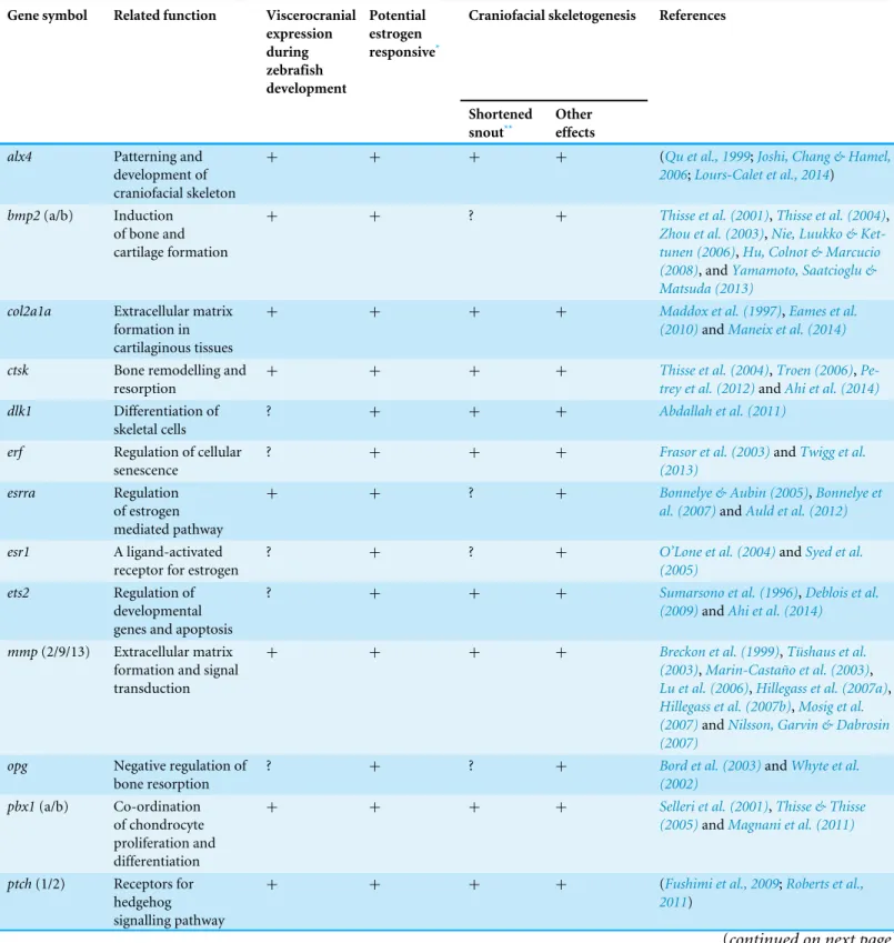
Table 1 Selected putative estrogen-regulated candidate genes, and available literature indicating their role in craniofacial development/skeletal formation in zebrafish or other vertebrates.
Gene symbol Related function Viscerocranial expression during zebrafish development Potential estrogen responsive*
Craniofacial skeletogenesis References
Shortened snout**
Other effects
alx4 Patterning and
development of craniofacial skeleton
+ + + + (Qu et al., 1999;Joshi, Chang & Hamel,
2006;Lours-Calet et al., 2014)
bmp2(a/b) Induction
of bone and cartilage formation
+ + ? + Thisse et al. (2001),Thisse et al. (2004),
Zhou et al. (2003),Nie, Luukko & Ket-tunen (2006),Hu, Colnot & Marcucio (2008), andYamamoto, Saatcioglu & Matsuda (2013)
col2a1a Extracellular matrix
formation in cartilaginous tissues
+ + + + Maddox et al. (1997),Eames et al.
(2010)andManeix et al. (2014)
ctsk Bone remodelling and
resorption
+ + + + Thisse et al. (2004),Troen (2006),
Pe-trey et al. (2012)andAhi et al. (2014)
dlk1 Differentiation of
skeletal cells
? + + + Abdallah et al. (2011)
erf Regulation of cellular
senescence
? + + + Frasor et al. (2003)andTwigg et al.
(2013)
esrra Regulation
of estrogen mediated pathway
+ + ? + Bonnelye & Aubin (2005),Bonnelye et
al. (2007)andAuld et al. (2012)
esr1 A ligand-activated
receptor for estrogen
? + ? + O’Lone et al. (2004)andSyed et al.
(2005)
ets2 Regulation of
developmental genes and apoptosis
? + + + Sumarsono et al. (1996),Deblois et al.
(2009)andAhi et al. (2014)
mmp(2/9/13) Extracellular matrix formation and signal transduction
+ + + + Breckon et al. (1999),Tüshaus et al.
(2003),Marin-Castaño et al. (2003),
Lu et al. (2006),Hillegass et al. (2007a),
Hillegass et al. (2007b),Mosig et al. (2007)andNilsson, Garvin & Dabrosin (2007)
opg Negative regulation of
bone resorption
? + ? + Bord et al. (2003)andWhyte et al.
(2002)
pbx1(a/b) Co-ordination
of chondrocyte proliferation and differentiation
+ + + + Selleri et al. (2001),Thisse & Thisse
(2005)andMagnani et al. (2011)
ptch(1/2) Receptors for hedgehog signalling pathway
+ + + + (Fushimi et al., 2009;Roberts et al.,
2011)
Table 1(continued)
Gene symbol Related function Viscerocranial expression during zebrafish development Potential estrogen responsive*
Craniofacial skeletogenesis References
Shortened snout** Other effects rankl Osteoclast differentiation and activation
? + ? + Bord et al. (2003)andLézot et al.
(2015)
rarab A receptor for
retinoic acid signalling pathway
+ + + + (Lohnes et al., 1994;O’Lone et al., 2004;
Linville et al., 2009)
runx2b Osteoblast
differentiation and skeletal morphogenesis
+ + + + Sears et al. (2007),McCarthy et al.
(2003)andFlores et al. (2006)
sfrp1a A soluble modulator
of Wnt signalling pathway
? + + + Satoh et al. (2006),Trevant et al.
(2008),Yokota et al. (2008),Fukuhara et al. (2013)andAhi et al. (2014)
Shh(a/b) Activators
of hedgehog signalling pathway
+ ? ? + Hu & Helms (1999)andSwartz et al.
(2012)
sox9b Chondrocyte
differentiation
+ + ? + Yan et al. (2005),Bonnelye et al. (2007)
andLee & Saint-Jeannet (2011)
sparc Extracellular matrix
synthesis and regulation of cell growth
+ + + + Lehane et al. (1999),Renn et al. (2006)
andRotllant et al. (2008)
spp1 Attachment of
osteoclasts to ECM in bone
+ + + + Craig & Denhardt (1991),Vanacker et
al. (1998)andVenkatesh et al. (2014)
timp2a Inhibition of
mmps and regulation of tissue homeostasis
? + + ? Dew et al. (2000),Lam et al. (2009),
Letra et al. (2012),Wang & Ma (2012)
andAhi et al. (2014)
Notes.
*The estrogen responsiveness indicates either transcriptional regulation or transactivation and the related information are mainly obtained from different model vertebrates, such as human and mouse, than teleost fishes.
**The shortened snout indicates the skeletal effects resulted from decrease in the length or changes in morphology of viscerocranial skeletal elements in different vertebrate species. This could bear a resemblance to an estrogen mediated shorter snout and flatter face phenotype in zebrafish.
hypothesized that these genes may be critical to the estrogen modulation of craniofacial skeletogenesis. We first identified the most stably expressed reference genes in developing heads of zebrafish treated with two doses of estrogen (2µM and 5µM) across five stages in
METHODS
Fish husbandry, treatment and sampling
Adult zebrafish were fed a diet of live brine shrimp supplemented with Ziegler zebrafish diet (Pentair) and maintained on a 14/10 day/night cycle. Embryos were raised in E3B (5 mM NaCl, 0.17 mM KCl, 0.33 mM CaCl2, 0.33 mM MgSO4, 0.00025% methylene blue). Embryos were treated with estrogen (17β-estradiol,E2, Sigma) dissolved in ethanol and diluted in E3B for a final ethanol concentration of 0.1%. Control fish were treated with 0.1% ethanol with no developmental malformations as described previously (Cohen et al., 2014). For each treatment group (estrogen concentration), zebrafish larva were raised in Petri dishes, and treatment solutions were refreshed daily until the stages indicated (3, 4, 5, 6 and 7 days post fertilization, dpf). Three biological replicates of 30 larva were collected at each time-point (3–7 dpf) and for each treatment group (control, 2µME2, and 5µM
E2) for a total of 90 larva at each time-point and treatment. The fishes were anesthetized with 0.4% tricaine (MS-222, Sigma). Isolated heads (anterior to the yolk sac) were placed into RNAlater (Qiagen) and stored frozen until RNA isolation. Zebrafish experiments were performed under the Roanoke College IRB protocol #14BIO76.
RNA isolation and cDNA synthesis
Around 30 heads of zebrafish from each treatment group and larval stage were pooled in TRI Reagent (Sigma) and homogenized with a disposable Kontes Pellet Pestle Cordless Motor tissue grinder (Kimble Kontes). RNA was prepared according to manufacturer’s instructions and dissolved in 50µl RNase-free water. RNA samples were treated with DNase
(New England Biolabs) to remove contaminating DNA. Quantity of the resulting RNA samples was assessed using a NanoDrop ND-1000 UV/Vis-Spectrophotometer (NanoDrop Technologies). The quality of the RNA samples was evaluated by agarose gel electrophoresis and all samples displayed intact 28 S and 18 S rRNA without noticable high molecular weight genomic DNA contamination. cDNA was prepared from 1000 ng of RNA using the High capacity cDNA Reverse Transcription kit (Applied Biosystems), according to manufacturer’s protocol. Several samples without addition of reverse transcriptase (-RT samples) were prepared to confirm the absence of genomic DNA. cDNA was diluted 3 fold in water for further use in quantitative real-time PCR.
Gene selection, Primer design and real-time qPCR
In order to validate suitable reference genes for accurate measurement of the transcriptional changes of candidate genes by qPCR, we selected 7 potential reference genes based on published studies in zebrafish (Table S1) (McCurley & Callard, 2008;Pelayo et al., 2012;
reliability, we filtered the genes by setting the mutual rank (MR) to the top-ranked 2000 and the Supportability score of minimum 1 (as described byObayashi & Kinoshita, 2011). This yielded 338 candidate genes, and from them, we selected 11 genes with reported craniofacial expression during zebrafish development according to the ZFIN database (http://zfin.org) (Bradford et al., 2011) (Table S1).
Locations overlapping exon boundaries of the genes in zebrafish were determined by NCBI Spidey software (www.ncbi.nlm.nih.gov/spidey) and annotated genome sequences in the Ensembl database (http://www.ensembl.org/Danio_rerio). The qPCR Primers were designed on exon boundaries using Primer Express 3.0 software (Applied Biosystems, Foster City, CA, USA) and checked for self-annealing, hetero-dimers and hairpin structures with OligoAnalyzer 3.1 (Integrated DNA Technology) (Table S1).
Real-time PCR was performed in 96 well-PCR plates on an ABI 7500 real-time PCR System (Applied Biosystems) using Maxima SYBR Green/ROX qPCR Master Mix (2X) as recommended by the manufacturer (Thermo Fisher Scientific, St Leon-Rot, Germany). Each biological replicate was run in duplicate together with no-template control (NTC) in each run for each gene and experimental set-up per run followed the preferred sample maximization method (Hellemans et al., 2007). The qPCR was run with a 2 min hold at 50◦
C and a 10 min hot start at 95◦
C followed by the amplification step for 40 cycles of 15 sec denaturation at 95◦
C and 1 min annealing/extension at 60◦
C. A dissociation step (60 ◦
C–95◦
C) was performed at the end of the amplification phase to identify a single, specific product for each primer set (Table S1). Primer efficiency values (E) were calculated with the LinRegPCR v11.0 programme (http://LinRegPCR.nl) (Ramakers et al., 2003) analysing the background-corrected fluorescence data from the exponential phase of PCR amplification for each primer-pair and those with E less than 0.9 were discarded and new primers designed (Table S1).
Data analysis
To detect the most stably expressed reference genes, three ranking algorithms; BestKeeper (Pfaffl et al., 2004), NormFinder (Andersen, Jensen & Ørntoft, 2004) and geNorm
For the analysis of qPCR data, the difference betweenCqvalues (1Cq) of the reference genes and the target genes was calculated for each gene;1Cqtarget=Cqtarget−Cqreference. The geometric mean of Cq values of three best ranked reference genes,ppia2,rpl8andtbp(see the ranking algorithms above), was used as Cqreferencein the1Cqcalculations. All samples
were then normalized to the 1Cqvalue of a calibrator sample to obtain a11Cqvalue (1Cqtarget−1Cqcalibrator). For each primer pair a biological replicate in the control group at 3dpf was selected as the calibrator sample. Relative expression quantities (RQ) were calculated based on the expression level of the calibrator sample (E−11Cq) (Pfaffl, 2001).
The RQ values were then transformed to logarithmic base 2 values (or fold differences; FD) for statistical analysis (Bergkvist et al., 2010). A two-way analysis of variance (ANOVA) followed by post hoc Tukey’s honest significant difference (HSD) test was implemented for each reference or target gene with larval stages and treatment groups as categorical variables. To assess similarities in expression patterns of the genes Pearson correlation coefficients (r) were calculated for all gene pairs using the data from 3 treatments at 5 larval stages (degree of freedom=13). R (http://www.r-project.org) was used for all statistical analysis.
RESULTS
tbp,ppia2 andrpl8 are the most suitable reference genes
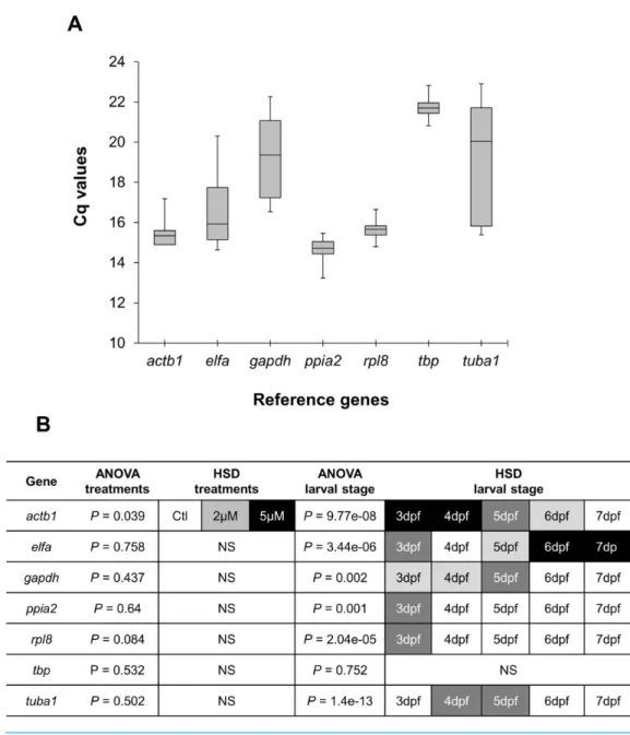
Real-time quantitative PCR for the 7 reference gene candidates was performed on cDNA generated from zebrafish head homogenates in three treatment groups at five larval stages. The expression levels of the candidates varied from ppia2, with the highest expression (lowest Cq) (Fig. 1A), totbpwith the lowest expression (highestCq). Statistical analysis revealed that all of the candidates exceptactb1are stably expressed between the treatment groups (Fig. 1B). However, onlytbpshowed constant expression during the larval stages examined. Two genes,ppia2andrpl8, were also stably expressed in developing heads of zebrafish larvae except for the first stage (3dpf). Based on these resultstbpfollowed by ppia2andrpl8 were found to be the overall most stable reference genes both over time and between the treatment groups. The candidate reference genes were ranked using three algorithms, i.e., BestKeeper, geNorm and NormFinder, and based on standard deviation (SD) as described inAhi et al. (2013)(Table 2). In all of the analyses three genes;ppia2,rpl8 andtbp, were the three highest ranking candidates, however their order varied between the rankings (Table 2). Furthermore, geNorm suggested the use of the three best ranked candidate genes as sufficient for accurate normalisation (Fig. S1). The data reflect the high expression stability of the best ranked candidate genes and suggests the combination of ppia2,rpl8andtbpas a suitable and sufficient normalization factor to accurately quantify small differences in gene expression in developing heads of zebrafish larvae across theE2 treatment groups.
Components of different signalling pathways and
skeletogenesis-associated genes are affected by estrogen during larval head development
Figure 1 Expression analysis of candidate reference genes in developing heads of zebrafish larvae across control andE2treated groups. (A) Expression profiles of candidate reference genes in raw Cq
val-ues for all samples (3 treatments for each of 5 larval stages and with 3 biological replicates). The middle line denotes the median and boxes indicate the 25/75 percentiles. (B) Expression differences of candidate reference genes in the head of zebrafish during the larval development and threeE2treatment groups. Fold changes in expression calculated from the qPCR data, were subjected to ANOVA and Tukey’s HSD anal-ysis to test the expression differences amongst three treatment groups (control, 2µM and 5µM) and
across five larval stages (3 to 7dpf). White boxes represent low expression, while black boxes represent high expression. Two or more steps of shade differences in the boxes represent significantly different ex-pression between the samples (alpha=0.05). NS, not significant.
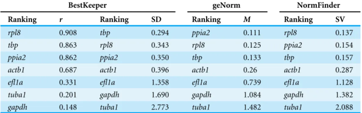
Table 2 Ranking and statistical analyses of candidate reference genes using BestKeeper, geNorm and NormFinder.
BestKeeper geNorm NormFinder
Ranking r Ranking SD Ranking M Ranking SV
rpl8 0.908 tbp 0.294 ppia2 0.111 rpl8 0.137
tbp 0.863 rpl8 0.343 rpl8 0.125 ppia2 0.154
ppia2 0.862 ppia2 0.350 tbp 0.133 tbp 0.157
actb1 0.687 actb1 0.396 actb1 0.26 actb1 0.287
efl1a 0.331 efl1a 1.358 efl1a 0.739 efl1a 1.128
tuba1 0.201 gapdh 1.690 gapdh 1.084 gapdh 1.382
gapdh 0.148 tuba1 2.773 tuba1 1.482 tuba1 2.088
Notes.
Abbreviations: SD, Standard deviation;r, Pearson product-moment correlation coefficient; SV, Stability value;M,Mvalue of stability.
associated with shortened snout morphogenesis in vertebrates (col2a1a,ctsk,mmp2/9/13, sparc,spp1andtimp2a); and (V) other potential targets of estrogen pathways with diverse functions which are also involved in viscerocranial skeletogenesis (alx4,dlk1,erf,ets2, pbx1a/b,rarabandsfrp1a). The expression levels of all candidates were measured in the three treatment groups during larval head development (Figs. 2,3,4and5). We found effects of differentE2concentrations on the expression of most of the target genes, except col2a1aandpbx1a, the effects, however, were highly variable among the genes (Figs. 2,3,
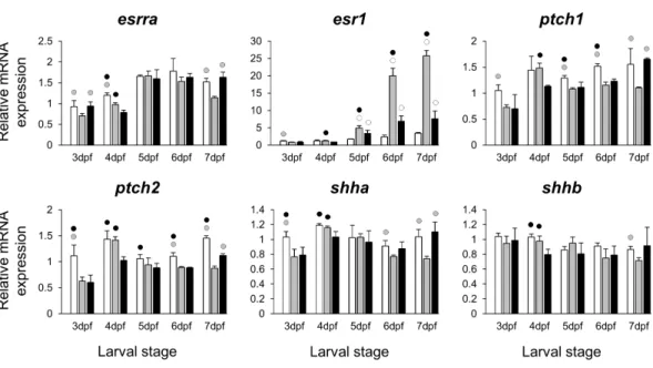
4and5). For instance, while some genes, i.e.,esr1,ptch1/2andrarabdisplayed differential expression between the treatment groups at most of the larval stages, other genes such as alx4,bmp2b,ctsk,ets2,opg, etc., showed expression differences at only one stage. Among the more highly affected genes,erf,esrra,mmp9,rankl,shha,sfrp1a,sparc andtimp2a were differentially expressed in at least three larval stages (Figs. 2,3,4and5). Although significant, most differences in expression levels of the target genes were slight between the treatment groups (RQ < 0.5), except foresr1at the last larval stages (Fig. 2). Moreover, for all of the affected genes, exceptesr1andmmp13, the differentE2 treatments had mainly repressive effects on transcription. These repressive effects were not, however, increased by higherE2concentration particularly at the last two stages when the lowerE2dose (2µM)
repressed expression of many of the genes more than the higher dose. At the last three stages, the expression of esr1was induced at highest levels for 2µM treatment groups
(Fig. 2). The transcriptional repression byE2was also variable between the genes and it was more pronounced forerf andptch2showing higher expression in control groups than both E2treated groups at three larval stages. Taken together, these results show significant effects of lowE2concentrations on the expression of a variety of genes involved in skeletogenesis and/or craniofacial development.
Figure 2 Expression differences of two estrogen receptors and components of hedgehog signaling pathway in developing heads of zebrafish larvae across control andE2treated groups. Expression of
esrra,esr1,ptch1,ptch2,shhaandshhbwas examined with qPCR and normalised using three highest
ranked reference genes (ppia2,rpl8andtbp). For analysis of relative expression levels for each target gene a replicate of the control group at 3dpf was set to one. The white, grey, and black bars in each graph represent expression levels for control, 2µME2treated and 5µME2treated groups respectively. Statistical differences of each treatment group versus the others are shown in white, grey, and black circles representing higher expressed than control, 2µME2treated and 5µME2treated groups respectively (P < 0.05). Error bars represent standard deviation calculated from three biological replicates. Each biological replicate is from a homogenate of 30 heads.
correlated expression. Negatively correlated expression was only seen betweenesr1and sfrp1a, and betweenshhband six genes includingesr1,ets2,mmp13,opg,pbx1aandspp1 (red shadings inFig. S2).
A co-expressed network of genes shows higher expression induction in lower E2treatment groups
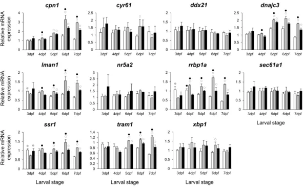
The stronger transcriptional response of esr1to the lowerE2 treatment (Fig. 2) could indicate a distinct regulatory mechanism associated with slight increase in estrogen concentration during zebrafish larval head development. In order to identify additional genes showing similar expression dynamics, we selected 11 candidate genes constructing a co-expression network withesr1using co-expression data for zebrafish in COXPRESdb (Obayashi & Kinoshita, 2011) (Table S2). These candidates are also known to have craniofacial skeletal expression during zebrafish development based on data submitted to the ZFIN database (http://zfin.org) (Bradford et al., 2011). Strikingly, we found stronger inductive effects of the lower E2 concentration on the expression of six genes, i.e.,cpn1, dnajc3,lman1,rrbp1a,ssr1andtram1(Fig. 6). The expression of these six genes followed a similar pattern and their higher expression levels were more pronounced at the last three stages of 2 µM treatment groups. Moreover, the gene showing strongest coexpression
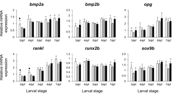
Figure 3 Expression differences of six potential skeletogenic targets of estrogen pathway in developing heads of zebrafish larvae across control andE2treated groups.Expression ofbmp2a,bmp2b,opg,
rankl,runx2bandsox9bwas examined with qPCR and normalised using three highest ranked reference
genes (ppia2,rpl8andtbp). For analysis of relative expression levels for each target gene a replicate of the control group at 3dpf was set to one. The white, grey, and black bars in each graph represent expression levels for control, 2µME2treated and 5µME2treated groups respectively. Statistical differences of each treatment group versus the others are shown in white, grey, and black circles representing higher expressed than control , 2µME2treated and 5µME2treated groups respectively (P<0.05). Error bars represent standard deviation calculated from three biological replicates. Each biological replicate was made from a homogenate of 30 heads.
at the last four stages of 2µM treatment groups (Table S2andFig. 6). Finally, we also
demonstrated positive expression correlations between the six candidates andesr1, but not the rest of the non-differentially expressed genes (blue shadings inFig. S3).
DISCUSSION
Estrogen signaling, through both canonical nuclear estrogen receptors and G-protein coupled receptors, is important in embryonic development (Griffin et al., 2013;Shi et al., 2013). Estrogens can act at autocrine, paracrine, and endocrine distances in the embryo and the adult (Boon, Chow & Simpson, 2010). Aromatase, the enzyme that synthesizes estrogens, is present in the developing brain of many species, including zebrafish (Lassiter & Linney, 2007) and would be a local source of the hormone during head development. In fact, the teleost brain produces relatively high levels of estrogen compared to other vertebrates (Forlano et al., 2001). Estrogens are thus present in the cranium of developing embryos and modulate viscerocranial development (Fushimi et al., 2009;Marquez Hernandez et al., 2011;Cohen et al., 2014). Estrogen signalling has been implicated in the sexual dimorphism of cranial bones in the Anolis lizard (Sanger et al., 2014). Hence, it may play a role in craniofacial morphological divergence among species and within sexes of the same species.
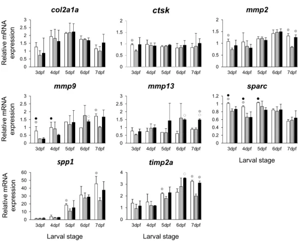
Figure 4 Expression differences of eight potential targets of estrogen pathway involved in skeletal ECM formation examined during zebrafish larval head development across control andE2treated
groups. Expression ofcol2a1a,ctsk,mmp2,mmp9,mmp13,sparc,spp1andtimp2was examined with qPCR and normalised using three highest ranked reference genes (ppia2,rpl8andtbp). For analysis of relative expression levels for each target gene a replicate of the control group at 3dpf was set to one. The white, grey, and black bars in each graph represent expression levels for control, 2µME2treated and 5µME2treated groups respectively. Statistical differences of each treatment group versus the others are shown in white, grey, and black circles representing higher expressed than control, 2µME2treated and 5µME2treated groups respectively (P<0.05). Error bars represent standard deviation calculated from three biological replicates. Each biological replicate was made from a homogenate of 30 heads.
estradiol (10µM) giving rise to major disruptions of chondrogenesis followed by severe
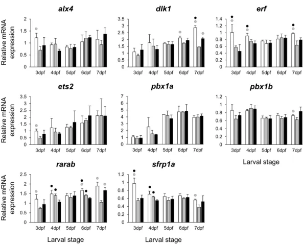
Figure 5 Expression differences of eight other potential targets of estrogen pathway involved in jaw skeletal elongation examined during zebrafish larval head development across control andE2treated
groups. Expression ofalx4,dlx1,erf,ets2,pbx1a,pbx1b,rarabandsfrp1awas examined with qPCR and normalised using three best ranked reference genes (ppia2,rpl8andtbp). For analysis of relative expression levels for each target gene a replicate of the control group at 3dpf was set to one. The white, grey, and black bars in each graph represent expression levels for control, 2µME2treated and 5µME2 treated groups respectively. Statistical differences of each treatment group versus the others are shown in white, grey, and black circles representing higher expressed than control, 2µME2treated and 5µM
E2treated groups respectively (P<0.05). Error bars represent standard deviation calculated from three biological replicates. Each biological replicate was made from a homogenate of 30 heads.
Figure 6 Expression differences ofesr1coexpressed genes in developing heads of zebrafish larvae
across control andE2treated groups. Expression levels of eleven candidate genes coexpresed withesr1,
based on data from COXPRESdb in zebrafish, were examined with qPCR and normalised using three best ranked reference genes (ppia2,rpl8andtbp). For analysis of relative expression levels for each target gene a replicate of the control group at 3dpf was set to one. The white, grey, and black bars in each graph represent expression levels for control, 2µME2treated and 5µME2treated groups respectively. Statistical differences of each treatment group versus the others are shown in white, grey, and black circles representing higher expressed than in control, 2µME2treated and 5µME2treated groups respectively (P < 0.05). Error bars represent standard deviation calculated from three biological replicates. Each biological replicate is based on a homogenate of 30 heads.
cells and some others such ascol2a1a,ctsk,mmp2/9/13,spp1andsparcare critical for the formation of ECM in craniofacial skeletal structures (seeTable 1).
The treatments with the two different doses ofE2(2 and 5µM) resulted in differential
expression of many of the candidates during the zebrafish larval head development (Figs. 2,
3,4and5). Consistent with a previous study in zebrafish using higherE2concentration (10µM) (Fushimi et al., 2009), we also found significant down-regulation ofptch1andptch2
limitations such as the use of a semi-quantitative method that is unable to reveal small differences in gene expression (Fushimi et al., 2009). In this study we found small and yet significant down-regulation ofshha, but notshhb, in E2treated groups, as well as positive co-expression of onlyshhawith the two Hh receptors. An important role of theshhin craniofacial skeletogenesis through activation of Hh signalling has been described (Hu & Helms, 1999), but it is not clear whether estrogen directly regulates its expression during development. The small reduction ofshhatranscripts in developing larval heads might be a result of a decreased number of cells expressingshhaand not a direct estrogen mediated transcriptional regulation.
Extracellular matrix remodelling is a critical process in the developmental program of bone and cartilage differentiation and morphogenesis (Werb & Chin, 1998). The spatio-temporal expression of genes encoding matrix metalloproteinases and their tissue inhibitors plays a pivotal role in orchestrating the ECM remodelling process (Werb & Chin, 1998;Page-McCaw, Ewald & Werb, 2007). Moreover, many ECM remodelling genes are downstream targets of pathways mediated by nuclear receptors, including estrogen signalling (Cox & Helvering, 2006;Heldring et al., 2007;Ganesan et al., 2008). The selected ECM remodelling factors (mmp2/9/13,timp2aandsparc) were all reported to be regulated by estrogen signalling (Lehane et al., 1999;Tüshaus et al., 2003;Marin-Castaño et al., 2003;
Lu et al., 2006;Nilsson, Garvin & Dabrosin, 2007;Lam et al., 2009;Wang & Ma, 2012) and play role in craniofacial skeletal morphogenesis (Dew et al., 2000;Renn et al., 2006;Hillegass et al., 2007a;Hillegass et al., 2007b;Mosig et al., 2007;Rotllant et al., 2008;Letra et al., 2012;
Ahi et al., 2014). Our results revealed slight but significant effects of the estrogen treatments on expression of the selected ECM remodelling genes during larval head development (Fig. 4). It is interesting to note that previous investigations have shown association between differential expression of these genes and craniofacial phenotypes with flatter face and shorter snout (Hillegass et al., 2007a;Hillegass et al., 2007b;Ahi et al., 2014). The mechanism by which estrogen regulates the expression of ECM remodelling genes is not well understood. The estrogen dependent regulation might be exerted through interaction between estrogen-receptors and transcription factors that regulate ECM remodelling genes such as members of Ap-1 complex and ETS factors (Lu et al., 2006;Ahi et al., 2014;
important to emphasize that the selected ECM genes can be expressed in other tissues of the head (though at considerably lower levels), thus their expression differences in other tissues might affect the overall changes in expression.
TheE2 treatments caused small and variable repressive effects on expression of other selected target genes (Figs. 2,3,4and5). The genes,bmp2aandrankl, are well characterized skeletogenic markers (Nie, Luukko & Kettunen, 2006;Hu, Colnot & Marcucio, 2008;Lézot et al., 2015) and their regulation by estrogen signalling has been reported in other vertebrate species (Bord et al., 2003;Zhou et al., 2003). It has been shown that treatment with high doses ofE2can reduce the number of skeletal cells in the craniofacial skeleton (Cohen et al., 2014), hence the small changes in transcript levels of skeletogenic markers (e.g.,sox9b) may again be caused by a decreased proportion of skeletal cells in the heads. We also found components of retinoic acid and Wnt/β-catenin signalling patways, raraband sfrp1a, to be transcriptionally affected byE2treatment indicating the potential crosstalk of these pathways with estrogen signalling during larval head development (Lohnes et al., 1994;O’Lone et al., 2004;Trevant et al., 2008;Yokota et al., 2008). Although, the selected components of the pathways and transcription factors in this study (Fig. 5) are known to have markedly high levels of expression in the craniofacial skeleton, they might also be expressed to a lesser extent in other tissues within the larval head. Therefore, the observed small changes in expression can not be readily attributed to viscerocranial skeletal elements and further gene expression studies using dissected skeletal elements are essential to confirm this.
In addition to skeletogenic genes, we were interested in investigating the effects of different doses of E2 on the expression of estrogen receptors. Therefore, we assessed the expression of two estrogen receptors, esrraandesr1, that could mediate estrogen signal during the development of skeletal tissues (Bonnelye & Aubin, 2005;Bonnelye et al., 2007;Auld et al., 2012). While theE2 treatments had small and variable repressive effects on expression ofesrra, the increased expression ofesr1was observed in bothE2 treated groups. Strikingly, the lowerE2concentration (2µM) resulted in higher induction
of esr1expression. This suggests that the distinct effects of lower doses of estrogen on craniofacial skeletogenesis, described byCohen et al. (2014), might be mediated byesr1, however further functional studies are required to demonstrate such a role. To identify genes sharing regulatory mechanisms in response to slight increases in estrogen levels, we further explored the expression of 11 genes constructing a co-expression network withesr1(Table S2andFig. 6). These candidate genes were selected by using a vertebrate co-expression database (Obayashi & Kinoshita, 2011) which we have successfully used for identification of gene networks associated with subtle craniofacial morphological divergence in another teleost (Ahi et al., 2014;Ahi et al., 2015). Our results indicate higher transcriptional induction of six genes, i.e., cpn1,dnajc3,lman1,rrbp1a,ssr1andtram1 in the lower (2 µM), than the moderate (5µM) treatment groups, during craniofacial
Such a mechanism might be involved in distinct regulation of estrogen receptors by different concentrations of estrogen hormone, which in turn could lead to recruitment of the receptors to distinct genomic binding sites and/or with different binding affinity (Stender et al., 2010). Among the six genes onlydnajc3, a gene encoding protein kinase inhibitor P58 (P58IPK), has been shown to be involved in skeletogenesis through regulation of a cytokine-dependent cartilage degradation (Gilbert et al., 2014). Although all of the six genes have recorded developmental expression patterns in zebrafish craniofacial elements based on data in the ZFIN zebrafish database (Thisse et al., 2001;Thisse et al., 2004), their roles in craniofacial morphogenesis have yet to be investigated. Finally, an unbiased approach such as transcriptome sequencing rather than candidate gene-based study would be warranted to provide better knowledge of estrogen mediated effects on expression of genes with unknown roles in craniofacial morphogenesis as well as links between already identified genes and molecular pathways involved.
CONCLUSIONS
In this study we quantitatively assessed the effects of two doses of estrogen (2 µM and
5µM) on gene expression during zebrafish larval head development. We performed a highly
sensitive and specific qPCR analysis and carefully validated reference genes. We assessed the expression of a selected set of genes involved in craniofacial skeletal development as well as genes coexpressed with esr1, an estrogen receptor showing stronger inductive response to 2 µM than 5µM estrogen concentration. The results implicate estrogen in
the expressional regulation of genes belonging to distinct signalling pathways such as hedgehog and retinoic acid pathways, as well as genes involved in ECM remodelling during craniofacial development. Furthermore, estrogen mediated transcriptional changes in a few tested major skeletogenic factors (e.g.,bmp2aandrankl), and a transcription factor,erf, with a demonstrated role in the formation of a shortened snout phenotype in human and mouse. Finally, we identified a gene network showing positive expression correlation with esr1and higher induction in response to treatment with 2µM than with 5µM estrogen.
This could suggest a co-regulated module of genes mediating the effects of low doses of estrogen during craniofacial development which is required to be further investigated at functional level.
ACKNOWLEDGEMENTS
The authors would like to acknowledge Rebecca Hudon, Sean Ryan, and Alexander Kramer for assistance with zebrafish husbandry and sample isolation.
ADDITIONAL INFORMATION AND DECLARATIONS
Funding
Grant Disclosures
The following grant information was disclosed by the authors: The University of Iceland Research Fund.
The Eimskip University Fund and Roanoke College, USA.
Competing Interests
The authors declare there are no competing interests.
Author Contributions
• Ehsan Pashay Ahi conceived and designed the experiments, performed the experiments,
analyzed the data, contributed reagents/materials/analysis tools, wrote the paper, prepared figures and/or tables, reviewed drafts of the paper.
• Benjamin S. Walker conceived and designed the experiments, performed the
experiments, contributed reagents/materials/analysis tools, wrote the paper, reviewed drafts of the paper.
• Christopher S. Lassiter contributed reagents/materials/analysis tools, wrote the paper,
reviewed drafts of the paper.
• Zophonías O. Jónsson conceived and designed the experiments, contributed
reagents/materials/analysis tools, wrote the paper, reviewed drafts of the paper.
Animal Ethics
The following information was supplied relating to ethical approvals (i.e., approving body and any reference numbers):
Zebrafish experiments were performed under the Roanoke College IRB protocol # 14BIO76.
Data Availability
The following information was supplied regarding data availability: The raw data has been supplied asData S1.
Supplemental Information
Supplemental information for this article can be found online athttp://dx.doi.org/10.7717/ peerj.1878#supplemental-information.
REFERENCES
Abdallah BM, Ditzel N, Mahmood A, Isa A, Traustadottir GA, Schilling AF,
Ruiz-Hidalgo M-J, Laborda J, Amling M, Kassem M. 2011.DLK1 is a novel regulator of
bone mass that mediates estrogen deficiency-induced bone loss in mice.Journal of Bone and Mineral Research26:1457–1471DOI 10.1002/jbmr.346.
Ahi EP, Guðbrandsson J, Kapralova KH, Franzdóttir SR, Snorrason SS, Maier VH,
Jónsson ZO. 2013.Validation of reference genes for expression studies during
craniofacial development in arctic charr.PLoS ONE8:e66389
Ahi EP, Kapralova KH, Pálsson A, Maier VH, Gudbrandsson J, Snorrason SS, Jónsson
ZO, Franzdóttir SR. 2014.Transcriptional dynamics of a conserved gene expression
network associated with craniofacial divergence in Arctic charr.EvoDevo5:1–19
DOI 10.1186/2041-9139-5-40.
Ahi EP, Steinhäuser SS, Pálsson A, Franzdóttir SR, Snorrason SS, Maier VH, Jónsson
ZO. 2015.Differential expression of the Aryl hydrocarbon receptor pathway
associates with craniofacial polymorphism in sympatric Arctic charr.EvoDevo 6:1–18DOI 10.1186/s13227-015-0022-6.
Albertson RC, Yan Y-L, Titus TA, Pisano E, Vacchi M, Yelick PC, Detrich HW,
Postlethwait JH. 2010.Molecular pedomorphism underlies craniofacial skeletal
evolution in Antarctic notothenioid fishes.BMC Evolutionary Biology10:4
DOI 10.1186/1471-2148-10-4.
Allgood OE, Hamad A, Fox J, Defrank A, Gilley R, Dawson F, Sykes B, Underwood
TJ, Naylor RC, Briggs AA, Lassiter CS, Bell WE, Turner JE. 2013.Estrogen
prevents cardiac and vascular failure in the ‘‘listless’’ zebrafish (Danio rerio) developmental model.General and Comparative Endocrinology189:33–42
DOI 10.1016/j.ygcen.2013.04.016.
Altmann S, Rebl A, Kühn C, Goldammer T. 2015.Identification and de novo sequencing
of housekeeping genes appropriate for gene expression analyses in farmed maraena whitefish (Coregonus maraena) during crowding stress.Fish Physiology and Biochem-istry 41:397–412DOI 10.1007/s10695-014-9991-y.
Andersen CL, Jensen JL, Ørntoft TF. 2004.Normalization of real-time quantitative
reverse transcription-PCR data: a model-based variance estimation approach to identify genes suited for normalization, applied to bladder and colon cancer data sets.Cancer Research64:5245–5250DOI 10.1158/0008-5472.CAN-04-0496. Auld KL, Berasi SP, Liu Y, Cain M, Zhang Y, Huard C, Fukayama S, Zhang J, Choe S,
Zhong W, Bhat BM, Bhat RA, Brown EL, Martinez RV. 2012.Estrogen-related
receptorαregulates osteoblast differentiation via Wnt/β-catenin signaling.Journal of Molecular Endocrinology48:177–191DOI 10.1530/JME-11-0140.
Bergkvist A, Rusnakova V, Sindelka R, Garda JMA, Sjögreen B, Lindh D, Forootan
A, Kubista M. 2010.Gene expression profiling–clusters of possibilities.Methods
50:323–335DOI 10.1016/j.ymeth.2010.01.009.
Bonnelye E, Aubin JE. 2005.Estrogen receptor-related receptor alpha: a mediator of
estrogen response in bone.The Journal of Clinical Endocrinology and Metabolism 90:3115–3121DOI 10.1210/jc.2004-2168.
Bonnelye E, Zirngibl RA, Jurdic P, Aubin JE. 2007.The orphan nuclear estrogen
receptor-related receptor-αregulates cartilage formationin vitro: implication of sox9.Endocrinology148:1195–1205DOI 10.1210/en.2006-0962.
Boon WC, Chow JDY, Simpson ER. 2010.The multiple roles of estrogens and the
enzyme aromatase.Progress in Brain Research181:209–232
Bord S, Ireland D, Beavan S, Compston J. 2003.The effects of estrogen on osteopro-tegerin, RANKL, and estrogen receptor expression in human osteoblasts.Bone 32:136–141DOI 10.1016/S8756-3282(02)00953-5.
Bradford Y, Conlin T, Dunn N, Fashena D, Frazer K, Howe DG, Knight J, Mani P, Martin R, Moxon SAT, Paddock H, Pich C, Ramachandran S, Ruef BJ, Ruzicka L, Bauer Schaper H, Schaper K, Shao X, Singer A, Sprague J, Sprunger B, Van Slyke
C, Westerfield M. 2011.ZFIN: enhancements and updates to the zebrafish model
organism database.Nucleic Acids Research39:D822–D829DOI 10.1093/nar/gkq1077. Breckon JJW, Papaioannou S, Kon LWM, Tumber A, Hembry RM, Murphy G,
Reynolds JJ, Meikle MC. 1999.Stromelysin (MMP-3) synthesis is up-regulated in
estrogen-deficient mouse osteoblastsin vivoandin vitro.Journal of Bone and Mineral Research14:1880–1890DOI 10.1359/jbmr.1999.14.11.1880.
Bronner ME, LeDouarin NM. 2012.Development and evolution of the neural crest: an
overview.Developmental Biology 366:2–9DOI 10.1016/j.ydbio.2011.12.042.
Bustin S. 2000.Absolute quantification of mRNA using real-time reverse transcription
polymerase chain reaction assays.Journal of Molecular Endocrinology25:169–193
DOI 10.1677/jme.0.0250169.
Callewaert F, Sinnesael M, Gielen E, Boonen S, Vanderschueren D. 2010.Skeletal
sexual dimorphism: relative contribution of sex steroids, GH-IGF1, and mechanical loading.The Journal of Endocrinology207:127–134DOI 10.1677/JOE-10-0209.
Cao P, Feng F, Dong G, Yu C, Feng S, Song E, Shi G, Liang Y, Liang G. 2015.Estrogen
receptorαenhances the transcriptional activity of ETS-1 and promotes the prolifer-ation, migration and invasion of neuroblastoma cell in a ligand dependent manner. BMC Cancer15:491 DOI 10.1186/s12885-015-1495-3.
Casadei R, Pelleri MC, Vitale L, Facchin F, Lenzi L, Canaider S, Strippoli P, Frabetti F.
2011.Identification of housekeeping genes suitable for gene expression analysis in
the zebrafish.Gene Expression Patterns11:271–276DOI 10.1016/j.gep.2011.01.003.
Cohen SP, LaChappelle AR, Walker BS, Lassiter CS. 2014.Modulation of estrogen
causes disruption of craniofacial chondrogenesis in Danio rerio.Aquatic Toxicology 152:113–120DOI 10.1016/j.aquatox.2014.03.028.
Cox DA, Helvering LM. 2006.Extracellular matrix integrity: a possible mechanism
for differential clinical effects among selective estrogen receptor modulators and estrogens?Molecular and Cellular Endocrinology247:53–59
DOI 10.1016/j.mce.2005.12.020.
Craig AM, Denhardt DT. 1991.The murine gene encoding secreted phosphoprotein 1
(osteopontin): promoter structure, activity, and inductionin vivoby estrogen and progesterone.Gene100:163–171DOI 10.1016/0378-1119(91)90362-F.
Deblois G, Hall JA, Perry M-C, Laganière J, Ghahremani M, Park M, Hallett M, Giguère
V. 2009.Genome-wide identification of direct target genes implicates
Dew G, Murphy G, Stanton H, Vallon R, Angel P, Reynolds JJ, Hembry RM. 2000. Localisation of matrix metalloproteinases and TIMP-2 in resorbing mouse bone.Cell and Tissue Research299:385–394DOI 10.1007/s004410050036.
Dufty A. 2002.Hormones, developmental plasticity and adaptation.Trends in Ecology &
Evolution17:190–196DOI 10.1016/S0169-5347(02)02498-9.
Eames BF, Singer A, Smith GA, Wood ZA, Yan Y-L, He X, Polizzi SJ, Catchen JM,
Rodriguez-Mari A, Linbo T, Raible DW, Postlethwait JH. 2010.UDP xylose
synthase 1 is required for morphogenesis and histogenesis of the craniofacial skeleton.Developmental Biology341:400–415DOI 10.1016/j.ydbio.2010.02.035.
Eberhart JK, Swartz ME, Crump JG, Kimmel CB. 2006.Early hedgehog signaling from
neural to oral epithelium organizes anterior craniofacial development.Development 133:1069–1077DOI 10.1242/dev.02281.
Elbaradie KBY, Wang Y, Boyan BD, Schwartz Z. 2013.Sex-specific response of rat
costochondral cartilage growth plate chondrocytes to 17β-estradiol involves
differential regulation of plasma membrane associated estrogen receptors.Biochimica et Biophysica Acta1833:1165–1172DOI 10.1016/j.bbamcr.2012.12.022.
Flores MV, Lam EYN, Crosier P, Crosier K. 2006.A hierarchy of runx transcription
factors modulate the onset of chondrogenesis in craniofacial endochondral bones in zebrafish.Developmental Dynamics235:3166–3176DOI 10.1002/dvdy.20957.
Forlano PM, Deitcher DL, Myers DA, Bass AH. 2001.Anatomical distribution and
cel-lular basis for high levels of aromatase activity in the brain of teleost fish: aromatase enzyme and mRNA expression identify glia as source.The Journal of Neuroscience 21:8943–8955.
Frasor J, Danes JM, Komm B, Chang KCN, Lyttle CR, Katzenellenbogen BS. 2003. Profiling of estrogen up- and down-regulated gene expression in human breast cancer cells: insights into gene networks and pathways underlying estrogenic control of proliferation and cell phenotype.Endocrinology 144:4562–4574
DOI 10.1210/en.2003-0567.
Fuentes EN, Safian D, Valdés JA, Molina A. 2013.Isolation and selection of suitable
reference genes for real-time PCR analyses in the skeletal muscle of the fine flounder in response to nutritional status: assessment and normalization of gene expression of growth-related genes.Fish Physiology and Biochemistry39:765–777
DOI 10.1007/s10695-012-9739-5.
Fujita T, Ohtani J, Shigekawa M, Kawata T, Kaku M, Kohno S, Tsutsui K, Tenjo K,
Motokawa M, Tohma Y, Tanne K. 2004.Effects of sex hormone disturbances
on craniofacial growth in newborn mice.Journal of Dental Research83:250–254
DOI 10.1177/154405910408300313.
Fukuhara K, Kariya M, Kita M, Shime H, Kanamori T, Kosaka C, Orii A, Fujita
J, Fujii S. 2013.Secreted frizzled related protein 1 is overexpressed in uterine
leiomyomas, associated with a high estrogenic environment and unrelated to pro-liferative activity.The Journal of Clinical Endocrinology & Metabolism87:1729–1736
Fushimi S, Wada N, Nohno T, Tomita M, Saijoh K, Sunami S, Katsuyama H. 2009. 17beta-Estradiol inhibits chondrogenesis in the skull development of zebrafish embryos.Aquatic Toxicology95:292–298 DOI 10.1016/j.aquatox.2009.03.004.
Ganesan K, Tiwari M, Balachandran C, Manohar BM, Puvanakrishnan R. 2008.
Estro-gen and testosterone attenuate extracellular matrix loss in collaEstro-gen-induced arthritis in rats.Calcified Tissue International83:354–364DOI 10.1007/s00223-008-9183-9. Gilbert SJ, Meakin LB, Bonnet CS, Nowell MA, Ladiges WC, Morton J, Duance VC,
Mason DJ. 2014.Deletion of P58(IPK), the cellular inhibitor of the protein kinases
PKR and PERK, causes bone changes and joint degeneration in mice.Frontiers in Endocrinology5:1–13DOI 10.3389/fendo.2014.00174.
Griffin LB, January KE, Ho KW, Cotter KA, Callard GV. 2013.Morpholino-mediated
knockdown of ERα, ERβa, and ERβb mRNAs in zebrafish (Danio rerio) embryos reveals differential regulation of estrogen-inducible genes.Endocrinology
154:4158–4169DOI 10.1210/en.2013-1446.
Gunter HM, Koppermann C, Meyer A. 2014.Revisiting de Beer’s textbook example
of heterochrony and jaw elongation in fish: calmodulin expression reflects hete-rochronic growth, and underlies morphological innovation in the jaws of belonoid fishes.EvoDevo5:1–13DOI 10.1186/2041-9139-5-8.
Hall JM, Couse JF, Korach KS. 2001.The multifaceted mechanisms of estradiol and
estrogen receptor signaling.Journal of Biological Chemistry276:36869–36872
DOI 10.1074/jbc.R100029200.
Heldring N, Pike A, Andersson S, Matthews J, Cheng G, Hartman J, Tujague M,
Ström A, Treuter E, Warner M, Gustafsson J-A. 2007.Estrogen receptors: how
do they signal and what are their targets.Physiological Reviews87:905–931
DOI 10.1152/physrev.00026.2006.
Hellemans J, Mortier G, De Paepe A, Speleman F, Vandesompele J. 2007.qBase
relative quantification framework and software for management and automated analysis of real-time quantitative PCR data.Genome Biology8:1–14
DOI 10.1186/gb-2007-8-2-r19.
Hillegass JM, Villano CM, Cooper KR, White LA. 2007a.Matrix metalloproteinase-13 is
required for zebra fish (Danio rerio) development and is a target for glucocorticoids. Toxicological Sciences100:168–179DOI 10.1093/toxsci/kfm192.
Hillegass JM, Villano CM, Cooper KR, White LA. 2007b.Glucocorticoids alter
cran-iofacial development and increase expression and activity of matrix metallopro-teinases in developing zebrafish (Danio rerio).Toxicological Sciences102:413–424
DOI 10.1093/toxsci/kfn010.
Hu Y, Albertson RC. 2014.Hedgehog signaling mediates adaptive variation in a
dynamic functional system in the cichlid feeding apparatus.Proceedings of the National Academy of Sciences of the United States of America111:8530–8534
DOI 10.1073/pnas.1323154111.
Hu D, Colnot C, Marcucio RS. 2008.Effect of bone morphogenetic protein signaling
on development of the jaw skeleton.Developmental Dynamics237:3727–3737
Hu D, Helms J. 1999.The role of sonic hedgehog in normal and abnormal craniofacial morphogenesis.Development 126:4873–4884.
Jenei-Lanzl Z, Straub RH, Dienstknecht T, Huber M, Hager M, Grässel S, Kujat R,
Angele MK, Nerlich M, Angele P. 2010.Estradiol inhibits chondrogenic
differenti-ation of mesenchymal stem cells via nonclassic signaling.Arthritis and Rheumatism 62:1088–1096DOI 10.1002/art.27328.
Jia M, Dahlman-Wright K, Gustafsson J-Å. 2015.Estrogen receptor alpha and beta in
health and disease.Best Practice & Research Clinical Endocrinology & Metabolism 29:557–568DOI 10.1016/j.beem.2015.04.008.
Joshi PA, Chang H, Hamel PA. 2006.Loss of Alx4, a stromally-restricted homeodomain
protein, impairs mammary epithelial morphogenesis.Developmental Biology 297:284–294DOI 10.1016/j.ydbio.2006.05.032.
Kubista M, Andrade JM, Bengtsson M, Forootan A, Jonák J, Lind K, Sindelka R,
Sjöback R, Sjögreen B, Strömbom L, Ståhlberg A, Zoric N. 2006.The real-time
polymerase chain reaction.Molecular Aspects of Medicine27:95–125
DOI 10.1016/j.mam.2005.12.007.
Kuratani S, Matsuo I, Aizawa S. 1997.Developmental patterning and evolution of
the mammalian viscerocranium: genetic insights into comparative morphology. Developmental Dynamics209:139–155
DOI 10.1002/(SICI)1097-0177(199706)209:2<139::AID-AJA1>3.0.CO;2-J. Lam K-K, Cheng P-Y, Hsiao G, Chen S-Y, Shen H-H, Yen M-H, Lee Y-M. 2009.
Estrogen deficiency-induced alterations of vascular MMP-2, MT1-MMP, and TIMP-2 in ovariectomized rats.American Journal of Hypertension22:27–34
DOI 10.1038/ajh.2008.306.
Lassiter CS, Linney E. 2007.Embryonic expression and steroid regulation of
brain aromatase cyp19a1b in zebrafish (Danio rerio).Zebrafish4:49–57
DOI 10.1089/zeb.2006.9995.
Lee Y-H, Saint-Jeannet J-P. 2011.Sox9 function in craniofacial development and disease.
Genesis49:200–208DOI 10.1002/dvg.20717.
Lehane DB, McKie N, Russell RG, Henderson IW. 1999.Cloning of a fragment of
the osteonectin gene from goldfish, Carassius auratus: its expression and poten-tial regulation by estrogen.General and Comparative Endocrinology 114:80–87
DOI 10.1006/gcen.1998.7237.
Letra A, Silva RM, Motta LG, Blanton SH, Hecht JT, Granjeirol JM, Vieira AR.
2012.Association of MMP3 and TIMP2 promoter polymorphisms with
nonsyn-dromic oral clefts.Birth Defects Research. Part A, Clinical and Molecular Teratology 94:540–548DOI 10.1002/bdra.23026.
Lézot F, Chesneau J, Navet B, Gobin B, Amiaud J, Choi Y, Yagita H, Castaneda
B, Berdal A, Mueller CG, Rédini F, Heymann D. 2015.Skeletal consequences
of RANKL-blocking antibody (IK22-5) injections during growth: mouse strain disparities and synergic effect with zoledronic acid.Bone73:51–59
Lin C, Spikings E, Zhang T, Rawson D. 2009.Housekeeping genes for cryopreservation studies on zebrafish embryos and blastomeres.Theriogenology 71:1147–1155
DOI 10.1016/j.theriogenology.2008.12.013.
Linville A, Radtke K, Waxman JS, Yelon D, Schilling TF. 2009.Combinatorial roles
for zebrafish retinoic acid receptors in the hindbrain, limbs and pharyngeal arches. Developmental Biology325:60–70DOI 10.1016/j.ydbio.2008.09.022.
Liu C, Xin N, Zhai Y, Jiang L, Zhai J, Zhang Q, Qi J. 2014.Reference gene selection
for quantitative real-time RT-PCR normalization in the half-smooth tongue sole (Cynoglossus semilaevis) at different developmental stages, in various tissue types and on exposure to chemicals.PLoS ONE9:e91715
DOI 10.1371/journal.pone.0091715.
Lohnes D, Mark M, Mendelsohn C, Dolle P, Dierich A, Gorry P, Gansmuller A,
Cham-bon P. 1994.Function of the retinoic acid receptors (RARs) during development
(I). Craniofacial and skeletal abnormalities in RAR double mutants.Development 120:2723–2748.
Loth SR, Henneberg M. 2001.Sexually dimorphic mandibular morphology in the
first few years of life.American Journal of Physical Anthropology 115:179–186
DOI 10.1002/ajpa.1067.
Lours-Calet C, Alvares LE, El-Hanfy AS, Gandesha S, Walters EH, Sobreira DR, Wotton KR, Jorge EC, Lawson JA, Kelsey Lewis A, Tada M, Sharpe C, Kardon G,
Dietrich S. 2014.Evolutionarily conserved morphogenetic movements at the
verte-brate head-trunk interface coordinate the transport and assembly of hypopharyngeal structures.Developmental Biology390:231–246DOI 10.1016/j.ydbio.2014.03.003.
Lu T, Achari Y, Sciore P, Hart DA. 2006.Estrogen receptor alpha regulates matrix
metalloproteinase-13 promoter activity primarily through the AP-1 transcriptional regulatory site.Biochimica et Biophysica Acta1762:719–731
DOI 10.1016/j.bbadis.2006.06.007.
Maddox BK, Garofalo S, Horton WA, Richardson MD, Trune DR. 1997.Craniofacial
and otic capsule abnormalities in a transgenic mouse strain with a Col2a1 mutation. Journal of Craniofacial Genetics and Developmental Biology18:195–201.
Magnani L, Ballantyne EB, Zhang X, Lupien M. 2011.PBX1 genomic pioneer function
drives ERαsignaling underlying progression in breast cancer.PLoS Genetics 7:e1002368DOI 10.1371/journal.pgen.1002368.
Maneix L, Servent A, Porée B, Ollitrault D, Branly T, Bigot N, Boujrad N, Flouriot
G, Demoor M, Boumediene K, Moslemi S, Galéra P. 2014.Up-regulation of
type II collagen gene by 17β-estradiol in articular chondrocytes involves Sp1/3, Sox-9, and estrogen receptorα.Journal of Molecular Medicine92:1179–1200
DOI 10.1007/s00109-014-1195-5.
Marazita ML. 2012.The evolution of human genetic studies of cleft lip and cleft palate.
Annual Review of Genomics and Human Genetics13:263–283
DOI 10.1146/annurev-genom-090711-163729.
Marin-Castaño ME, Elliot SJ, Potier M, Karl M, Striker LJ, Striker GE, Csaky KG,
estrogens in human retinal pigment epithelium.Investigative Ophthalmology & Visual Science44:50–59DOI 10.1167/iovs.01-1276.
Marquez Hernandez RA, Ohtani J, Fujita T, Sunagawa H, Kawata T, Kaku M,
Mo-tokawa M, Tanne K. 2011.Sex hormones receptors play a crucial role in the control
of femoral and mandibular growth in newborn mice.The European Journal of Orthodontics33:564–569DOI 10.1093/ejo/cjq124.
McCarthy TL, Chang W-Z, Liu Y, Centrella M. 2003.Runx2 integrates estrogen
activity in osteoblasts.The Journal of Biological Chemistry 278:43121–43129
DOI 10.1074/jbc.M306531200.
McCurley AT, Callard GV. 2008.Characterization of housekeeping genes in zebrafish:
male–female differences and effects of tissue type, developmental stage and chemical treatment.BMC Molecular Biology 9:102DOI 10.1186/1471-2199-9-102.
Mosig RA, Dowling O, DiFeo A, Ramirez MCM, Parker IC, Abe E, Diouri J, Aqeel A Al, Wylie JD, Oblander SA, Madri J, Bianco P, Apte SS, Zaidi M, Doty SB,
Majeska RJ, Schaffler MB, Martignetti JA. 2007.Loss of MMP-2 disrupts skeletal
and craniofacial development and results in decreased bone mineralization, joint erosion and defects in osteoblast and osteoclast growth.Human Molecular Genetics 16:1113–1123DOI 10.1093/hmg/ddm060.
Ng KP, Datuin JP, Bern HA. 2001.Effects of estrogensin vitroandin vivoon cartilage
growth in the tilapia (Oreochromis mossambicus).General and Comparative Endocrinology121:295–304DOI 10.1006/gcen.2001.7598.
Nie X, Luukko K, Kettunen P. 2006.BMP signalling in craniofacial development.The
International Journal of Developmental Biology50:511–521.
Nilsson UW, Garvin S, Dabrosin C. 2007.MMP-2 and MMP-9 activity is regulated
by estradiol and tamoxifen in cultured human breast cancer cells.Breast Cancer Research and Treatment 102:253–261 DOI 10.1007/s10549-006-9335-4.
Obayashi T, Kinoshita K. 2011.COXPRESdb: a database to compare gene
coex-pression in seven model animals.Nucleic Acids Research39:D1016–D1022
DOI 10.1093/nar/gkq1147.
Oginni FO, Adenekan AT. 2012.Prevention of oro-facial clefts in developing world.
Annals of Maxillofacial Surgery 2:163–169DOI 10.4103/2231-0746.101346.
O’Lone R, Frith MC, Karlsson EK, Hansen U. 2004.Genomic targets of nuclear estrogen
receptors.Molecular Endocrinology 18:1859–1875DOI 10.1210/me.2003-0044.
Page-McCaw A, Ewald AJ, Werb Z. 2007.Matrix metalloproteinases and the regulation
of tissue remodelling.Nature Reviews. Molecular Cell Biology8:221–233. Pelayo S, Oliveira E, Thienpont B, Babin PJ, Raldúa D, André M, Piña B. 2012.
Triiodothyronine-induced changes in the zebrafish transcriptome during the eleutheroembryonic stage: implications for bisphenol A developmental toxicity. Aquatic Toxicology110–111:114–122DOI 10.1016/j.aquatox.2011.12.016. Petrey AC, Flanagan-Steet H, Johnson S, Fan X, De la Rosa M, Haskins ME, Nairn
AV, Moremen KW, Steet R. 2012.Excessive activity of cathepsin K is associated
Pfaffl MW. 2001.A new mathematical model for relative quantification in real-time RT-PCR.Nucleic Acids Research29:e45DOI 10.1093/nar/29.9.e45.
Pfaffl MW, Tichopad A, Prgomet C, Neuvians TP. 2004.Determination of stable
housekeeping genes, differentially regulated target genes and sample integrity: BestKeeper–Excel-based tool using pair-wise correlations.Biotechnology Letters 26:509–515DOI 10.1023/B:BILE.0000019559.84305.47.
Powder KE, Milch K, Asselin G, Albertson RC. 2015.Constraint and diversification
of developmental trajectories in cichlid facial morphologies.EvoDevo6:1–14
DOI 10.1186/s13227-015-0020-8.
Qu S, Tucker SC, Zhao Q, DeCrombrugghe B, Wisdom R. 1999.Physical and genetic
interactions between Alx4 and Cart1.Development126:359–369.
Ramakers C, Ruijter JM, Deprez RHL, Moorman AFM. 2003.Assumption-free analysis
of quantitative real-time polymerase chain reaction (PCR) data.Neuroscience Letters 339:62–66DOI 10.1016/S0304-3940(02)01423-4.
Renn J, Schaedel M, Volff J-N, Goerlich R, Schartl M, Winkler C. 2006.Dynamic
expression of sparc precedes formation of skeletal elements in the Medaka (Oryzias latipes).Gene372:208–218DOI 10.1016/j.gene.2006.01.011.
Roberts RB, Hu Y, Albertson RC, Kocher TD. 2011.Craniofacial divergence and
ongoing adaptation via the hedgehog pathway.Proceedings of the National Academy of Sciences of the United States of America108:13194–13199
DOI 10.1073/pnas.1018456108.
Rotllant J, Liu D, Yan Y-L, Postlethwait JH, Westerfield M, Du S-J. 2008.Sparc
(Osteonectin) functions in morphogenesis of the pharyngeal skeleton and inner ear. Matrix Biology27:561–572DOI 10.1016/j.matbio.2008.03.001.
Sanger TJ, Seav SM, Tokita M, Langerhans RB, Ross LM, Losos JB, Abzhanov A. 2014. The oestrogen pathway underlies the evolution of exaggerated male cranial shapes in Anolis lizards.Proceedings of The Royal Society B Biological Sciences281: 20140329
DOI 10.1098/rspb.2014.0329.
Satoh W, Gotoh T, Tsunematsu Y, Aizawa S, Shimono A. 2006.Sfrp1 and Sfrp2
regulate anteroposterior axis elongation and somite segmentation during mouse embryogenesis.Development133:989–999 DOI 10.1242/dev.02274.
Schiller V, Wichmann A, Kriehuber R, Schäfers C, Fischer R, Fenske M. 2013.
Tran-scriptome alterations in zebrafish embryos after exposure to environmental estrogens and anti-androgens can reveal endocrine disruption.Reproductive Toxicology
42:210–223DOI 10.1016/j.reprotox.2013.09.003.
Sears KE, Goswami A, Flynn JJ, Niswander LA. 2007.The correlated evolution of Runx2
tandem repeats, transcriptional activity, and facial length in carnivora.Evolution & Development 9:555–565DOI 10.1111/j.1525-142X.2007.00196.x.
Selleri L, Depew MJ, Jacobs Y, Chanda SK, Tsang KY, Cheah KS, Rubenstein JL,
O’Gorman S, Cleary ML. 2001.Requirement for Pbx1 in skeletal patterning