i
DigiScope
Collector
–
Unobtrusive
Collection and Annotating of Auscultations
in Real Hospital Environments
Daniel Cláudio Pereira
OCT|2010
iii 4ª ed
Daniel Cláudio Pereira
Ricardo João Cruz Correia CIDES | FMUP Miguel Tavares Coimbra IT | FCUP
OCT|2010
DigiScope
Collector
–
Unobtrusive
Collection and Annotating of Auscultations
in Real Hospital Environments
v
Acknowledgements
I wish to thank my wife, Bebiana. She supports me every day and she believes in me.
I wish to thank my parents. You are the best. Thank you for all.
I wish to thank to my supervisor, Professor Ricardo Correia, and my co-supervisor, Professor Miguel Coimbra, to their orientation, motivation, organization, patience and for believing in me. And their invitation to join this project.
I wish to thank all the DigiScope team: Miguel Coimbra, Ricardo Correia, Inês Dutra, Pedro Ferreira, Filipa Almeida, Marina Fernandes, Sandra Mattos and her team. And last but not least Fabio Hedayioglu. Thank you for having imagined this project and all your help and dedication.
I would like to thank all the people who make up CINTESIS, but would like to highlight two people without that would have been impossible, Eduardo Burnay and José Almeida. Thank you for everything you did.
vi
Abstract
Keywords: Decision Support Systems, Clinical; Heart Auscultation;
Stethoscopes; Data Collection; User-Computer Interface
Introduction: Digital stethoscopes are medical devices that can collect,
store and sometimes transmit acoustic auscultation signals in a digital format. These can then be replayed, sent to a colleague for a second opinion, studied in detail after an auscultation, used for training or, as we envision it, can be used as a cheap powerful tool for screening cardiac pathologies. In this work, we present the design, development and deployment of a prototype for collecting and annotating auscultation signals within real hospital environments.
Aim: Our main objective is creating a prototype that can gather a large
annotated database for cardiology signal processing and machine learning. As such, accomplishing the overall goal of requires the accomplishment of the following objectives: (1)Define a database model for all the collected data, including not only audio data but also patient record information; (2)Devise effective data collection systems that do not hinder typical routine hospital work; (3)Study solutions for fast and simple annotation of the audio samples collected.
Methods: The presented prototype revolves around a digital stethoscope
that can stream the collected audio signal to a nearby tablet PC. Interaction with this system is based on two models: a data collection model and a data annotation model. A specific data model was created for the repository.
Results: The contribution of this work is the presentation of the design,
development and deployment of a prototype for collecting and annotating auscultation signals within real hospital environments. This prototype is operational and has been deployed in two hospitals (Centro Hospitalar do Alto Ave, Guimarães, Portugal, and Real Hospital Português, Recife, Brazil).
vii
Sumário
Palavras-chave: Sistemas de apoio à decisão clínica; Auscultação cardíaca;
Estetoscópios; Colecta de dados; interface homem-maquina.
Introdução: Os estetoscópios electrónicos são aparelhos médicos que
podem colectar, armazenar e por vezes transmitir batimentos cardíacos de uma auscultação acústica num formato digital. Pode então ser reproduzido, enviado para um colega para uma segunda opinião, estudado ao pormenor depois de uma auscultação, usado para ensino ou, tal como encaramos isso, pode ser utilizado como uma ferramenta barata e poderosa para triagem de patologias cardíacas. Neste trabalho, apresentamos o desenho, desenvolvimento e implementação de um protótipo para colectar e anotar os batimentos cardíacos em ambientes hospitalares reais.
Objectivo: O nosso principal objectivo é criar um protótipo que possa
reunir uma grande base de dados anotada para o processamento de sinal cardíaco e o machine learning. Como tal, cumprir o objectivo principal requer a realização dos seguintes objectivos: (1) Definir um modelo de base de dados para todos os dados colectados que incluirá, além dos dados de áudio, as informações do registo do paciente, (2) Conceber sistemas eficazes de colecta de dados que fazem não perturbem a rotina hospitalar; (3) Estudar soluções para a anotação rápida e simples das amostras de áudio colectadas.
Métodos: O protótipo apresentado gira em torno de um estetoscópio
electrónico que pode transmitir o sinal de áudio colectado para um tablet PC. Interacção com este sistema é baseado em dois modelos: um modelo de colecta de dados e um modelo de anotação de dados. Um modelo de dados específico foi criado para o repositório.
viii
Resultados: A contribuição deste trabalho é a apresentação do desenho,
desenvolvimento e implementação de um protótipo para colectar e anotar os batimentos cardíacos em de ambientes hospitalares reais. Este protótipo está operacional e já foi implementado em dois hospitais (Centro Hospitalar do Alto Ave, Guimarães, Portugal, e Real Hospital Português, Recife, Brasil).
ix
Prologue
“When you open your mind to the impossible, sometimes you find the truth” (Walter
Bishop)
In 2009, when I joined the master's degree in medical informatics, I was a professor of computer science at a secondary school and had never been in contact with medical informatics. However, the master gave me to know the surrounding areas of medical informatics. When it came time to choose the theme of the thesis, I thought initially in an area linked to a medical emergency, but which was not possible to advance. I spoke with Professor Ricardo Correia and he proposed that I enter the Digiscope project, a project that had been presented during the master. After a first meeting with Professor Ricardo Correia Professor and Professor Miguel Coimbra (project leader), I decided to embark on this project. I joined the data collection team led by Professor Ricardo Correia, I had the responsibility to develop a prototype for the collection and annotation of heartbeat. I started my work in October 2010 on the premises of the CINTESIS, in the Medicine Faculty of the University of Porto, becoming a member of this service in June 2011. In parallel, I continued to teach computer science at a secondary school at night. I left this work in September 2011 when I started a research grant for this project.
Do to my academic training was focused on information systems, the whole process of design and application development allowed me to acquire new knowledge, particularly in the area of programming (including object-oriented programming and particularly Java) and realize the importance of usability in this area.
This project allowed me to learn not only from an IT point of view but also better understand the perceptive health professionals and their needs in this area.
Besides all the knowledge I gained with this project, I got a PhD grant from FCT to continue this project.
x
Index
Acknowledgements ... 5 Abstract ... 6 Sumário ... 7 Prologue ... 9 Index ... 10Abbreviations and acronyms ... 13
Figures list ... 14
Tables list ... 15
Thesis organization ... 16
Scientifics and financial results ... 18
1. Introduction ... 21
1.1. Introduction ... 21
1.2. DigiScope project ... 23
1.2.1. Audio Processing for Cardiology ... 25
1.2.2. Pathology Detection ... 26
1.3. Objective ... 27
2. State of art ... 29
2.1. Electronic Stethoscopes ... 30
2.2. Clinical decision support system ... 32
2.3. Signal processing ... 35
3. DigiScope Collector system ... 37
xi 3.2. System architecture... 44 3.2.1. System model ... 44 3.2.2. Conceptual model ... 46 3.2.3. Data model ... 48 3.3. Implementation ... 53 3.3.1. Prototype technology ... 53 3.3.2. User interface ... 55 3.3.3. Problems ... 58 4. System evaluation ... 61 4.1. Methods ... 61 Observational study ... 61 Questionnaire ... 61 4.2. Results ... 63 4.3. Interpretation/Discussion... 65 5. Discussion ... 67 5.1. Conclusion ... 67 5.2. Future work ... 68 6. References ... 69 Appendix ... 75
Appendix 1 – Paper entitled "DigiScope – Unobtrusive Collection and Annotating of Auscultations in Real Hospital Environments", presented at the 33rd Annual International IEEE EMBS Conference, at Boston (USA) in 2011 ... 75
Appendix 2 – Paper entitled "DigiScope – Unobtrusive Collection and Annotating of Auscultations in Real Hospital Environments", presented at the the 17th edition of the Portuguese Conference on Pattern Recognition (RecPad), at Porto (Portugal) in 2011 ... 80 Appendix 3 – Appendix 2 – Paper entitled "The DigiScope Auscultation Data: First Explorations", presented at the the 17th edition of the
xii
Portuguese Conference on Pattern Recognition (RecPad), at Porto (Portugal) in 2011 ... 83 Appendix 4 – Screenshots of DigiScope Collector representing the storyboard part 1 ... 86 Appendix 5 – Screenshots of DigiScope Collector representing the storyboard part 2 ... 88 Appendix 6 – Screenshots of DigiScope Collector representing the storyboard part 3 ... 89 Appendix 7 – Observational study form ... 90 Appendix 8 – Questionnaire for User Interaction Satisfaction ... 94
xiii
Abbreviations and acronyms
CDSS – Clinical Decision Support SystemUML – Unified Modeling Language XML – Extensible Markup Language API – Application Programming Interface
xiv
Figures list
Figure 1 – The three areas of the DigiScope project and theirs tasks ... 24
Figure 2 - DigiScope logo ... 25
Figure 3 - Welch Allyn Master Elite Electronic Stethoscope ... 31
Figure 4 - Heartbeat graph from the auscultation screen of the DigiScope Collector ... 37
Figure 5 - UML Sequence diagram of a patient auscultation using the DigiScope Collector and the Littmann 3200 stethoscope, and the introduction of patient data. ... 38
Figure 6 - UML Sequence diagram of the introduction of patient data in the DigiScope Collector ... 40
Figure 7 - UML Sequence diagram of the playback of an auscultation record with the DigiScope Collector and the Littmann 3200 stethoscope. And introduction of the diagnosis ... 42
Figure 8 - UML Use case diagram for the storyboard ... 43
Figure 9 - The DigiScope system model ... 44
Figure 10 - Images of the examination room at Centro Hospitalar do Alto Ave ... 45
Figure 11 - UML Activity diagram of the data collection model ... 47
Figure 12 - UML Activity diagram of the data annotation model ... 48
Figure 13 - XML of the patient data form ... 50
Figure 14 - Preview of the data model for the DigiScope server ... 52
Figure 15 – Littmann 3200 stethoscope ... 53
Figure 16 - Image of the DigiScope Collector hardware prototype ... 54
Figure 17 - The DigiScope Collector technologies ... 55
Figure 18 - Screenshot showing the menu after the process number introduction ... 56
Figure 19 - The five areas of the patient data form ... 57
xv
Tables list
Table 1 – Results of the observational study: Patient age and duration of the consultation, application utilization, pressure measurement and auscultation. . 64
xvi
Thesis organization
This thesis is divided into six chapters, Introduction, State of art, DigiScope Collector system, System evaluation, Discussion and References.
The first chapter – Introduction, present a brief description of the importance of the stethoscope in medicine and how it is possible to increase their capacities using the audio processing. The DigiScope project is detailed; the objectives and the five functional tasks are described. And the expected results are presented. Finally, an approach to the main objective of this work and a description of the necessary tasks to achieve it are made.
The second chapter – State of the art, presents a large part of the bibliographic research performed prior to the development of the system. The three focus areas for the literature search are: electronic stethoscopes, clinical decision support system and processing signal.
The third chapter – DigiScope Collector system, describes the idealization process of the system, its features and how it should interact with the different users. This chapter presents also a comprehensive description of the system architecture as well as UML diagrams created during previous development. The conceptual model shows the functions and mechanism of interaction with the various users. The data model defined for these propose is also presented. Finally, the implementation shows an explanation for the prototype technology chose, a large description of the user interface, and for concluding, the encountered problems and possible solutions are explains.
The fourth chapter, System evaluation, is divided into three sections: Evaluation Methodology, Results and Interpretation/Discussion. For better understanding and organization of the thesis, we opted for this division of the chapter, the reader can well fit in the overall satisfaction, and the details of the methodology used to assess the implementation of the system, the results obtained with this assessment and thus better understand the interpretation of these results.
xvii
The fifth chapter - Discussion, presents the final conclusions the main results of this work, the limitations it presents, and simultaneously makes proposals for future work.
xviii
Scientifics and financial
results
Publications in proceedings of scientific meetings
Daniel Pereira, Fabio Hedayioglu, Ricardo Correia, Tiago Silva, Inês Dutra, Filipa Almeida, Sandra Mattos, Miguel Coimbra, "DigiScope – Unobtrusive Collection and Annotating of Auscultations in Real Hospital Environments", 33rd Annual International IEEE EMBS Conference, 2011. (Appendix 1)
Fabio Hedayioglu, Felipe Mourato, Daniel Pereira, Miguel Coimbra, Sandra Mattos - "DigiScope: Uma ferramenta para coleta e anotação de auscultas em ambientes clínicos e hospitalares", XXI Congresso Pernambucano de Cardiologia, Agosto 2011, submitted.
Daniel Pereira, Fabio Hedayioglu, Ricardo Correia, Tiago Silva, Inês Dutra, Filipa Almeida, Sandra Mattos, Miguel Coimbra, "DigiScope – Unobtrusive Collection and Annotating of Auscultations in Real Hospital Environments", RecPad 2011, the 17th edition of the Portuguese Conference on Pattern Recognition.. (Appendix 2)
Pedro Ferreira, Daniel Pereira, Inês Dutra, Fabio Hedayioglu, Miguel Coimbra, “The DigiScope Auscultation Data: First Exploration”, RecPad 2011, the 17th edition of the Portuguese Conference on Pattern Recognition. (Appendix 3)
Others publications
Press release entitled: "New sound synchronisation technology holds the key to earlier diagnosis of heart disease", EPSRC, 02 June 2011, available at: http://www.epsrc.ac.uk/newsevents/news/2011/Pages/earlierdiagnosisofhear tdisease.aspx
xix
Journal article entitled "Novo estetoscópio digital pode revolucionar diagnóstico cardíaco", CiênciaHoje, June 17, 2011, available at: http://www.cienciahoje.pt/index.php?oid=49658&op=all
Press release entitled: "Um estetoscópio que faz bem ao coração",
noticias.up.pt, June 13, 2011, available at:
http://noticias.up.pt/catalogo_noticias.php?ID=7804
Press release entitled: "New Sound Synchronization Technology Holds the Key to Earlier Diagnosis of Heart Disease", ScienceDaily, June 3, 2011,
available at:
http://www.sciencedaily.com/releases/2011/06/110602095414.htm?utm_sour ce=feedburner&utm_medium=feed&utm_campaign=Feed:%20sciencedaily%2 0%28ScienceDaily:%20Latest%20Science%20News%29
Press release entitled: "New Sound Synchronization Technology Holds The Key To Earlier Diagnosis Of Heart Disease", ScienceDaily, June 2, 2011,
available at:
http://www.redorbit.com/news/health/2058049/new_sound_synchronization _technology_holds_the_key_to_earlier_diagnosis/index.html
Press release entitled: "New technology holds the key to earlier diagnosis of
heart disease", 2 June 2011, available at:
http://www.qmul.ac.uk/media/news/items/se/49371.html
Presentation of the DigiScope project, 3rd Medical Informatics Symposium, October 29-30, 2010, Faculty of Sciences, University of Porto.
Presentation of the DigiScope project, BEST Days On Technology'11 simposium, April 11-13, 2011.
Presentation of the DigiScope project, 4th Medical Informatics Symposium, October 8, 2011, Faculty of Sciences, University of Porto.
21 Introduction
1. Introduction
1.1.
Introduction
Used by an experienced physician, a stethoscope provides important clinical information which can help a first assessment of a patient‟s health, thereby driving the need for more specific tests. This is particularly true for cardiology and pneumology, and why the stethoscope still holds a key position in modern medicine. However, listening to an auscultation is a difficult skill to master. The heart sounds are low frequency and the intervals between events are in the order of milliseconds, requiring a lot of training for the human ear to distinguish the differences between a normal and a pathological heart sound. The use of a digital stethoscope, adequate for training inexperienced physicians, or as a tool for worldwide screening of specifics cardiac diseases, are just some examples where advanced technology can be used to benefit society. It is based on this motivation that we define the main objective of the DigiScope project: developing a prototype of a digital stethoscope, capable of automatically extracting clinical features from the collected heart sounds, combine them with other available patient information, in order to provide a second medical opinion about specific cardiac pathologies.
Traditional stethoscopes depend solely on acoustics to amplify and transmit the heart sounds to the physician. The concept of electronic stethoscope arrived when electronic components were first used to amplify, filter and transmit the sound (Durand and Pibarot 1995). Several electronically enhanced and digital stethoscopes have been developed and described in literature (Tavel, Brown et al. 1994; Brusco and Nazeran 2005; Hedayioglu, Mattos et al. 2007). Introducing a digital stethoscope in clinical practice can bring several advantages, all focused on its capability of recording and possibly transmitting heart sounds. Access to such sounds allows several tasks such as sending the sound to a colleague for a second opinion, using recorded sounds as a teaching tool or, more ambitiously, learning patterns of normal or abnormal heart beats
Introduction 22
so such systems can be used as a cheap powerful tool for cardiac pathology screening. The contribution of this thesis is the presentation and the evaluation of the design, development and deployment of a prototype for collecting and annotating auscultation signals within real hospital environments. This prototype is operational and has been deployed in two hospitals (Centro Hospitalar do Alto Ave, Guimarães, Portugal, and Real Hospital Português, Recife, Brazil), with sounds being collected mostly in a primary care environment.
Previous research on audio processing for cardiology (Hedayioglu, Coimbra et al. 2009) has shown that it is vital to collect large amounts of data from real clinical situations, all of which must be manually registered by cardiology specialists. Such a simple task becomes quite complex when confronted with the reality of current Hospitals where information systems are complex and highly heterogeneous, or where clinicians have very busy schedules and cannot be hindered by obtrusive audio data collection systems. As such, accomplishing the overall goal of creating a prototype that can gather a large annotated database for cardiology signal processing and machine learning requires the accomplishment of the following objectives:
Define a database model for all the collected data, including not only audio data but also patient record information;
Devise effective data collection systems that do not hinder typical routine Hospital work;
Study solutions for fast and simple annotation of the audio samples collected;
Extract information from complex heterogeneous Hospital information systems.
23 Introduction
1.2.
DigiScope project
The DigiScope project (DIGItally enhanced stethoSCOPE for clinical usage) is a nationally funded project (FCT – Fundação para a Ciência e Tecnologia) that involves Portuguese academic (Instituto de Telecomunicações, CINTESIS/FMUP, INESC-Porto) and medical institutions (Centro Hospitalar Alto Ave, Guimarães) but also cooperating international academic (Queen Mary, University of London) and medical institutions (Real Hospital Português, Recife, Brazil) (See Figure 1). It started in February 2010 and has duration of 3 years. The principal objective of the DigiScope project is the creation of an advanced clinical tool, capable of recording, processing and analyzing heart sounds to aid physicians in screening different cardiac pathologies. Previous literature shows this objective cannot be accomplished from a purely technological perspective (Tavel, Brown et al. 1994; Durand and Pibarot 1995; Brusco and Nazeran 2005; Hedayioglu, Mattos et al. 2007; Hedayioglu, Coimbra et al. 2009). A close collaboration between physicians and medical signal processing scientists is vital so that the experience from the clinical examination of the patient can be used into building the new device. Otherwise we risk ending up with a very sophisticated tool with limited clinical application. Our experience with multi-disciplinary projects, especially between clinicians and computer scientists, helps us in devising a convincing research plan that not only stimulates the dialogue between researchers but also layers the objectives of each task, so that we guarantee that the project will accomplish its principal objective, even if some novel research ideas prove to be unsuccessful in practice.
Introduction 24
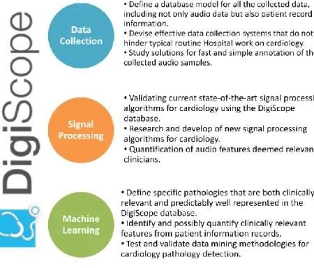
Figure 1 – The three areas of the DigiScope project and theirs tasks
The DigiScope project is structured into five functional tasks. Tasks 1 (Data Management) and 4 (Prototype) are essentially development tasks that will guarantee the viability of the final prototype. This thesis is essentially based on the work developed in these tasks. Tasks 2 (Audio Processing) and 3 (Data Mining) are the core research of the project. They are responsible for exploring novel ideas that will add new functionalities to the prototype. Task 5 (Clinical Validation) will make sure that the device is clinically useful and predict the conforming to the high clinical requirements of routine Hospital usage. All tasks constantly followed by clinical partners that provide consultation on the clinical usefulness of the targeted research objectives, and validate all resulting technology.
25 Introduction
Figure 2 - DigiScope logo
Expected key results of the project are:
Produce, deploy and evaluate a DigiScope prototype;
Design, implement and populate a massive clinically annotated database for research on this topic;
Publish all novel research in high-impact journals and conferences.
1.2.1. Audio Processing for Cardiology
The research has given a clear ideas regarding what has been accomplished in this topic so far. Based on this, three research lines have defined, that will be explored in this task:
Field validation of the robustness of published Heart Sound Segmentation algorithms. Although these results seem quite impressive (Liang, Lukkarinen et al. 1997; Liang and Hartimo 1998; Omran and Tayel 2003), the interest is in testing them in a real environment where circumstances are much harder to control (resisting patients, noisy environments) and understanding if they are robust enough for clinical routine;
Deeper exploration of Aortic Pulmonary Signal Decomposition. Based on current research, the interest is in improving the performance of these algorithms since they provide direct clinical features to a physician's examination. Furthermore, it‟s expectable to validate the relationship between the second heart sound signal and its associated pulmonary blood pressure, hopefully creating robust ways to screen the generic population for pulmonary hyper-pressure;
Extraction of relevant clinical features. By working directly with cardiologists, it is possible to gain a deeper understanding of what it is considered relevant in an examination. This will lead to the
Introduction 26
definition of new challenges for signal processing. Examples are the presence of a third heart sound, or the instability of some heart sound features (e.g. volume, duration, etc.). Furthermore, that will interact directly with the pathology detection task, for providing statistical features that might be interesting for data mining algorithms.
1.2.2. Pathology Detection
Following previous successful work done on extracting relevant information for other medical data such as mammographies, the DigiScope project intends to continue exploring better ways of reducing false positives in pathology screening algorithms. In previous work, elements of the DigiScope team applied inductive logic programming and Bayesian networks (Davis, Burnside et al. 2005) to predict malignant findings in mammograms. The approach resulted in two interesting achievement: First, the language used by inductive logic programming and Bayesian networks was well understood by medical professionals, validating the possibility of generating rules that make sense for clinical specialists, and secondly, interesting results were published in regarding advances in classification methods and in medical analysis of mammographies (Burnside, Davis et al. 2005; Davis, Burnside et al. 2005; Davis, Burnside et al. 2007).
The DigiScope team aims to continue on this successful track by applying the same principles of this previous research to heart pathology detection. The methods will be based on what has already been used for detecting malignant findings in mammographies, and on the improvement of the classification algorithms in order to give better diagnosis and prediction models. An example is to investigate how to better combine different classification approaches as evaluation functions, in the same way it was done in SAYU (Davis, Burnside et al. 2005). Another possibility is to investigate how the integration of medical reports with data produced by the digital stethoscopes can help uncover new relevant knowledge. Another track to follow is based on the work started by Ong et al. (Ong, Dutra et al. 2005) on better connecting related objects and paths to improve the quality of extracted knowledge, and the work of Salvini et al. (Salvini, Aguilar et al. 2007) on improving quality and efficiency of the classification algorithms.
27 Introduction
1.3.
Objective
The principal objective of this thesis, it is the presentation of a working prototype of a system capable of recording and annotating heart sounds, and his implementation and evaluation. To accomplish this goal, we have split this problem into two distinct tasks:
Data Collection and Management;
Prototype design, development, deployment and evaluation.
As we have explained previously, research on audio processing for cardiology has shown that it is essential to collect large amounts of data from real clinical situations, all of which must be manually annotated by cardiology specialists. Such a simple task becomes quite complex when confronted with the reality of current hospitals where information systems are complex and highly heterogeneous, or where clinicians have very busy schedules and cannot be hindered by non-transparent audio data collection systems. Furthermore, we can think of several smaller specific problems such as how to correlate extracted audio samples with stethoscope body position or how can clinicians perform fast manual annotations of such samples. As such, accomplishing the objective of creating a very large clinically annotated database for cardiology signal processing has required the following sub-tasks:
Define a database model for all the collected data, including not only audio data but also patient record information;
Design and develop a technological solution to collect audio samples;
Study solutions for fast and simple annotation of the collected audio samples.
After understanding how to collect and annotate data as transparently as possible, we need to build a DigiScope prototype that can integrate such knowledge with the automatic processing modules (Signal processing and Data mining) produced by the others members the DigiScope team. The accomplishment of this task has been divided in three parts:
Study and implement an user-centered interaction system for the clinician, which is both simple and effective;
Introduction 28
Creation of the software platform of the prototype, which can integrates the processing modules (Signal processing and Data mining).
29 State of art
2. State of art
The DigiScope project proposes to collect patient data but this objective presuppose two fundamental tasks:
Gathering clinical data directly from clinicians. That is considered one of the greatest challenges for the successful implementation of Electronic Health Records (EHR) (Dick, Steen et al. 1997).
Auscultate. The auscultation is a fundamental type of data for medicine and there is a growing awareness that a technological leap that allows its integration with EHR is now possible (Tavel 2006). In this state of art, we proposed to analyze the published literature about three fundamental topics:
Electronic stethoscopes;
Clinical Decision Support System (CDSS); Signal processing.
The first one, electronic stethoscopes, it is the most obvious, because it is the principal tool to auscultate and to record the heartbeat.
The second topic is the CDSS. We choose this one because the main objective of the DigiScope project it is to be a system that can help the physician to detect cardiac pathologies. In this topic, we approach also the machine learning because we consider them as part of the CDSS.
The last topic is the signal processing because it is a fundamental part of the project. Without the signal processing it is impossible to obtain results and consequently potential diagnosis suggestion.
State of art 30
2.1.
Electronic Stethoscopes
The stethoscope has a special place in medicine, being closely bound up with the doctor´s image. Since the invention of the first stethoscope by the French physician René Laennec (Necker hospital, Paris) in 1816, auscultation via a stethoscope is widely used by physicians as a simple, non-invasive and patient-friendly diagnostic method of chest diseases, where the sounds heard are correlated with the underlying pulmonary pathology (Andres, Brandt et al. 2008). It is one of the most simple and practical diagnostic tools used in medicine (Tavel 2006). Actually, two different types of stethoscopes are available on the market: acoustic and electronic. The main advantages of acoustic stethoscopes are their robustness and ergonomic designs. However, they are not ideal because they attenuate sound transmission proportional to frequency, their frequency response shows maxima and minima at very specific frequencies due to tubular resonance effects, and differences in the transmission properties are observed between different models (Ertel, Lawrence et al. 1969; Kindig, Beeson et al. 1982; Charbonneau and Sudraud 1985; Abella, Formolo et al. 1992).
Traditional stethoscopes depend solely on acoustics to amplify and transmit the heart sounds to the physician. The concept of electronic stethoscope arrived when electronic components were first used to amplify, filter and transmit the sound (Durand and Pibarot 1995). The US Military Aircraft Command recommended in 1966 that an electronic stethoscope be developed for use aboard aircraft because listening procedures were extremely difficult, if not impossible, with regular stethoscopes (Brogan, Collins et al. 1967).
We can thus think of digital stethoscopes as an evolution of the later, since we exploit the advantages of converting the audio signal to the digital domain, whether these are storage, transmission, analysis or simply visualization. Bredesen and Schmerle (Bredesen and Schmerler 1988) have patented an intelligent stethoscope designed for performing auscultation and for automatically diagnosing abnormalities by comparing digitized sounds to reference templates using a signature analysis technique. Several other electronic stethoscopes have been developed and described in literature (Tavel, Brown et al. 1994; Brusco and Nazeran 2005; Hedayioglu, Mattos et al. 2007).
There is, however, very little published data comparing conventional and electronic stethoscopes. An early study in 1998 comparing some of the more
31 State of art
primitive electronic stethoscopes with standard devices, concluded that the acoustic stethoscopes were preferable, however, they proposed that an ideal device would feature a combination of both (Grenier, Gagnon et al. 1998). More recently, a Norwegian study randomized third year medical students to either a traditional or an electronic stethoscope (Welch Allyn see Figure 3)and found no difference in terms of diagnostic accuracy when assessed by a cardiac auscultation test (Hoyte, Jensen et al. 2005). A Danish study comparing a standard stethoscope with a „cardiology‟ stethoscope also found no difference (Iversen, Sogaard Teisner et al. 2006). Experiences with electronic stethoscopes when teaching medical students has been a positive one based primarily on the ability to amplify sounds and reduce background noise. The ability to record the abnormal auscultatory findings and immediate facility to replay that sound has been particularly useful (Asghar, Alam et al. 2010).
Figure 3 - Welch Allyn Master Elite Electronic Stethoscope
Besides early attempts to use electronic stethoscopes for computer assisted decision, some studies have shown that the cardiac auscultation skills of undergraduate medical students were not negatively influenced by the use of an electronic sensor-based stethoscope (Hoyte, Jensen et al. 2005; Sverdrup, Jensen et al. 2010). This reinforces our belief that this learning could then benefit from a system that includes an electronic stethoscope, if an adequate interactive solution is researched, implemented and deployed (Hedayioglu, Coimbra et al. 2009).
State of art 32
We can conclude that there is scarce literature on this topic. The few comparative studies are few comprehensive, and show there are no diagnostic differences between acoustic and electronic stethoscopes. Here is an interesting opportunity to make a large comparison between acoustic and electronic stethoscopes, and publish a paper. Another interesting point is that studies show that there is no negative influence to the medical students and on the contrary that the electronic stethoscope can be a great learning tool. This is one of the tasks that we propose as future work. We can finally conclude that the electronic stethoscope offers possibilities such as recording, filtering, etc... that are not available to of traditional stethoscopes.
2.2.
Clinical decision support system
Before analyzing the importance of clinical decision support system, it‟s essential to define what a Clinical Decision Support System (CDSS) is. Studying the literature we see that there are different definitions proposed by various authors.
Wyatt defines a CDSS as an active knowledge systems which uses two or more items of patient data to generate case-specific advice (Wyatt and Spiegelhalter 1992). A computer program that provides reminders, advice or interpretation specific to a given patient at a particular time (Wyatt 2000).
Any mechanical, paper, or electronic aid that collects or processes data from an individual patient to generate output that aids clinical decisions during the doctor-patient encounter. Examples include decision support systems, paper or computer reminders and checklists, which are potentially useful tools in public health informatics, as well as other branches of medical informatics (Wyatt and Liu 2002).
Osheroff suggest that a clinical decision support is defined as providing clinicians or patients with computer-generated clinical knowledge and patient-related information, intelligently filtered or presented at appropriate times, to enhance patient care (Osheroff, Pifer et al. 2005).
CDSS had a high point in the seventies with the first experiments using Bayesian techniques and rules chaining. The results were promising. But expert systems stagnated, continued only as an object of academic study, but without
33 State of art
extensive practical use. One of the reasons given for this fact was the fear that the decision support software producers could be responsible for medical errors caused, even indirectly, by use, or misuse of systems. With the US tradition of insurance and significant compensation for medical errors, software vendors judged risky to take risks in this market. The subject seemed forgotten, but in recent years, interest in clinical support decision systems is being taken up by the path of integration with other tools of electronic medical records and greater coverage.
CDSS are increasingly important in primary care for the practice of evidence-based medicine and the development of shared general practitioner-patient decision making (Short, Frischer et al. 2004). But integrating computerized decision aids into routine care has been shown to be difficult (Eccles, McColl et al. 2002). A range of reasons have been identified. These include a reluctance by general practitioners to use systems because of limitations in their information technology skills and, consistent with previous research, difficulties in finding the time to use a support system in consultation (Sullivan and Mitchell 1995). Others factors include „„a lack of agreed national standards, a failure of systems to examine the needs of users adequately, and the profusion of different systems that do not communicate with each other‟‟ (Delaney, Fitzmaurice et al. 1999). Research specifically into the adoption of prognostic models in practice has proposed a lack of clinical credibility and uncertainty concerning the evidence as potential reasons (Wyatt and Altman 1995). But, may be the most important factor is the time. First, if systems are to be used in a consultation, designers must ensure that the system is practical within limited time available to general practitioners. Any guidance or information must be accessible to the user quickly and clearly. Given the importance of time, practitioners are less likely to use a system if the process to access the information is complex or time consuming (Short, Frischer et al. 2004).
After defining the factors that hinder the growth of CDSS, let us see what the main functions that they should have are. Clinical decision support systems are typically designed to integrate a medical knowledge base, patient data and an inference engine to generate case specific advice. Four key functions of electronic clinical decision support systems are outlined in (Perreault and Metzger 1999):
State of art 34
Administrative: Supporting clinical coding and documentation, authorization of procedures, and referrals;
Managing clinical complexity and details: Keeping patients on research and chemotherapy protocols; tracking orders, referrals follow-up, and preventive care;
Cost control: Monitoring medication orders; avoiding duplicate or unnecessary tests;
Decision support: Supporting clinical diagnosis and treatment plan processes; and promoting use of best practices, condition-specific guidelines, and population-based management.
It is important to show some of the most respected of diagnosis systems: DXplain: it‟s a decision support system which uses a set of clinical findings (signs, symptoms, laboratory data) to produce a ranked list of diagnoses which might explain (or be associated with) the clinical manifestations. DXplain provides justification for why each of these diseases might be considered, suggests what further clinical information would be useful to collect for each disease, and lists what clinical manifestations, if any, would be unusual or atypical for each of the specific diseases (Barnett, Cimino et al. 1987). DXplain includes 2,200 diseases and 5,000 symptoms in its knowledge base and was developed by Laboratory of Computer Science, Massachusetts General Hospital, and Harvard Medical School. Despite its usage in clinician training, similar to other clinical decision support systems, DXplain has not expanded beyond the research laboratory or medical training setting, due in part to a lack of support by clinicians in real-world settings (Coiera 2003).
QMR (Quick Medical Reference): A diagnostic decision-support system with a knowledge base of diseases, diagnoses, findings, disease associations and lab information. With information from the primary medical literature on almost 700 diseases and more than 5,000 symptoms, signs, and labs. Developed in 1980 by the University of Pittsburgh and First Databank, California. QMR was designed for 3 types of use:
as an electronic textbook;
as an intermediate level spreadsheet for the combination and exploration of simple diagnostic concepts;
35 State of art
Here are some of the most respected information systems laboratories: PUFF system: automatic interpretation of pulmonary function testing (OpenClinical 1983).
GermWatcher: police hospital infections, comparing national and local criteria (Doherty, Noirot et al. 2006).
PEIRS (Pathology Expert Interpretative Reporting System): interpret 80 to 100 laboratory tests per day, with a diagnostic accuracy of approximately 95% (OpenClinical 1991).
The studies reveal that the CDSS has grown because it has been integrated with other tools. The doctors are still reluctant to adopt the CDSS because it often perturb their routine work and are unreliable. To develop a CDSS, we will have to include the following factors: fast, user-friendly, quick to learn, intelligent and reliable. More importantly, do not disturb the routine of the physician. Finally we can take another conclusion is that the CDSS is inseparable from machine learning.
2.3.
Signal processing
Realization of the potential of computer-aided auscultation is supported by decades of research in heart sound analysis, clinical studies employing phonocardiography, and advances in signal processing methods applied to heart sounds.
A significant amount of literature discusses clinical phonocardiography, of which the early encyclopedic work of McKusick (McKusick 1958)serves as an outstanding example, and of which the works of Tavel (Tavel 1985),Leatham (Leatham 1975), and Harris (Harris, Sutton et al. 1976) are representative. These and numerous other studies have helped in identification of the spectral and temporal properties of heart sounds and murmurs, as well as their association with physiological events and diseases. Echocardiography has done much to illuminate and further clarify the process through which heart sounds and murmurs are generated, although problems remain to be solved and processes must be better understood.
State of art 36
Recent works in heart sound analysis have taken advantage of advanced digital signal processing methods to characterize heart sounds and murmurs (for reviews, see Durand (Durand and Pibarot 1995)Lin (Lin and Chen 1996) Obaidat (Obaidat 1993) and Rangayyan (Rangayyan and Lehner 1988)). Specifically, advanced time-frequency methods, especially wavelets, have been identified as having excellent properties for heart sound analysis (Khadra, Matalgah et al. 1991; Bulgrin and Rubal 1994) and have been applied to analysis of the first heart sound (Yoganathan, Gupta et al. 1976; Durand, Chen et al. 1997; Durand, Chen et al. 1997; Durand, Chen et al. 1997), the second heart sound (Yoganathan, Gupta et al. 1976; Xu, Durand et al. 2000), and murmurs (van Vollenhoven, van Rotterdam et al. 1969; Debiais, Durand et al. 1997; Debiais, Durand et al. 1997).
Advanced analysis of heart sounds and murmurs has provided a useful substrate for the design of algorithms for the automatic detection and identification of these sounds. Early approaches used linear classification methods (Iwata, Ogawa et al. 1979) and reported excellent clinical results in murmur detection (Rangaraj and Murthy 1979) and in distinguishing innocent from pathological murmurs (Ninova, Dascalov et al. 1978).
The signal processing is essential to obtain an efficient CDSS. There is significant amount of literature about this topic. Recent studies show significant results in the heartbeat characterization and murmurs detection. The investigation in this area will be fundamental to obtain good results with the DigiScope system. It will be essential to collect a maximum of auscultation to allow researchers to have a relevant database for the investigations in this area and also for the machine learning and CDSS.
37 DigiScope Collector system
3. DigiScope Collector system
3.1.
Requirements
Actually, according to the literature search conducted in the first phase of this investigation there is no system to collect and annotate cardiac and pulmonary auscultation to that presented in this research.
We present the following storyboard:
Storyboard part 1
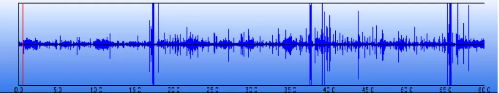
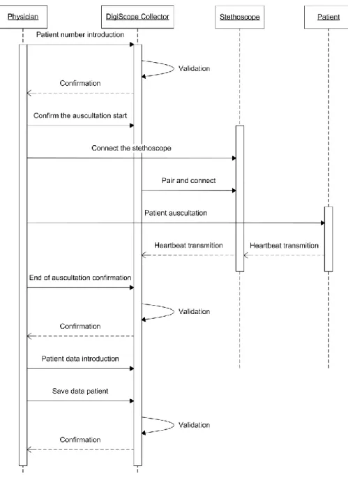
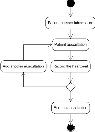
Miss Rosetta Stone came to a consultation with the cardiologist, Jean-François Champollion. The cardiologist used the DigiScope Collector to record all details of the consultation (including heartbeat). He measures the systemic pressure and using the Littmann 3200 stethoscope, the cardiologist auscultates the patient. With this system he can see in real time the heartbeat graph (Figure 4). The cardiologist introduces in the DigiScope Collector the systemic pressure of the patient.
DigiScope Collector system 38
Figure 5 - UML Sequence diagram of a patient auscultation using the DigiScope Collector and the Littmann 3200 stethoscope, and the introduction of patient data.
39 DigiScope Collector system
The cardiologist introduces the unique number of the patient, generally the internal process number of the hospital;
The system validates the number. It verifies if the number already exists in the existing process. If the process number exists a pop-up appears to inform the cardiologist.
The cardiologist press the button Begin to indicates to the system the begin of the auscultation. A popup asking to turn on the stethoscope and confirm if the Bluetooth is blinking. The cardiologist turn on the stethoscope and press the button OK. The system searches the stethoscope and when it founds, connects
with it.
A message appears on the stethoscope display asking to the cardiologist to press the M button of the stethoscope when he will begin the auscultation.
When the cardiologist press the M button, the stethoscope begins the transmission of heartbeat signal and the cardiologist can see a graph of the signal on the screen of the computer (Figure 4). After the auscultation, two buttons appears: Add another
auscultation and End auscultation. The cardiologist chooses to indicate the end of the auscultation.
The appendix 4 shows the part 1 of the storyboard illustrated with screenshots of the DigiScope Collector.
DigiScope Collector system 40
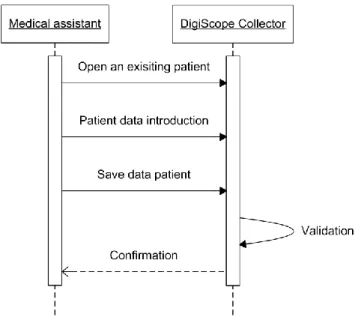
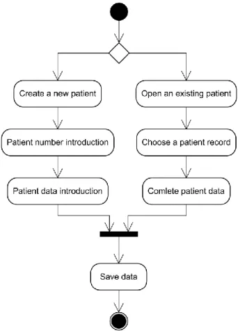
Storyboard part 2
Due to the high number of patients who have to attend, the cardiologist asks at his medical assistant to complete some basic data (birth, weight, height...) which can be found in the patient record.
Figure 6 - UML Sequence diagram of the introduction of patient data in the DigiScope Collector
The Figure 6 shows the sequence of actions of the part 2 of the storyboard:
The medical assistant in the initial menu, select he button Existing patient.
A table appears with the patient already created. The medical assistant can see for each patient: number id, name, numbers of auscultation recorded and the state of the patient data form (empty, incomplete or complete). The medical assistant selects the name of Rosetta Stone.
Using the internal information system of the hospital, the medical assistant introduces in the DigiScope Collector form the basic data (birth, weight, height...).
41 DigiScope Collector system
Finally she presses Save button. The system validates the data in the fields and confirms the existence of any anomaly.
The appendix 5 shows the part 2 of the storyboard illustrated with screenshots of the DigiScope Collector.
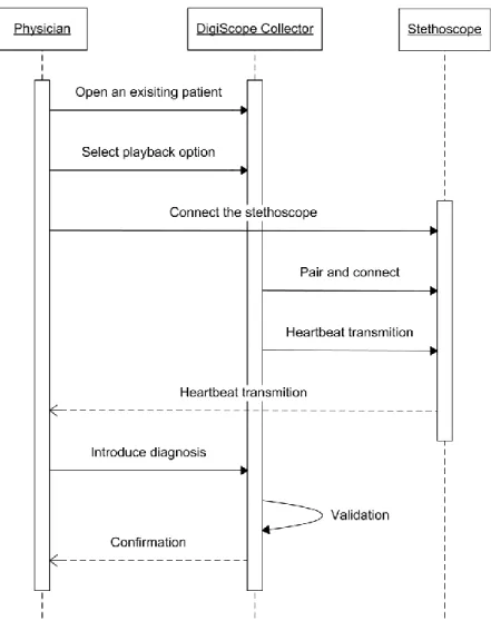
Storyboard part 3
At the end of the day, the cardiologist decides to review the case of Miss Rosetta Stones to confirm the diagnosis. He access at the patient form in the DigiScope Collector and decides to hear again the heartbeat using the stethoscope playback function. He confirms the previous diagnosis and records in the form the cardiac pathology (Arterial hypertension).
DigiScope Collector system 42
Figure 7 - UML Sequence diagram of the playback of an auscultation record with the DigiScope Collector and the Littmann 3200 stethoscope. And introduction of the
diagnosis
The Figure 7 shows the sequence of actions of the part 3 of the storyboard:
The medical assistant in the initial menu, select he button Existing patient.
A table appears with the patient already created. The cardiologist can see for each patient: number id, name, numbers of
43 DigiScope Collector system
auscultation recorded and the state of the patient data form (empty, incomplete or complete). He presses the name of Rosetta Stone. The form appears with some data introduced. The cardiologist presses the Playback button.
He is invited by the system to connect the stethoscope.
After the connection between the stethoscope and the system, a message appears in the display of the stethoscope inviting the cardiologist to press the M button when he is ready to listen the heartbeat recorded.
After the playback, the cardiologist presses the form button to introduce the diagnosis.
Finally he presses the Save button.
The appendix 6 shows the part 3 of the storyboard illustrated with screenshots of the DigiScope Collector.
DigiScope Collector system 44
3.2.
System architecture
3.2.1. System model
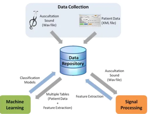
Figure 9 shows the main components of our system and their respective interactions. The system has four main components:
• Data Collection; • Signal Processing; • Machine Learning; • Data Repository.
Figure 9 - The DigiScope system model
The proposed system was designed to be implemented in hospitals and help physicians incorporate it in their routine work with minimal disruption. Some generic usability requirements were established (Norman 2007):
Minimize disruption - The system should be easy to use and the equipment should adapt well to the normal routine work of the
45 DigiScope Collector system
physician with minimum interference. It should also accommodate different ways to perform data collection, namely allowing the collection of patient data after each auscultation or after a set of auscultations;
Minimize errors - The flow of interactions should be adequately constrained in order to minimize the number of errors done by the physician and its associated reduction in data quality;
Easy to learn - The interface should be intuitive and guide the physician through the use of the application, reducing the adaptation time and difficulty to the system. This is essential for increasing the number of physicians that are willing to adopt and test this new technology.
User studies were performed in the two cooperating hospitals (Centro Hospitalar do Alto Ave, Guimarães, Portugal, and Real Hospital Português, Recife, Brazil). The objectives were:
Learn and model the auscultation procedure typically applied in hospitals;
Identify hospital environments where such a system is both adequate and useful;
Define a set of clinically useful and viable to annotate metadata to be associated with each auscultation.
Figure 10 - Images of the examination room at Centro Hospitalar do Alto Ave
Contextual studies methodologies were used, which mainly involved a combination of observation sessions and semi-structured interviews. As a
DigiScope Collector system 46
result, two hospital environments were selected, namely the Emergency Room and Primary Care to deploy the future DigiScope system for clinical decision support. The reasons for this choice are that the typical physician present is not a cardiologist (and can thus benefit from a system that can provide a first level of screening of cardiac pathologies), auscultation is performed in nearly every situation (it simply implies changing from a normal stethoscope to a digital one), there is a strong influx of new patients (maximizing the potential benefits of the screening process), and it is simple to deploy a tablet-type PC within enough communication range to receive the signals transmitted by a digital stethoscope. An environment that was discarded although an initual intuition might say otherwise was the Cardiology service. The main reason for this is that there is convincing evidence that it is theoretically possible to screen pathologies using signal processing and machine learning methodologies, namely the ones that cardiologists can identify but other physicians can‟t. We can argue that there are enough differences in the signal itself that allow for this distinct performance. We can‟t, however, say the same for pathologies that even cardiologists can‟t identify using auscultation alone.
3.2.2. Conceptual model
Two functional conceptual models were produced for the DigiScope Collector and can be observed in Figure 11 and Figure 12. User studies have shown us that the interaction mechanism must be very simple in order to be used within an Emergency Room. Very rarely the physician will have time to do anything besides its conventional auscultation, so we only require the bare minimum as additional effort. In this model, after starting the application, the physician simply needs to introduce a new patient (a number is exclusively assigned to each one). After that, the physician can start the auscultation using the digital stethoscope. This device is connected to a computer or computing base through wireless connection (Bluetooth). The heart sounds are then transmitted by streaming. The physician has the possibility to add more than one heartbeat record for each patient, using a simple button press in the stethoscope itself. After the auscultation is finished, the physician can start a session for a new patient or move to the annotation procedure, defined by the annotation conceptual model. Here, the physician can complete the patient‟s data, or this can be also done by a nurse or a medical assistant.
47 DigiScope Collector system
DigiScope Collector system 48
Figure 12 - UML Activity diagram of the data annotation model
3.2.3. Data model
Given the significant amount of data that can be captured each day by this prototype, it is important to define exactly what information to store and in which format. In the medical area, it is common to use specific terminologies when describing a certain sub-specialty (for example, in the area of breast cancer, the terminology is based on the BIRADS – Breast Imaging Reporting and Data System – lexicon (Radiology 2007)). We then define what patient metadata is interesting for pathology screening, and is viable to be annotated in this context. Contributions to this definition also came from the HL7 standard (Health Level Seven 2011) and openEHR publicly available archetypes (Foundation 2011).
49 DigiScope Collector system
We had the possibility to choose between saving the data in a database or in a XML file. Choosing a database provides a more traditional, fast to query and easy way to store data. XML is easier to use, offers an unparalleled portability and allows to read the file without any specific application. Bearing in mind that we anticipate the evolution of the system is easier using XML A new version of the DigiScope Collector can read older version of the XML, two XML versions can coexist. The choice of XML seemed to us the most pertinent.
Figure 13 shows the XML file created from the data patient entered on the form. It is strutured as follows:
The red tags represent the form tabs;
The blue tags correspond to each field or button of the form.; The values of the fields (elements) appears in black.
And respect theses rules:
If a field is not filled or a button is not selected, the tag appears like that: <Telesystolic />;
If a field or a button of the form can not be filled or selected, the
tag appears with a NA (Not Available):
<Protodiastolic>NA</Protodiastolic>.
All units of measurement were added to the name of the corresponding field in the XML tag, for example: <Weight-kg
>90</Weight-kg>. For each auscultation episode a XML is created with the correspondent
patient id. The XML file is stored in a folder named with the patient id. As for each patient there is only one XML file, this file is equaly named with the patient id.
DigiScope Collector system 50
<?xml version="1.0" encoding="UTF-8"?> <Patient id="12345">
<General>
<Name>John Doe</Name> <Weight-kg>90</Weight-kg> <Height-cm>200</Height-cm> <Day>12</Day>
<Month>10</Month> <Year>1979</Year> <Sex>Male</Sex>
<PressurePosition>Sit</PressurePosition> <AuscultationPosition>Sit</AuscultationPosition> </General>
<SystemicPressure>
<SystemicPressureMethod>Manometry</SystemicPressureMethod> <SystolicSystemicPressure-mmHg>145</SystolicSystemicPressure-mmHg> <DiastolicSystemicPressure-mmHg>60</DiastolicSystemicPressure-mmHg> </SystemicPressure>
<PulmonaryPressure>
<PulmonaryPressureMethod>Echocardiogram</PulmonaryPressureMethod> <SystolicPulmonaryPressure-mmHg>20</SystolicPulmonaryPressure-mmHg> <DiastolicPulmonaryPressure-mmHg>8</DiastolicPulmonaryPressure-mmHg> <CatheterismSimultaneousMeasurement> NA</CatheterismSimultaneousMeasurement> <EchocardiograSameConsultation>NA</EchocardiograSameConsultation> </PulmonaryPressure> <Murmur>
<Cycle>Systolic</Cycle> <Protosystolic />
<Mesosystolic>Yes</Mesosystolic> <Telesystolic /> <Holosystolic /> <Protodiastolic>NA</Protodiastolic> <Mesodiastolic>NA</Mesodiastolic> <Telediastolic>NA</Telediastolic> <Holodiastolic>NA</Holodiastolic> <Grading>3</Grading> </Murmur> <S1>
<S1Status>Normal</S1Status> </S1>
<S2>
<S2Status>Normal</S2Status> <IfAbnormal>NA</IfAbnormal>
<PulmonaryComponent>Normal</PulmonaryComponent> </S2> <S3> <S3Exist>No</S3Exist> </S3> <S4> <S4Exist>No</S4Exist> </S4> <Diagnosis>
<CardiacPathology>Yes</CardiacPathology> <PulmonaryHypertension />
<ArterialHypertension>Yes</ArterialHypertension> <ValvularAorticDisease>Yes</ValvularAorticDisease> <IntraventricularCommunication />
<OtherCardiacPathology /> </Diagnosis>
<FormStatus>
<StatusForm>Complete</StatusForm> </FormStatus>
</Patient>
51 DigiScope Collector system
However, being aware that for the next version of the system it will be necessary to use a server to keep the data. It becomes essential to use a database to the server. In Figure 14, we propose a database model. A strong emphasis was given to collecting data that might be helpful in the near future for machine learning research on cardiac pathology detection. We defined our data model with attributes that could be relevant to uncover new knowledge about:
The history of exams of patients (one patient can have several episodes of auscultation). This information can be useful to learn temporal diseases relations.
Differences between normal and abnormal cases (several attributes, in particular, the characteristic of the second heart sound (S2) can be very important do distinguish between normal and abnormal findings)
Multiple diseases for the same patient (multi-labeling (Ghamrawi and McCallum 2005)).
DigiScope Collector system 52
53 DigiScope Collector system
3.3.
Implementation
3.3.1. Prototype technology
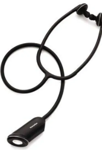
The electronic stethoscope chosen is a Littmann Model 3200. This stethoscope has three frequency response modes such as bell, diaphragm, and external range. The bell mode amplifies from 20 to 1000Hz, the diaphragm mode: amplifies from 20 to 2000Hz and the extended range mode amplifies sounds from 20 to 2000Hz. It can amplify the heart and lung sounds up to 24 times (2010). The LCD screen allows some information to be shown to the physician. This model can transmit signals wirelessly using Bluetooth technology. In fact, this was the only widespread commercial stethoscope that we could find with this transmission capability. Other available options did not have the traditional shape of conventional stethoscopes, which was considered essential for the first usability requirement defined in section 3.2.1 (minimize disruption). Figure 16 shows an image of the final version of the hardware prototype.
Figure 15 – Littmann 3200 stethoscope
The computer chosen is an Asus Eee PC with touch screen T101MT. This choice is motivated not only by the simplicity of a touch-based solution, but mainly by the fact that it has a rotating screen, effectively allowing the physician
DigiScope Collector system 54
to work in the two modes defined by the conceptual models. In the collection mode the tablet PC form is used (physician standing up, prototype on his hand), while the annotation mode allows the use of the keyboard (physician sitting at a desk, faster and more conformable typing).
Figure 16 - Image of the DigiScope Collector hardware prototype
As can be seen in Figure 17, the heart sounds are recorded in WAV format, and the patient data (see section 3.2.3) is saved in a XML format (Figure 13). The data are kept in a folder for each patient. At the end of each day, all the data is sent to a database on a secure server. The application was developed using Netbeans IDE 6.9.1 (Oracle 2010) on a Java platform version 6. The Bluecove API (2008) was used to establish the communication between the system and the stethoscope.
55 DigiScope Collector system
Figure 17 - The DigiScope Collector technologies
3.3.2. User interface
As motivated previously, our conceptual design is focused on the three generic usability requirements (Wyatt and Wright 1998) described in Section 3.2.2. The physician uses the system in two distinct phases.
First, when he is standing and executes the patient auscultation, he will use the electronic stethoscope and the computer in touch mode (Figure 11). During this phase, the proposed system suggests only two options (Figure 18), which can be selected using large buttons placed in the center of the screen, thus heavily constraining the user to simplify his understanding and reduce possible errors. The secondary options are proposed with smaller buttons and placed in the top of the screen.
DigiScope Collector system 56
Figure 18 - Screenshot showing the menu after the process number introduction
In a second phase (Figure 12), the physician is sitting and can complete patient data. This does not need to be done immediately after the auscultation and can also be partially performed by a nurse. The data form is clearly more complex than in the previous model, benefiting from the undivided attention of the user. Data fields are split in tabs: general, systemic pressure, pulmonary pressure, murmurs, S1, S2, S3 and S4, and Diagnosis. Some of these data fields can actually be completed before the auscultation itself. As essential function was thus the ability to only provide partial information, which can be completed at a later stage. During this phase (Figure 19 and Figure 20), typing errors are inevitable, we have used toggle buttons whenever possible to minimize them. For all the text fields, except the patient name, physicians can use a numeric virtual keyboard. For the patient name, an alphanumeric virtual keyboard is displayed, although this is clearly a sub-optimal choice, which is hidden whenever it is not needed. When the button “Save & Quit” is pressed, the fields are validated, that limit the numbers of errors. If an error is detected, for example an invalid date or letters in a numeric field, the tab where is localized the error is selected and the label field where is the error appears in red.
From the main menu, the physician can access a table which lists all patients who have already been introduced in the application. In this table he can see: