CHARACTERISATION OF THE OLIGOMERISATION-DEPENDENT
SIGNALLING FUNCTION OF THE LD DOMAIN OF THE TARP
VIRULENCE FACTOR FROM Chlamydia
by
Ana Celeste Teixeira Nogueira
CHARACTERISATION OF THE OLIGOMERISATION-DEPENDENT SIGNALLING FUNCTION OF THE LD DOMAIN OF THE TARP VIRULENCE
FACTOR FROM Chlamydia
Thesis presented to Escola Superior de Biotecnologia of the Universidade Católica
Portuguesa to fulfil the requirements of Master of Science degree in Applied
Microbiology
by
Ana Celeste Teixeira Nogueira
Place: Imperial College London
Supervision: Dr Rey Carabeo
iii
Resumo
Espécies do género Chlamydia são caracterizadas como bactérias intracelulares obrigatórias, Gram-negativas capazes de causar diversas doenças em humanos e animais. Estes patogénicos possuem um ciclo de vida bifásico que se inicia com o corpo elementar (CE), a forma extracelular, e metabolicamente inerte de Chlamydia, com a capacidade de induzir a sua internalização. Após a entrada na célula hospedeira, o CE diferencia-se em corpo reticulado (CR), forma replicativa e metabolicamente activa. O processo pelo qual o CE invade as células é dependente do efector secretado pelo sistema secretor tipo III, designado por TarP (de Translocated actin-recruiting phosphoprotein). Este efector tem a capacidade de reestruturar o citoesqueleto de actina por um mecanismo de sinalização promovendo a invasão bacteriana. Sendo FAK (de Focal Adhesion kinase) uma molécula importante na regulação da dinâmica de actina, investigou-se a existência de um possível mecanismo de activação e recrutamento de FAK, por Chlamydia sp.. Utilizando duas técnicas diferentes de indução de sinalização a partir do TarP e o dos seus derivados, mostrou-se que uma nova região nesta proteína tem a capacidade de recrutar FAK, vinculina, e Arp2/3, culminando no recrutamento de actina. Mostrou-se igualmente que o recrutamento destas moléculas é induzido com a oligomerização da nova região de TarP, designada de LD. Concluindo, no presente trabalho, descreve-se um novo mecanismo de sinalização utilizado por várias espécies de Chlamydia.
v
Abstract
Chlamydia species are Gram-negative obligate intracellular bacteria capable of
causing several diseases in humans and animals. These pathogens have a unique biphasic development cycle that initiates with the elementary body (EB), the extracellular and metabolically inert form capable of inducing its internalization. Once inside the host cell, the EBs differentiate into a different form, the reticulate body (RB), which are the replicative and metabolically active form. The process by which the EBs invade cells is dependent on a type III secretion system effector called TarP (Translocated actin-recruiting phosphoprotein). TarP is known to modulate actin cytoskeleton by a signalling mechanism to promote bacterial invasion. Since FAK (Focal Adhesion kinase) is an important molecule in the regulation of actin dynamics, it was hypothesised that Chlamydia species have a pathway to induce activation and recruitment of FAK. Using two different approaches of inducing signalling from TarP and its derivatives, we show that a new motif within TarP is capable of recruiting FAK, vinculin and Arp2/3 leading to actin recruitment. We presented, as well, that the recruitment of these molecules are enhanced upon oligomerisation of the LD domain. Altogether, we described a new signalling pathway used by several chlamydial species.
vii
Acknowledgements
I would like to give my sincere gratitude to my supervisor, Dr. Rey Carabeo, who gave me this amazing opportunity (that gave more work than expected, with the amount of paperwork needed) to belong to an ideal environment to learn. Also, I’d like to thank you for the guidance through my thesis, in special, the times I reached a brick wall.
Furthermore, I am grateful to all my colleagues in Carabeo’s group in CMMI. Specifically Tristan Thwaites for all the guidance in lab work, Denise Malcolm for the support every time it was needed and António T. Pedrosa for discussion and the sharing of protocols. Thank you all Carabeo’s group elements (in particular to António, Juli, Rahul, Rey and Tristan) for the fun moments outside work.
I’m grateful for being an ESB student because my training contributed for the foundation of my career. There, I met staff (in particular Mónica Coutinho and Drª Vânia Fernandes) and teachers who guide me through my graduation and master courses and left a mark on me. A special thank you to Prof. Drª Célia Manaia.
To my family and friends: Thank you so much! My sincere apologize for those I do not mention by name.
Um especial e carinhoso obrigada aos meus pais por toda a ajuda. Sem vocês este sonho não se teria concretizado. ―Mamacita‖, obrigada pelas longas conversas; os teus constantes conselhos e incentivos ajudaram-me sempre a enfrentar as dificuldades como desafios e a aproveitar esta experiência na sua totalidade. Sérgio, no one like you gives me the most tragic news like they are the best thing in the world. Thank you so much for the updating news that continuously contributes to see my best options in life. Teresa, I appreciate the care that you showed at the time of my departures and arrivals.
Joana, besides the distance, you were always present in the moments I needed a friend. Our conversations feel always like fresh air, a special thank you. Gafa, you are always there for me, thank you for everything.
António, you always helped me when everything became chaotic, making a stress-free environment around me. Your support and a familiar face in this new journey were precious. Thank you for the adventures we had in London.
ix
Contents
Page Resumo iii Abstract v Acknowledgements vii List of figures xiList of tables xiii
List of abbreviations xv
Introduction 1
Materials and Methods 7
Results and Discussion 14
I - Identification of specific TarP domain
responsible for FAK recruitment 14
II - Functional determination of LD domain
Oligomerisation 20 General conclusions 37 Future work 41 Appendix xvii Annex xix References xxi
xi
List of figures
Page 1. A schematic representation of the chlamydial TarP orthologs 3 2. EPEC system as a model to reproduce membrane-localised TarP
clustering 15
3. Phospho-active FAK and actin are recruited by Tarp from
C. caviae GPIC clustered at the plasma membrane 16 4. TarP orthologs of C. trachomatis serovar L2 and C. caviae GPIC 17
5. TarP LD domain is functional 18
6. TarP LD domain is functional 19
7. pHom-Mem1 vector as a model to replicate membrane-localised
TarP clustering 21
8. Model to replicate membrane-localised TarP clustering 21 9. Amplification of TARP inserts for cloning into pHom-Mem1 22 10. Analysis of plasmid digestion to confirm the functionality of SpeI 23
11. Colony PCR of transformants of pHom-Mem1 24
12. LD domain of TarP oligomerised in the membrane is sufficient to
recruit phospho-active FAK 26
13. B/B homodimerizer does not interfere with subcellular localisation
of phospho-active FAK 27
14. LD domain oligomerised recruits Arp2/3 complex. Cells
transfected with or without B/B treatment. 29
15. B/B homodimerizer does not interfere with subcellular localisation
of Arp2/3 complex 30
16. LD domain oligomerised recruits vinculin 32
17. B/B homodimerizer does not interfere with subcellular localisation
of vinculin 33
18. LD domain oligomerised specifically binds to the tips of actin
filaments 35
19. B/B homodimerizer does not interfere with subcellular localisation
xiii
List of tables
Page
Plasmids generated in this study 7
Primers used in PCR reactions in this study 8
xv
List of abbreviations
ABD Actin binding domain Abi1 Abl-interactor 1
Abl Abelson tyrosine-protein kinase Arp2/3 Actin-related protein 2/3
CFU Colony forming-unit
DMEM Dulbecco’s Modified Eagle Media DmrB Dimerization domain
DMSO Dimethyl Sulfoxide
EB Elementary body
E. coli Escherichia coli
EHEC Enterohaemorrhagic E. coli
EPEC Enteropathogenic Escherichia Coli EspFu EHEC Secreted Protein FU
FAT Focal Adhesion Targeting domain FAK Focal Adhesion Kinase
FBS Fetal bovine serum
Fyn Member of proto-oncogene tyrosine-protein kinase GPIC Guinea pig inclusion conjunctivitis
HA-tag Hemagglutinin epitope tag
IFN Interferon
IMDM Iscove’s Modified DMEM medium
L2 Chlamydia trachomatis serovar L2
LB Luria-Bertani
xvi
N-WASP Neural Wiskott-Aldrich syndrome protein NCK Cytoplasmic protein NCK1
MEK/ERK MEK/ Extracellular signal-regulated kinase PBS Phosphate buffered saline
PCR Polymerase chain reaction
PDGFR Platelet-derived growth factor receptor
PFA Paraformaldehyde
PIP3 Phosphor-inositol 3,4,5-P3 Rho Ras homolog (family)
RT Room temperature
SHC1 Src homology 2 domain-containing-transforming protein C1 SOB Super optimal broth
SOS-1 Son of sevenless homolog 1
Src Member of proto-oncogene tyrosine-protein kinase Syk Member of the Syk family of tyrosine kinases TarP Translocated Actin-recruiting Protein
Tir Translocated intimin receptor TTSS Type Three Secretion System
Tyr Tyrosine
Vav2 Guanine nucleotide exchange factor
WAVE-2 Wiskott-Aldrich syndrome protein family member 2
WH2 WASP homology domain 2
1
Introduction
Chlamydiae sp. are obligate intracellular bacterial pathogens of humans and animals,
and comprise of several species. They undergo a unique biphasic developmental cycle consisting of infectious and replicative forms, beginning with elementary bodies (EBs) which are the extracellular and metabolically inert forms. EBs have the ability to attach and invade susceptible cells making them responsible for dissemination of infection. The attachment to the host cell occurs in a two-stage process. The first stage involves a reversible electrostatic interaction between the bacteria and heparin sulphate-containing glycosaminoglycans present on the host cell surface (Zhang and Stephens, 1992). The second, irreversible, stage occurs upon interaction between the EB and an unidentified receptor (Carabeo and Hackstadt, 2001). The entry of bacteria involves the recruitment of actin leading to pedestal formation at the site of attachment (Carabeo et al., 2002; Carabeo et al., 2004). Once inside the host cell within a membrane-bound vacuole called an inclusion, EBs differentiate into reticulate bodies (RBs), which are the metabolically active and replicative, but non-infectious forms. This primary differentiation is followed by repeated cycles of binary fission, which is followed a few hours later by the secondary differentiation back to EBs. At the end of the cycle, the host cell lyses freeing the EBs to infect neighbouring cells. Inducers such IFN-gamma, iron starvation, or antibiotic treatment can lead to persistence and/or abnormal growth distinguished by enlarged and pleomorphic morphology. These abnormal RBs undergo neither binary fission nor differentiation to EBs, but still maintain, to some extent the replication of their genomes (AbdelRahman and Belland, 2005).
The taxonomy of Chlamydiae has experienced numerous revisions (Everett et al., 1999) relying on 16S rRNA gene phylogenetic to distinguish Chlamydia from other bacterial species (Stephens et al., 2009). The most recent revision recognises nine species within the
Chlamydia genus: Chlamydia trachomatis (C. trachomatis), Chlamydia muridarum (C. muridarum), Chlamydia pneumoniae (C. pneumoniae), Chlamydia abortus (C. abortus), Chlamydia abortus (C. suis), Chlamydia felis (C. felis), Chlamydia psittaci (C. psittaci), Chlamydia caviae (C. caviae), and Chlamydia pecorum (C. pecorum) (Marsh et al., 2011).
The most studied species are C. caviae, C. pneumoniae, and C. trachomatis, where the last two are responsible for the majority of human Chlamydia infections.
2
C. caviae (GPIC) causes inclusion conjunctivitis in guinea pigs and respiratory
infection in newborn animals (Frazer et al., 2012). In humans, C. pneumoniae is a respiratory pathogen that causes acute respiratory diseases like pneumonia, bronchitis, and sinusitis (Campbell et al., 2002; CDC, 2005).
The species C. trachomatis is the causative agent of several distinct human diseases. Depending on the serovar, C. trachomatis can cause blinding trachoma (serovars A-C), sexually transmitted diseases (serovars D-K) and lymphogranuloma venereum (serovars L1, L2 and L3), which are also sexually transmitted (Schachter, 1999). C. trachomatis is one of the most common bacterial sexually transmitted infections (Low, 2004) having around 92 million of cases estimated the world, in 1999 (WHO, 2001). In USA, there are around 2,8 million new cases of Chlamydia each year (Meyers et al. 2007). These numbers are increasing in many European countries and the USA in part due to the increased testing and the use of more sensitive tests for diagnosis. Also, these numbers are likely to be higher as the majority (~70 %) of infections remain asymptomatic. This makes Chlamydia a serious public health as individuals spread it unsuspectingly, and if left untreated can have long-term serious consequences such as infertility in women (Cates et al., 1991).
A key virulence factor of pathogenic Gram-negative bacteria is the Type III Secretion System (TTSS) enabling the bacteria to modulate host cell functions such as reorganizing the host cytoskeleton, modulating immune signalling or apoptosis by injecting effector proteins directly into the host cell (Hueck, 1998; Cornelis and Gijsegem, 2000). Interestingly, Matsumoto (1981) observed the presence of protruding structures on the surface of EBs, which is currently suspected as the TTSS apparatuses. Based on homology, it was possible to identify putative chlamydial TTSS components (Kim, 2001; Betts et al., 2008; Arnold et al., 2009; Samudrala et al., 2009; Stone et al., 2008; Markham et al., 2009). However, since
Chlamydia species are genetically intractable it has been a challenge to confirm and study the
functions of such proteins. The understanding of the structure and protein secretion of the
Chlamydia TTSS derives from recombinant chlamydial proteins studied in the context of
heterologous TTSS of other bacteria, such as Yersinia, E. coli and Salmonella (Subtil et al., 2001; Ho et al., 2005). One of the proteins discovered to be secreted by a type III-dependent mechanism into the host cell was CT456 or TarP (Translocated actin-recruiting phosphoprotein), which is pre-synthesised and found in EBs associated with its chaperone for immediate translocation (Brinkworth et al., 2011).
3
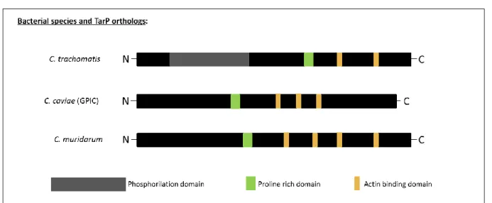
TarP orthologs are present in all Chlamydia species examined to the date (Jewett et al., 2010). TarP harbours at least three functionally distinct domains (Figure 1) - the N-terminal tyrosine rich repeat domain (also named phosphorylation domain or phosphodomain) (Clifton
et al., 2004), the actin binding domain (ABD) that is capable of nucleating actin in vitro
(Jewett et al., 2006; Jewett et al., 2010) and a proline rich domain essential for TarP oligomerisation (Jewett et al., 2010). However, the alignment analysis of the orthologs from
C. trachomatis serovars L2, D and A, C. muridarium, C. pneumoniae and C. caviae indicated that only Chlamydia trachomatis orthologs possessed the tyrosine-rich domain. Besides, additional studies showed that it is also the only domain in which phosphorylation occurs upon entry of EBs. While the tyrosine-rich domain may be necessary for C. trachomatis invasion (Lane et al., 2008), this mechanism of entry does not account for chlamydial species that lack this domain in their TarP ortholog (Clifton et al., 2004; Clifton et al., 2005; Jewett et
al., 2008; Jewett et al., 2010). Thus, there must be an alternative means of invading cells
utilised by the non-trachomatis species.
Figure 1: A schematic representation of the chlamydial TarP orthologs.
After the irreversible binding to the host cell, Chlamydia trachomatis translocates TarP across the host membrane via the TTSS where it is tyrosine phosphorylated (Carabeo et
al., 2004) by host cell tyrosine kinases, such as Src, Yes, Fyn, Syk and Ab1-1 kinase (Elwell et al., 2008; Jewett et al., 2008; Mehlitz et al., 2008). This phosphorylation of the
tyrosine-rich repeat induces the recruitment of guanine nucleotide exchange factors Sos-1 and Vav-2 (Carabeo et al., 2007; Lane et al., 2008), which form a complex with Abi-1 and PIP3
4
(phosphatidylinositol 3,4,5-P3), respectively to activate Rac (Lane et al. 2008). Rac induces actin recruitment in an Arp2/3-dependent manner via the activation of the WAVE-2/Abi-1 complex leading to actin polymerization and pedestal formation (Lane et al., 2008). TarP also has an invasion-independent function. It has been shown by Mehlitz et al. (2010) that TarP phosphorylation up regulates the host cell MEK/ERK pathway so activates the protein SHC1. This pathway is important for host cell survival, and its activation by the effector TarP prevents host cell apoptosis. The induction of anti-apoptotic mechanisms in the host by TarP soon after infection is necessary because the chlamydial EB becomes irreversibly committed to continue with the infection.
As mentioned above, TarP can induce actin remodelling through signalling from the phosphodomain, or the actin nucleating function of the WH2-like actin-binding domain. The actin-binding domain (Figure 1) is a domain contained within the C-terminal region of the TarP protein. The comparison of C. trachomatis TarP orthologs with three classes of known eukaryotic actin nucleation factors (such as Arp2/3, formins and Drosophila Spire protein) shows that TarP is a distinct and novel actin nucleator. Other than 9-aa sequence of actin binding domain of WH2-family proteins, TarP does not shown any similarity to known eukaryotic actin nucleators (Jewett et al., 2006). Actin nucleation in eukaryotes brings together three or four monomers of actin, a process that is thermodynamically unfavourable. This thermodynamic barrier can be overcome through the actions of protein nucleators. Their configuration involves either tandem repeats of WH2-like actin binding sites or the oligomerisation of multiple units of an actin-binding protein. The desired result is the formation of stable nucleating actin trimer or tetramer. Once this is established, subsequent addition of monomer to the growing actin filament becomes thermodynamically favourable, making the nucleator unnecessary at these later stages. TarP as a nucleator has the capacity to induce the formation of long unbranched actin filaments, does not require pre-existing actin filaments and does not require host factors like Arp2/3 complex; TarP binds monomeric actin in the absence of host proteins whereas formin does not. In contrast with the Drosophila Spire protein, TarP requires only one actin-binding domain to promote nucleation of actin filaments, provided that it remains capable of oligomerisation through the proline-rich domain (Jewett et al., 2006). Also, TarP of some Chlamydia strains and species contain more than one actin binding site arranged in tandem. This may function to bind multiple actin monomers simultaneously to a single peptide (TarP) (Figure 1). In this configuration, it was determined that the oligomerising proline-rich domain was dispensable (Jewett et al., 2010). Whilst
5
being a potent nucleator of actin polymerization, the actin-binding domain does not appear to account for the full invasion mechanism of non-trachomatis species. C. caviae and C.
pneumoniae, for example, still required the activation of signalling pathways to invade, in
spite of the presence of ABD function (Subtil et al. 2004, for C. caviae; Coombes and Mahony, 2002, for C. pneumoniae).
As mentioned before, due to the lack of the tyrosine-rich domain in the N-terminal of TarP in other Chlamydia species than C. trachomatis, alternative signalling pathways for the modulation of actin by other species of Chlamydia were suggested. Coombes and Mahony (2002) showed that rapid phosphorylation of Focal Adhesion kinase (FAK) occurred during
C. pneumoniae invasion. Also, it was shown, in vitro, that the depletion of FAK, as well the
downstream signalling components, decreased the infection of C. trachomatis serovar L2 (Elwell et al., 2008). The basis of FAK activation and functionality in Chlamydia invasion is still poorly understood. Because of the set of circumstantial evidence that points to a role in chlamydial infection, and its potent ability to induce actin remodelling at focal adhesion sites, FAK may indeed be involved in invasion.
FAK is a 125-kDa protein that plays a major role in different cellular functions such as adhesion, motility, survival, proliferation and cell cycle (reviewed in Parsons et al., 2000). FAK has several binding partners in the N-terminal, Central and terminal domains. The C-terminal domain of FAK (853-1012 a.a.) called FAT (Focal adhesion targeting domain) domain is necessary for targeting FAK to focal adhesion complexes by binding with different integrin-associated proteins such as paxillin and talin (Hildebrand et al., 1993; Tachibana et
al., 1995; Chen et al., 1995; reviewed in Hayashi et al., 2002).
Paxillin interacts with the FAT domain of FAK through a series of leucine-rich repeats named LD motifs present with the sequence L(D/E)xLLxxL (Brown et al. 1996; Brown et al., 1998). The activation of FAK by integrins clustering leads to the regulation of the actin cytoskeleton, particularly by Rho family GTPases (such as Rac1, Cdc42 and Rho) (Parsons et
al., 2000; Schaller, 2010). Therefore, if FAK is indeed involved, we should expect it to be
present at the sites of chlamydial entry, and such a colocalisation with invading EBs would imply interaction with TarP.
Despite the observation of phosphorylation, and presumably activation of the host Focal Adhesion kinase (FAK), upon bacterial invasion, its role in chlamydial invasion was not thoroughly explored. Given its potent ability to signal to and remodel the actin
6
cytoskeleton, we hypothesised that FAK could be involved in invasion through the action of TarP. Questions we would like to address include i) Is TarP involved in FAK recruitment; ii) What domain of TarP is required for FAK recruitment; iii) What effects on signalling do oligomerisation confer to TarP.
7
Materials and Methods
Bacterial strains
Chemically competent Escherichia coli (E. coli) Top10 were used for plasmid propagation and stocked in Luria-Bertani (LB) Broth containing 30 % of glycerol at –80 ºC. Initial overnight cultures were made in LB medium with the appropriate antibiotics (100 μg/ml of Penicillin) and incubated at 37 ºC with agitation (220 rpm).
Enteropathogenic E. coli (EPEC) Δtir and EPEC ΔescN (Garmendia et al., 2004) were grown in Luria-Bertani (LB) broth supplemented with kanamycin (50 µg/ml) in a shaking incubator at 37 °C.
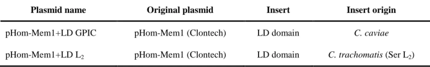
Plasmids
Plasmids generated in this study are listed in Table 1.
Table 1: Plasmids generated in this study
Plasmid name Original plasmid Insert Insert origin
pHom-Mem1+LD GPIC pHom-Mem1 (Clontech) LD domain C. caviae
pHom-Mem1+LD L2 pHom-Mem1 (Clontech) LD domain C. trachomatis (Ser L2)
Polymerase Chain Reaction (PCR)
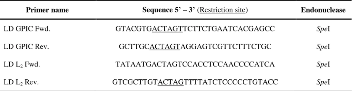
The primers used for the PCR reactions are listed in Table 2.
For cloning, different domains from Tarp (LD domain from C. trachomatis GPIC and
C. trachomatis L2) were obtained using DNA polymerase with proof reading Accuprime Pfx polymerase supermix (Invitrogen Life Technologies). The temperature used for the PCR cycle followed the instructions provided by the manufacturer. The cycle used for polymerase activation was 95 ºC, for 5 minutes, followed by 35 cycles with the following steps: denaturation at 95 ºC for 15 seconds, annealing at lowest melting temperature primer (calculated using Integrated DNA Technologies) minus 5 ºC for 30 seconds and elongation
8
phase at 68 ºC, 1 minute per kilobase (kb). After the 35 cycles were completed a final extension step was added, 68 ºC for 5 minutes, to prevent incomplete DNA sequences.
The PCR fragments were analysed by electrophoresis on 1,2 % agarose gel and SYBR Safe (Invitrogen, Life Technology) staining was performed in order to visualise the DNA fragments obtained.
To confirm the presence of positive colonies containing the different domains of TarP in pHom-Mem1 (Clontech), PCR assays were performed according to the instructions provided by the manufacturer for the PCR supermix (Invitrogen Life Technologies). The conditions used were polymerase activation at 95 ºC for 5 minutes followed by 35 cycles with the following steps: denaturation at 95 ºC for 20 seconds, annealing at lowest melting temperature primer (calculated using Integrated DNA Technologies) minus 5 ºC for 30 seconds and elongation phase at 72 ºC, 1 minute per kb. After the 31 cycles were completed a final extension set was added, 72 ºC for 5 minute, to prevent incomplete DNA sequences. The PCR fragments obtained were analysed as previously described.
Table 2: Primers used in PCR reactions in this study
Primer name Sequence 5’ – 3’ (Restriction site) Endonuclease
LD GPIC Fwd. GTACGTGACTAGTTCTTCTGAATCACGAGCC SpeI
LD GPIC Rev. GCTTGCACTAGTAGGAGTCGTTCTTTCTGC SpeI
LD L2 Fwd. TATAATGACTAGTCCACCTCCAACCCCATCA SpeI
LD L2 Rev. GTCGCTTGTACTAGTTTTATCTCCCCCTGTACC SpeI
PCR Fragments Purification or Extraction Kit
Purification of PCR DNA products obtained were achieved using QIAquick Kit (Qiagen) following the manufacturers recommendation in ―QIAquick PCR Purification Kit Protocol‖ or ―QIAquick Gel Extraction Kit Protocol‖ depending on whether the PCR product were purified directly or by gel extraction, respectively.
9
Plasmids purification
Plasmid DNA was purified from E. coli Top10 genetically modified strains using Qiaprep Spin Miniprep Kit, following manufactures instructions (Qiagen).
Restriction Enzyme Digestion
Restriction enzyme digestion was performed using 4 μl of NEB4 buffer 10x (New England Biolabs), 4 μl of BSA 10x, 4 μl of SpeI restriction enzyme and 28 μl of PCR insert or plasmid in a final volume of 40 μl. Restriction enzyme reaction was performed at 37 ºC for 2 hours, followed by 10 minutes at 65 ºC for deactivation of the enzymes. A small amount of cut plasmid and uncut were analysed using electrophoreses in a 1,2 % agarose gel to guarantee the functionality of the restriction enzymes.
Determination of Plasmids and Insert Concentration
DNA concentration was measured on NanoDrop UV-Vis spectrophotometer (NanoDrop®), using 2 μl of each sample (insert or plasmid).
Ligation
The ligation between the insert and the vector was made using T4 ligase (New England Biolabs). The ratio used was 50 ng of vector with 3-fold molar excess of insert. The volume was adjusted to 10 μl of water. To achieve ligation, 10 μl of 2X Quick Ligation buffer and 1 μl of Quick T4 Ligase enzyme were used. The reaction occurred for 5 minutes at room temperature (RT).
Bacterial transformation
Preparation of chemically competent E.coli Top10
Wild type strains of E. coli Top10 were inoculated, in 5 ml of Super Optimal Broth (SOB) medium (5 g of NaCl, 20 g of tryptone, 5 g yeast extract, 2,5 ml of 1 M KCl solution in 1 l of double distilled water (ddH2O) and incubated overnight at 18 ºC with agitation (200 rpm). The following day OD600 was measured, when it reached a value between 0,4-0,45 the
10
cultures were put on ice. Using pre-cooled 50 ml falcon tubes the cultures were centrifuge at 4000 rpm for 30 minutes at 4 ºC. Afterwards the supernatant was discard and the pellet was resuspended in 15 ml of ice-cold Inoue solution (10,9 g of MnCl2, 2,2 g of CaCl2, 18,7 g of KCl, 20 ml of 0,5 piperazine-1,2-bis-ethanesulfonic acid, in 1 l of ddH2O). Repeat the centrifugation; discard the supernatant and resuspension of the pellet in 20 ml of ice-cold Inoue Solution, twice. Without discarding the supernatant 1,5 ml of dimethyl sulfoxide (DMSO) was added to the 20 ml of cell suspension and incubated on ice for 10 min. The cell suspension was then divided in 100 μl aliquots in precooled, sterile 1,5 ml eppendorfs, and stored at -80 ºC until used.
Heat shock transformation
DNA (between 1 and 10 μl of insert-plasmid ligation product) was mixed with 100 μl of chemically competent bacterial cells and homogenized gently before incubating on ice for 30 minutes. A heat-shock was performed for 45 seconds at 42 ºC followed by 10 minutes on ice. Bacteria were then incubated with 500 μl of SOC medium, at 37 ºC with agitation (200 rpm) for 1 hour and 30 minutes. 100 μl of bacterial suspension was plated in LB agar with the appropriate antibiotic and incubated overnight at 37 ºC.
Tissue culture and transfection
Cos7 (ATCC) cells were cultivated in a humidified 5 % CO2 incubator at 37 °C in Iscove’s Modified DMEM medium (IMDM; Invitrogen) supplemented with 10 % fetal bovine serum (FBS) and L-glutamine (2 mM). Transfections were carried out following manufacturer’s instructions using Lipofectamine 2000 (Invitrogen). Cells were grown in 24-well to 70 – 80 % confluence. Transfections were always preceded at 37 ºC in a humidified incubator.
iDimerizeTM Inducible Homodimer System (Clontech)
Cos7 cells were transfected with 0,1 ng of pHom-Mem1 fused to GPIC LD or L2 LD using Lipofectamine 2000 (Invitrogen). Transfected cells were incubated at 37 ºC in a humidified incubator for 3 hours, washed twice with Phosphate buffered saline (PBS) and left
11
in supplemented IMDM for up to 11 hours. Cells were left overnight in cycloheximide (10µg/ml) to control expression levels of protein. To induce homodimerisation, IMDM containing cycloheximide (100µg/ml) and the B/B homodimerizer reagent (Clontech) was added to cells and incubated at 37 ºC in a humidified incubator.
Cells were rinsed three times with PBS and fixed with 4 % paraformaldehyde (PFA) for 15 minutes at room temperature. The fixed cells were rinsed with PBS and permeabilised with 0,2 % Triton X-100 in PBS for 5 minutes at RT (Room Temperature) with agitation. After three washes with PBS, cells were incubated with primary antibodies. Cells were rinsed 3 times with PBS and incubated with appropriate Alexa Fluor conjugated secondary
antibodies (Invitrogen).
Optimisation of the oligomerisation assay
After the constructs have been made and confirmed by nucleotide sequencing, they were used for their ability to localise to the plasma membrane and oligomerise in response to the addition of the B/B homodimerizer. It was important that we determined the level of sensitivity of each construct to oligomerisation to ensure that we achieved consistent recruitment of specific host signalling molecules. For that reason, the B/B homodimerizer was not kept constant through the different experiments because the responsiveness, as indicated by the recruitment of specific proteins, of each specific domain with regards to oligomerisation efficiency would have been different. In addition, we also had to account for the potential differences in the efficiency of detection of recruited signalling molecules by various antibodies. In other words, it was possible that recruitment may have occurred at similar levels of oligomerisation, but because of different antibodies were used, which may have had different affinities for their respective targets, the B/B concentrations were needed to be adjusted to compensate for any differences in detection efficiency. Also, untransfected cells were treated with B/B and compared with cells not treated with B/B to determine if unwanted effects of the dimerizer on the host cell, independent of the expression of the various fusion proteins, were present.
The different constructs (LD from GPIC and LD from L2) of TarP have already been demonstrated to be functional in the EPEC TirMC system (Thwaites et al., submitted). The constructs were transiently transfected and tested for dimerisation in response to B/B
12
Homodimerizer. Medium containing between 0,5 and 100 nM of B/B Homodimerizer was added to transfected cells in order to induce oligomerisation. Cos7 cells were chosen for these experiments as they are ideal for microscopy studies, and also provide ample surface area on the plasma membrane, where we expect the fusion proteins to be targeted and to form oligomeric patches.
One problem encountered was the overexpression of the fusion proteins, which resulted in insoluble aggregates, which at times confounded image analysis. To overcome these, we needed to limit the amount of fusion proteins expressed by inhibiting host translation with cycloheximide.
Confocal microscopy
Cos7 cells were transfected with 0,1 ng of pHom-Mem1 fused to GPIC LD or L2 LD or TirMC/TarP or TirMC/LD plasmid DNA using Lipofectamine 2000 (Invitrogen).
Cells grown on coverslips were rinsed three times with PBS and fixed with 4 % PFA for 15 minutes at room temperature. The fixed cells were rinsed with PBS and permeabilised with 0,2 % Triton X-100 in PBS for 5 minutes at RT with agitation. After three washes with PBS, cells were incubated with anti-HA.11 clone 16B12 1:500 (Covance) and anti-phospho-active FAK 1:500 (abcam), anti-vinculin 1:250 (Invitrogen) or anti-p34-Arc\Arpc2 1:500 (Millipore) primary antibodies; DAPI (1:1000) was used to stain DNA and actin was detected with phalloidin antibody (1:250 dilution) conjugated to Alexa Fluor 488 dye (Invitrogen). Cells were rinsed 3 times with PBS and incubated with appropriate Alexa Fluor conjugated
secondary antibodies (Invitrogen).
Coverslips were mounted onto microscope glass slides using mowiol. Sample visualisation was performed at RT on a laser scanning microscope (LSM 510; Carl Zeiss). Images were processed using Zeiss LSM Image Browser Version 4.2.0.121.
13 Table 3: Antibodies used in this study
Antibody Description Target Host Manufacturer
Anti-HA-tag HA.11 Clone 16B12
Monoclonal Antibody, purified HA-tag Mouse Covance Anti-phospho-active FAK Polyclonal to FAK (phospho
Y397), purified
Phospho-FAK Rabbit abcam Anti-vinculin Monoclonal antibody, purified Vinculin Rabbit Invitrogen Anti-p34-Arc\Arpc2 Polyclonal Antibody Purified
anti-p34-Arc\Arpc2 Rabbit
Upstate (Millipore) DAPI Blue-fluorescent DAPI nucleic
acid stain dsDNA - Invitrogen
Alexa Fluor 488 Phalloidin Probe conjugated Alexa Fluor®
488 dye. F-actin - Invitrogen
Alexa Fluor 594 goat anti— mouse IgG
Red-fluorescent Alexa Fluor 594 dye
Anti-mousse
antibody Goat Invitrogen Alexa Fluor 488 goat anti—
rabbit IgG
Green-fluorescent Alexa Fluor 488 dye
Anti-rabbit
antibody Goat Invitrogen
EPEC infection conditions
Cells grown in coverslips were transfected with 0,1 µg of TirMC/TarP or TirMC/LD plasmid DNA using Lipofectamine 2000 and were incubated at 37 ºC in a humidified incubator for 32 hours prior to infection. EPEC cultures were primed for infection by diluting the overnight bacterial culture with pre-warmed low glucose (1,000 mg/l) Dulbecco’s minimal Eagle medium (DMEM) (Sigma), in a dilution of 1:100, and incubated as static cultures at 37 °C in 5 % CO2 for 2 hours. The infected cells were washed with cold PBS to remove unattached EPEC. Gentamycin (200 µg/ml) was added to media to prevent the growth of adherent EPEC and the infection was allowed to continue for an additional 4 hours at 37 ºC, followed by three washes with PBS and PFA fixation. Then, cells were prepared for confocal microscopy like describe before.
14
Results and Discussion
Previous evidence highlighted a possible role of FAK in Chlamydia invasion due to its rapid phosphorylation during C. pneumoniae entry and depletion of this protein and downstream signalling components leading to a decrease in invasion in C. trachomatis serovar L2 (Coombes and Mahony, 2002; Elwell et al., 2008). However, this mechanism is still poorly understood. To understand the role of FAK in chlamydial invasion, Thwaites et al. (submitted) investigated the localisation of phospho-active FAK in HeLa cells upon infection with C. caviae GPIC or C. trachomatis serovar L2, showing that phospho-active FAK was recruited. These results led to several experiments to understand the role of FAK in
Chlamydia species invasion using confocal microscopy, which allows us to observe direct
localisation between FAK and its recruiting protein.
I - Identification of specific TarP domain responsible for FAK recruitment
TarP alone is sufficient to recruit phospho-FAK
Focal adhesion kinase was implicated in Chlamydia infection of cultured cells in an RNA interference screen (Elwell, et al. 2008). Furthermore, FAK was activated very early in
C. pneumoniae invasion, indicating that this event could be associated with the invasion
process (Coombes and Mahony, 2002). Since translocation of TarP is a critical event that mediates the uptake of Chlamydia by its ability to induce signalling to the actin remodelling machinery, we attempted to identify if TarP was capable of recruiting phospho-FAK.
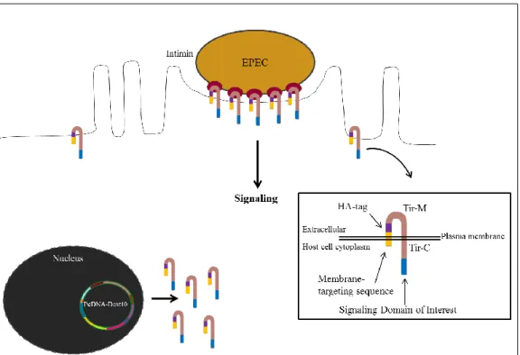
Using the EPEC (Δtir or ΔescN) Tir/MC system (Figure 2), a TarP ortholog was cloned with the transmembrane domain of Tir (Translocated intimin receptor) (Figure 2), which enabled us to replicate the usual membrane-localised clustering of TarP in the host membrane (Thwaites et al., submitted). Additionally, the use of this experimental system allowed an insight into the signalling role of TarP in isolation, overcoming the difficulty in mutating an obligate intracellular bacterium. Also, using a Δtir or ΔescN EPEC mutant strains ensured that the observed signalling could only be attributed to TirMC/TarP and not to EPEC type III effectors. In these strains the TTSS function related to actin recruitment was
15
deactivated either through deletion of the required ATPase (EscN), or the signalling molecule Tir. The dysfunctional TTSS in the ΔescN background necessitated the expression of the TirMC/TarP in the host cell itself, instead of depending on a translocation mechanism. To target the fusion proteins to the membrane efficiently, a membrane-targeting motif derived from a Newcastle Disease Virus glycoprotein was included in the constructs. The TirMC parental vector used to transfect cells to localise the TirMC on the eukaryotic cells was obtained with permission from Prof. John Leong (Tufts University Medical School) (Campellone et al. 2004) and adapted to Gateway cloning (Thwaites et al., submitted) (See vector diagram of pcDNA-Dest40 in Annex).
Figure 2: EPEC system as a model to reproduce membrane-localised TarP clustering. Schematic representation of the enteropathogenic E. coli (EPEC) heterologous system used to study the recruitment of signalling molecules by TarP. A signalling domain of interest is fused to TirMC. The fused protein is directed to the membrane via a membrane targeting sequence. The black box shows a scheme with the position of all domains of the protein, where the membrane-targeting sequence is yellow, HA-tag is purple and the signalling domain of interest is blue.
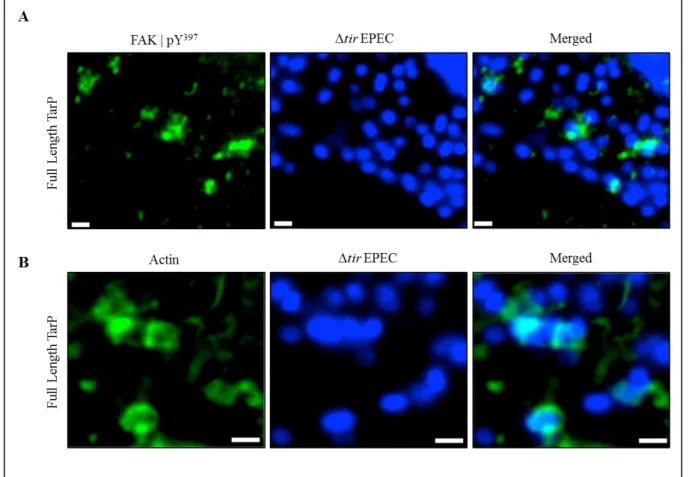
Upon adhesion of EPEC (Δtir) on to the surface of a TirMC/TarP-expressing cell, phospho-activated FAK localise directly underneath the bacteria (Figure 3A), as visualised by immunolabelling with an antibody specific for phospho-Y397-FAK (pY397-FAK) and
16
fluorescent confocal microscopy. Further analyses with phallodin staining also showed apparent actin recruitment (Figure 3B), demonstrating that in this context, GPIC TarP is sufficient to recruit activated FAK and actin. Functionality could be inferred from the phosphorylation state of FAK, with Tyr397 being the target of host tyrosine kinases, such as Src to initiate FAK activation (Westhoff et al., 2004). Interestingly, TarP of C. trachomatis L2 was also able to recruit phospho-active FAK and actin (Tristan et al., submitted). Both TarP orthologs shared the common characteristic of FAK recruitment indicating that a common motif may be responsible for the TarP-FAK interaction. The C-terminal domain of GPIC and L2 TarP are highly similar in amino acid sequence, and thus served as a good starting point for our search for a FAK-recruiting motif.
Figure 3: Phospho-active FAK and actin are recruited by Tarp from C. caviae GPIC clustered at the plasma membrane. A and B - Cos7 cells transfected with plasmids encoding TirMC/TarP were infected with EPEC Δtir to induce clustering. Localisation of phosphor-active FAK (A) and actin (B) are seen. Phospho-active FAK and actin (both green) were visualised with an anti-FAK (Y397) antibody and phalloidin, respectively. Bacteria were visualised by DAPI staining (blue). Scale bars = 1 µm.
17
Phospho-FAK recruitment is mediated by the LD domain of TarP
FAK contains a C-terminal domain termed FAT (Focal adhesion targeting) that interacts with the LD motif (LDxxLLxL) of signalling partners. Analysis of chlamydial TarP sequences showed a putative LD motif within the conserved C-terminal half of the protein downstream of the actin-binding WH2-like domains (ABDs) described by Jewett et al., (2010). Interestingly, Jewett et al. annotated this putative LD motif as a non-functional actin-binding domain because critical residues required for actin actin-binding have been altered to resemble an LD motif. For example, the GPIC ABD1 (Actin binding Domain 1) sequence (LQQILNNVREHL) differed in two amino acid positions from the GPIC LD (LExLLPxLRAHL), which were also critical in LD function. Letters in bold indicate the area of ABD1 that resembles the LD motif. Thus, it appears that the amino acid changes that abrogated actin binding led to a potentially new function - that of FAK binding (Figure 4).
Figure 4: TarP orthologs of C. trachomatis serovar L2 and C. caviae GPIC. Schematic representation of TarP orthologs. Phosphorylation domain (or tyrosine-rich domain) in grey, proline-rich domain in green, the putative actin binding domain in orange and the putative LD domain in red.
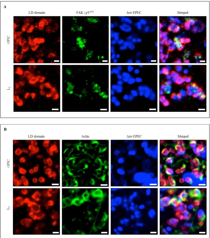
In order to demonstrate if this putative LD motif was responsible for the recruitment of FAK, EPEC (Δtir or ΔescN) Tir/MC system was used. Cos7 cells expressing TirMC/LD (LD domain from C. trachomatis L2 or C. caviae GPIC) were infected with EPEC, which upon adherence to the cell host, induced the clustering of TirMC/LD at the cell surface. After fixation, cells were stained for phospho-active FAK (Y397) and visualised for F-actin with phalloidin staining. Clustered TirMC/LD for either L2 or GPIC, for EPEC Δtir, showed that phospho-activated FAK and actin were strongly recruited (Figure 5 A and B). The same
18
results were observed for EPEC ΔescN (Figure 6 A and B).
Figure 5: TarP LD domain is functional. Cos7 cells expressing TirMC/LD, from GPIC, were infected with EPEC Δtir (A and B) to induce clustering. Localisation of phospho-active FAK (A) and actin (B) are seen. Phospho-active FAK and actin (both green) were visualised with an anti-FAK (Y397) antibody and phalloidin, respectively. Bacteria were visualised by DAPI staining (blue). Scale bars = 1 µm.
19
Figure 6: TarP LD domain is functional. Cos7 cells expressing TirMC/LD, from L2, were infected with EPEC ΔescN (A and B) to induce clustering. Localisation of phospho-active FAK (A) and actin (B) are seen. Phospho-active FAK and actin (both green) were visualised with an anti-FAK (Y397) antibody and phalloidin, respectively. Bacteria were visualised by DAPI staining (blue). Scale bars = 1 µm.
These results were consistent with the co-precipitation results (Thwaites et al., submitted), indicating that TarP LD domain alone was sufficient to induce the recruitment of phospho-activated FAK and actin.
20
II - Functional determination of LD domain oligomerisation
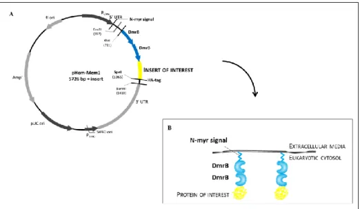
The EPEC system is an experimental approach to evaluate the recruitment to the membrane of signalling molecules involved in actin recruitment. However, despite our efforts to eliminate EPEC components by disabling either the TTSS function through deletion of the required ATPase (EscN), or the signalling molecule Tir, we could not discount existence of unknown or uncharacterised factors that may influence our experimental results. To address this concern another approach was utilised in order to investigate the recruitment of molecules such FAK directly from the LD domain. The system was the iDimerize™ Inducible Homodimer System (Clontech). The system included a vector called pHom-Mem1 that allows the localisation of a protein of interest beneath the plasma membrane through the N-terminal myristoylation (N-myr) motif for membrane targeting. The vector contains two sets of restriction sites that provided flexibility in regards to the placement of the dimerization domains (DmrB) - either at the N- or C-terminus of the protein of interest. Cloning into the
XbaI site places the DmrB domains at the C-terminus, whilst cloning into the SpeI site places
the DmrB domains at the N-terminus of the protein of interest.
The construct also included a hemagglutinin (HA) epitope tag (HA-tag) to enable monitoring of the expression of the fusion protein by immunofluorescence microscopy or Western blot (Figure 7). Also, a dimerization domain (DmrB) enables the formation of oligomers of the protein of interest by the addition of a small-molecule ―dimerizer‖ ligand (B/B homodimerizer) (Figure 7 and 8). The B/B homodimerizer is membrane permeant, and thus have access to the intracellular fusion proteins. The dimerizer is bivalent, allowing it to bind two different molecules of the fusion protein; and because each fusion protein has two dimerisation domains, the remaining DmrB domain could be crosslinked by the dimerizer to another molecule of the fusion protein, thus creating an oligomerised network of signalling proteins (Figure 8). This, in essence resembled the natural membrane-localised clustering of TarP during infection. Because oligomerisation depended on the addition of the B/B homodimerizer, this experimental system also provided control on the level of oligomerisation by varying the concentration of the dimerizer added and the time of incubation.
21
Figure 7: pHom-Mem1 vector as a model to replicate membrane-localised TarP clustering. A - Schematic representation of the pHom-Mem1 vector used to study the recruitment of signalling molecules by TarP. The protein of interest is directed to the membrane via N-terminal myristoylation (N-myr) and the localisation is possible by the hemagglutinin (HA) epitope tag (HA-tag) that is expressed at C-terminus of fusion proteins. B - Scheme with the fusion protein at the eukaryotic membrane.
Figure 8: Model to replicate membrane-localised TarP clustering. Schematic representation of fusion protein at the membrane. Upon addition of B/B homodimerizer induces formation of oligomers of the protein of interest. In the case of oligomerisation there is an activation of signal transduction pathway.
To generate the fusion proteins, the insert of interest was amplified by PCR using an appropriate template plasmid (C. trachomatis serovar L2 or C. caviae GPIC DNA). Both forward and reverse primers used for the PCR amplification were designed so that it contains a SpeI restriction sites (Table 2) at the 5’ and 3’ end to allow cloning of the recombinogenic PCR fragment in the pHom-Mem1 plasmid. The SpeI endonuclease was chose instead of XbaI to place the DmrB domains at the N-terminus of the protein of interest.
22
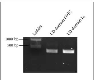
After amplification, the PCR products were analysed on a 1,2 % agarose gel and stained with SYBR safe. The expected sizes were 345 and 303 bp for LD domain of GPIC and LD domain of L2, respectively. Results are shown in Figure 9. In each lane (Figure 9) looks like doublets are present, but looking to the molecular weight we know that the band of interest is the brightest and the others can be justify by being artefacts of lane formation producing this type of band image.
Figure 9: Amplification of TARP inserts for cloning into pHom-Mem1. PCR-amplified inserts of TARP from two chlamydial species are displayed on a 1,2 % agarose gel, stained with SYBR safe.
PCR fragments (LD domain GPIC and LD domain L2) were then gel-purified. The pHom-Mem1 plasmid and the purified inserts were digested with SpeI restriction enzyme, which cuts the DNA to form sticky ends allowing ligation of DNA sequences. The success of the plasmid digestion was confirmed initially on a 1,2 % agarose gel of cut (linearized) and uncut (circular) plasmid shown in Figure 10 and from it is possible to confirm that the restriction enzyme worked correctly. In the cut plasmid one band was expected around 5726 bp whereas in the uncut plasmid three bands were expected due to the correspondence of the relaxed circular form, plasmid is linearized and the supercoiled circular DNA. However, in the uncut result one band because the plasmid only shown supercoiled form.
23
Figure 10: Analysis of plasmid digestion to confirm the functionality of SpeI. In the digested plasmid lane one band clearly appears, demonstrating a good cleavage by SpeI. In the undigested lane, one bands appeared due to the fact that the DNA had only the supercoiled form.
A ligation was performed between each insert and Mem1. Recombinant pHom-Mem1 vectors (pHom-pHom-Mem1+LD GPIC or pHom-pHom-Mem1+LD L2) were transformed into
E.coli Top10 strains. These strains allow high transformation efficiency (1 x 10⁹ CFU/µg supercoiled DNA) and are ideal for high-efficiency cloning and plasmid propagation.
The pHom-Mem1 vector has an Ampr cassette (Figure 7) allowing the selection of transformed bacteria in plates containing LB media with ampicillin (100 µg/ml). Several clones have been selected, tested by PCR for the presence of the insert and analysed in a 1,2 % agarose gel, stained with SYBR safe. The results obtained (Figure 11) show some positive colonies for the presence of the insert but others did not contain the insert of interest, which possibly arose from self-ligation to reconstitute the original vector. The expected sizes were 345 and 303 bp for LD domain of GPIC and LD domain of L2, respectively.
24
Figure 11: Colony PCR of transformants of pHom-Mem1. Colony PCR was performed on ampicillin-selected transformants and the 1,2% agarose gels displayed here show the presence of the inserts of each
Chlamydia species in the pHom-Mem1 vector, highlighted with a white box.
Overnight cultures were started in LB medium and incubated overnight for all the positive colonies. Plasmid DNA was extracted from all the overnight cultures using the miniprep kit (Qiagen) and sent for sequencing (GATC Biotech) to confirm the correct inserts.
The primers used for sequencing was CMV-F universal primer,
CGCAAATGGGCGGTAGGCGTG (5’ - 3’), and the respective forward primer of each species and domain (Table 2). Sequencing was a procedure required to make sure that the insert did not have any mutations and to assure that the insert was fused in the right direction within the plasmid. The sequencing results were analysed using Geneious Basic 5.5.3 (See insert sequencings in Appendix A1).
The colonies confirmed to have the insert in the correct orientation and location were stored at -80 ºC in 30 % glycerol. Also, these positive constructs were used for subsequent experiments.
25
LD domain oligomerisation mediates phospho-FAK recruitment
As mentioned previously, the EPEC system does not exclude unknown factors that may mediate the recruitment of phospho-FAK and/or actin. To address this question, the optimized iDimerize™ Inducible Homodimer System protocol was used.
To investigate the possible recruitment of phospho-FAK by the LD domain, Cos7 cells were transfected with pHom-Mem1+GPIC LD or pHom-Mem1+L2 LD vectors, 100 nM of B/B homodimerizer was added for 30 minutes to induce oligomerisation of the protein of interest, except in the controls where no B/B homodimerizer was added. Microscopic examination of transfected cells using an anti-HA-tag antibody revealed the recruitment of phospho-activated FAK directly underneath the non-uniform LD domain patches in the plasma membrane of cells treated with B/B, whereas in the controls, despite the uniform localisation of the LD domain at the plasma membrane, no recruitment was observed (Figure 12). Importantly, Cos7 cells in the absence of TarP LD domain (untransfected cells), but treated with the homodimerizer, showed no effect on the subcellular localisation of phospho-active FAK (Figure 13). This was important, as it demonstrated that the membrane localisation of phospho-FAK could be attributed to the oligomerised LD domain.
26
Figure 12: LD domain of TarP oligomerised in the membrane is sufficient to recruit phospho-active FAK. A and B - Transfected Cos7 cells were treated with 100 nM B/B homodimerizer ligand to induce the oligomerisation of fusion proteins containing LD domain of C. trachomatis L2 or C. caviae GPIC. Phospho-active FAK (green) recruitment was dependent on the addition of the B/B homodimerizer in both LD domains (red). Phospho-active FAK was visualised with an anti-FAK (Y397) antibody and LD motif with an anti-HA-tag antibody. Scale bar = 5 µm.
27
Figure 13: B/B homodimerizer does not interfere with subcellular localisation of phospho-active FAK. Untransfected Cos7 cells treated with 100 nM of B/B homodimerizer shown no influence on the subcellular localisation of phospho-active FAK. Phospho-active FAK was visualised with an anti-FAK (Y397). Scale bar = 5 µm.
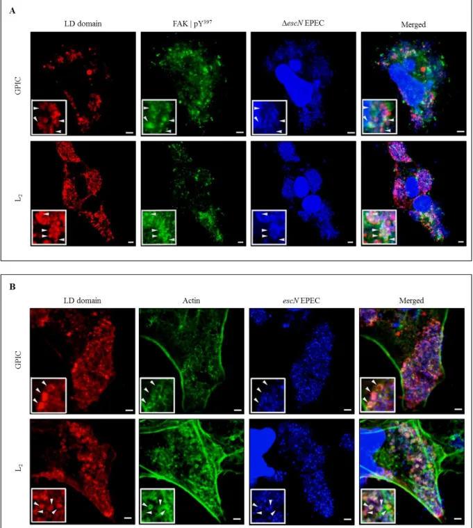
Overall, phospho-active FAK recruitment to TarP LD domain oligomerised indicates recruitment of FAK signalling and may provide a new mechanism for LD to recruit actin. However, this system was not suitable to investigate the recruitment of actin in response to the oligomerisation since signals associated with endogenous cortical actin were difficult to distinguish from the signal triggered by the LD domain.
LD domain oligomerisation promotes Arp2/3 complex recruitment
Eukaryotic cells have the ability to regulate the actin cytoskeleton, which is important for processes like cell locomotion, phagocytosis and intracellular motility of lipid vesicles. The cell motility is driven by key component that in the case of cell motility regulation is through a seven-subunit protein called Arp2/3, a complex localised in the lamellipodia (protrusion structure on the edge of the cell). This protein constitutes one of the most important nucleators of new actin filaments (Pollard, 2007). Arp2/3 binds to the side of an
28
existing actin filament and initiates polymerisation leading to branched F-actin network (Rouiller et al., 2008). Since the presence of Arp2/3 is related to dynamic actin remodelling, we tried to establish a link between the recruitment of Arp2/3 to the recruitment of actin.
In order to do so, LD domain of the strains GPIC or L2 were transfected into Cos7 cells to detect Arp2/3 colocalisation to LD oligomeric patches on the plasma membrane. Both proteins of interest, localised in the eukaryotic membrane linked to two dimerisation domains were oligomerised with 0,5 nM B/B ligand. Transfected cells stained with the anti-HA-tag antibody showed Arp2/3 complex underneath the LD domain in both GPIC and L2 strains. In the absence of the oligomerisation inducer, despite the presence of the LD domains in the membrane, no recruitment was seen (Figure 14). Again, Cos7 cells in the absence of TarP LD domain (untransfected cells) and with or without homodimerizer ligand showed no effects on the subcellular localisation of Arp2/3 (Figure 15).
29
Figure 14: LD domain oligomerised recruits Arp2/3 complex. Cells transfected with or without B/B treatment. A and B - Cos7 cells expressing LD domain from GPIC or L2 were treated or not with 0,5 nM of oligomerisation inducer. Arp2/3 complex was recruited only when LD domain (red) was oligomerised (arrow head) despite the some localisation of Arp2/3 staining (green) in the membrane of cells without oligomerisation ligand (arrow) due to the formation of lamellipodia. Arp2/3 was visualised with an Anti-p34-Arc\Arpc2 antibody and LD motif with an anti-HA-tag antibody. Scale bars = 5 µm.
30
Despite the clear difference between the cells with and without treatment with B/B homodimerizer in some areas of the membrane, it appears that even without B/B-induced oligomerisation, the Arp2/3 complex could be observed in some regions of the membrane (Figure 14, white arrows). Note that these areas show a uniform, rather than patched staining of the LD domain. The same localisation on the membrane was also seen in the untransfected cells (Figure 15). As described recently by Suraneni et al. (2012) Arp2/3 complex localises to the actin-rich lamellipodia of spreading or polarized cells and is a critical protein in the formation of these structures. Taking these findings into account we believe that the localisation of the Arp2/3 complex in cells treated without B/B is due to the presence of a lamellipodia formation at that particular location of the plasma membrane. To confirm this, in future experiments, a double staining for Arp2/3 complex and Rac or Wave2 proteins (important key components in the lamellipodia formation) of LD domain-expressing cells and their respective localisation at the membrane should support or refute this idea.
Figure 15: B/B homodimerizer does not interfere with subcellular localisation of Arp2/3 complex. Untransfected Cos7 cells treated with 0,5 nM of B/B homodimerizer shown no influence on the subcellular localisation of Arp2/3. Arp2/3 was visualised with an Anti-p34-Arc\Arpc2 antibody. Scale bar = 5 µm.
31
Our results showed that, phospho-active FAK and Arp2/3 complex were both recruited in cells transfected with C. caviae GPIC or C. trachomatis serovar L2 LD motif. These observations indicated that the chlamydial LD motif resembling LD domain of paxillin was able to recruit phospho-active FAK. Also, this observation confirmed the previous observations by Coombes and Mahony (2002) where rapid phosphorylation upon C.
pneumoniae entry was shown. In this report, we extended it by demonstrating TarP LD
motif-mediated recruitment. This potential interaction of FAK with the invasion-associated effector TarP further implicated FAK in invasion.
Additionally, our data demonstrating Arp2/3 recruitment to oligomerised LD domain molecules suggested subsequent actin recruitment, an activity of the LD domain already shown in the EPEC TirMC system. Therefore, it is highly likely that the LD domain and FAK constitute a signalling pathway to activate the Arp2/3 complex and thus induce actin polymerization and remodelling. The TarP LD motif could indeed be involved in a novel pathway to facilitate chlamydial invasion.
LD domain oligomerisation mediates vinculin recruitment
Integrins present at the membrane cell are linked to the actin cytoskeleton by the focal adhesion complex. This complex is composed of a number of molecules, which include FAK, vinculin, talin, and paxillin amongst others. Since the results above showed that TarP recruited activated FAK, we investigated the possibility of other focal adhesion-associated molecules, specifically vinculin, being associated with the LD domain oligomeric complexes at the plasma membrane. Vinculin has the ability to interact with integrins and is a critical control of cytoskeleton mechanisms. For this reason, we investigated the recruitment of vinculin from LD domain of TarP.
Vectors pHom-Mem1+GPIC LD or pHom-Mem1+L2 LD were transfected into Cos7 cells. Upon addition of the B/B homodimerizer to induce oligomerisation of the LD domain, transfected cells stained against HA-tag showed recruitment of vinculin and co-localising with the LD domain oligomeric patches on the plasma membrane. In the absence of oligomerisation, vinculin was not recruited by the LD domain (Figure 16). Untransfected cells (lacking TarP LD domain) treated with homodimerizer showed no effects on the subcellular localisation of vinculin (Figure 17). Taken together, the data indicate that the LD domain also
32
has the ability to recruit vinculin, suggesting that the signalling complex induced by this motif may resemble the signalling pathway originating from clustered integrin molecules.
Figure 16: LD domain oligomerised recruits vinculin. A and B - Cos7 cells expressing LD domain from GPIC or L2 were treated or not with 100 nM of oligomerisation inducer. Vinculin (green) was recruited only when LD domain (red) was oligomerised. Vinculin was visualised with an Anti-vinculin antibody and LD motif with an anti-HA-tag antibody. Scale bars = 5 µm.
33
Figure 17: B/B homodimerizer does not interfere with subcellular localisation of vinculin. Untransfected Cos7 cells treated with 100 nM of B/B homodimerizer shown no influence on the subcellular localisation of vinculin. Vinculin was visualised with an Anti-vinculin antibody. Scale bar = 5 µm.
Localisation of the LD domain to focal contacts
In the process to investigate the recruitment of actin by the LD domain of TarP, which was not possible due to the presence of pre-existing cortical actin in the membrane, we noticed an interesting subcellular localisation of the LD domain, when expressed in cells (Figure 18). Regardless of the presence of the B/B homodimerizer, a subset of the LD domain molecules could be observed consistently to localise to tips of the stress fibres; the same location where focal complexes (FAK, vinculin, talin, etc.) are regularly found. This phenomenon could be observed in the absence of the dimerizing molecule suggesting that this was not an artefact induced by the dimerizer (Figure 18). Looking at the untransfected cells, there were no indications of actin cytoskeleton abnormality, at least under these conditions when the LD domain expression was limited by treatment with cycloheximide (Figure 19).
What could be the significance of this localisation to the tips of the stress fibres? The results for the localisation of LD domain of GPIC or L2 in the extremity of actin imply that the LD motif may have a post-invasion role. C. trachomatis was described to disturb cell–cell
34
contacts between polarized epithelial cells in culture (Wyrick et al., 1989). This event was suggested to be part of C. trachomatis to decrease the protection of cells from anti-chlamydial antibodies, facilitating bacterial dissemination. More recently another study was consistent with these results where, Prozialeck et al. (2002), showed that cervical epithelial cells separate from each other as a result of C. trachomatis infection. Other bacteria have been reported to interfere with components of adhesion like Salmonella typhimurium. Salmonella infection decreased transepithelial electrical resistance and increased paracellular permeability. This process is thought to facilitate cell-to-cell spreading of these pathogens (Sears et al., 2000). Our results showed a specific localisation of LD domain molecules to the tips of actin stress filaments, which may indicate involvement in the cell extrusion process reported for Chlamydia-infected cells during infection of the ocular conjunctiva and genital mucosa (Rank et al., 2008). To address this question, we would need to observe structural disruptions of the focal contacts, stress fibres, and intercellular junctions. The latter has been reported for Chlamydia infected monolayers through the degradation of the tight junction protein, nectin-1 (Sun et al., 2008).
35
Figure 18: LD domain oligomerised specifically binds to the tips of actin filaments. A and B - Cos7 cells expressing LD domain from GPIC or L2 were treated or not with 100 nM of oligomerisation inducer. LD domain (red) was localised in the tip of actin filament (green) in the presence or absence of B/B homodimerizer. Actin was visualised with phalloidin antibody and LD motif with an anti-HA-tag antibody. Scale bars = 5 µm.
36
Figure 19: B/B homodimerizer does not interfere with subcellular localisation of actin. Untransfected Cos7 cells treated with 100 nM of B/B homodimerizer shown no influence on the subcellular localisation of actin. Actin was visualised with phalloidin antibody. Scale bar = 5 µm.